Fecal artifcats
 Call Kai
 Learn
Learn Practice Test
 Spaced Repetition
 Match
Match Flashcards
 Knowt Play
1/52

There's no tags or description
Looks like no tags are added yet.
Name | Mastery | Learn | Test | Matching | Spaced | Call with Kai | Chat |
|---|
No analytics yet
Send a link to your students to track their progress
53 Terms
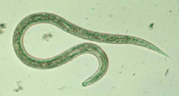
• ”ladder-like” appearance
• May be confused with helminth larva because of shape and size
• Lacks head or tail region
Vegetable Spiral


• May be confused with helminth larva (Hookworms and Strongyloides) because of shape and size
• Lacks head or tail region
• They are often broken at one end
• Have a refractile center
• Lack internal structures seen in true parasites
Plant Hair/Fiber


• Resemble Taenia other helminth eggs
• Has no little to no internal structures
• Lacks 6-hooked oncosphere lack of embryo wall
Pollen

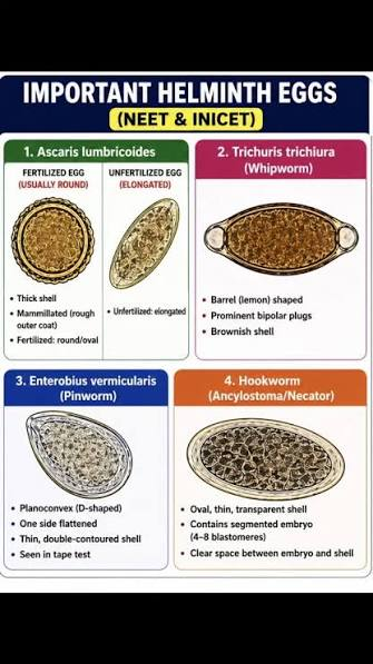
• May be confused with helminth eggs
• Appear in larger in sizes compared to helminth eggs
• Polygonal and flattened
• Interior portion is unorganized and contain large vacuoles
Vegetable Cells

are chains of elongated yeast cells that remain attached after budding
Pseudohyphae
Seen in improperly prepared wet mounts/smear

• Rectangular in shape
• With or without striations
• Normally present especially in high red-meat diet
May be confused with tapeworm proglottid
Muscle Fiber

Presence of too much muscle fibers
“creatorrhea”
Exception for high amounts of Muscle Fibers
seen in biliary obstruction and gastrocolic fistulas
in patients with pancreatic insufficiency, such as in cases of cystic fibrosis

• Spherical in shape
• Resemble protozoan cysts
• Large amounts may indicate presence of excess fecal fat (steatorrhea)
Normal samples contain 100 small droplets, <4 um in diameter per HPO
Fat globules

Condition for “Steatorrhea”
Observation of >60 large, 6 to 75 um large droplets per HPO = steatorrhea

May be confused with Entamoeba histolytica and Entamoeba dispar cysts due to size, segmentation, and similar trichrome staining characteristics
Size averages 14 μm (10 to 12 μm on permanent stained smears)
Displays a high nuclear-to-cytoplasmic ratio (1:1)
Contains 2 to 4 nuclear segments connected by narrow chromatin strands (which can mimic separate nuclei)
Features granular cytoplasm
PMNs


May be confused with E. histolytica / E. dispar trophozoites (size, trichrome staining)
Size: 30–60 μm (5–10 μm smaller on permanent smears)
Nuclear-to-cytoplasmic ratio: 1:4 to 1:6
Single, large, irregular nucleus (monocyte-like; may be absent)
Ingested material: Debris, PMNs, RBCs, red-staining round bodies
Macrophages
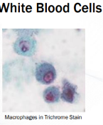

May be confused with macrophages during microscopic examination
Size ranges from 12 to 60 μm, averaging 20 μm (smaller on permanent stained smears)
Displays a nuclear-to-cytoplasmic ratio of 1:10 to 1:12
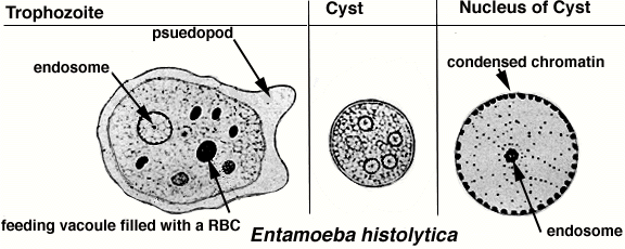
Entamoeba histolytica/dispar

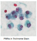
May be confused with PMNs during microscopic examination
Size averages 20 μm (smaller on permanent stained smears)
Displays a nuclear-to-cytoplasmic ratio of 1:2 to 1:3 for cysts (1:10 to 1:12 for trophozoites)
Characterized by a round nucleus with a central karyosome and peripheral chromatin
Contains uniform, agranular cytoplasm (may contain ingested RBCs)
Stains with green cytoplasm and dark red nuclear material on Trichrome stain
Entamoeba histolytica/dispar cyst
• These are formed from the break-down products of eosinophils
• Seen in stool or sputum
• Slender crystals with pointed ends “diamond-shaped”
• Stain red-purple with trichrome stain
• May indicate hypersensitivity/allergic reaction and parasitic helminth infections
Charcot Leyden Crystals

With large, refractile nucleus.
With distinct cell borders
• Look like amebic trophozoites
• Lack the typical interior structures of amebic trophozoites
Epithelial Cells Squamous epithelial cells (from anal mucosa) & Columnar epithelial cells (from intestinal mucosa


• Round to irregular round-shaped
• Very refractile
• Has no internal structure or nucleus
• May resemble protozoan cysts (E. hartmanni & E. nana)
Starch cells/ Granules


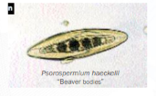
• Protistan parasites of crayfish
• Present in stool if crayfish is consumed in significant amounts
• Aka Corpora parasitica
• Resemble helminth ova
• No clinical significance
Beaver Bodies

Parasite or Artifact? Identify.
Artifact- Beaver Bodies

Parasite or Artifact? Identify.
Artifact- Beaver Bodies

Parasite or Artifact? Identify.
Artifact- Fat globules
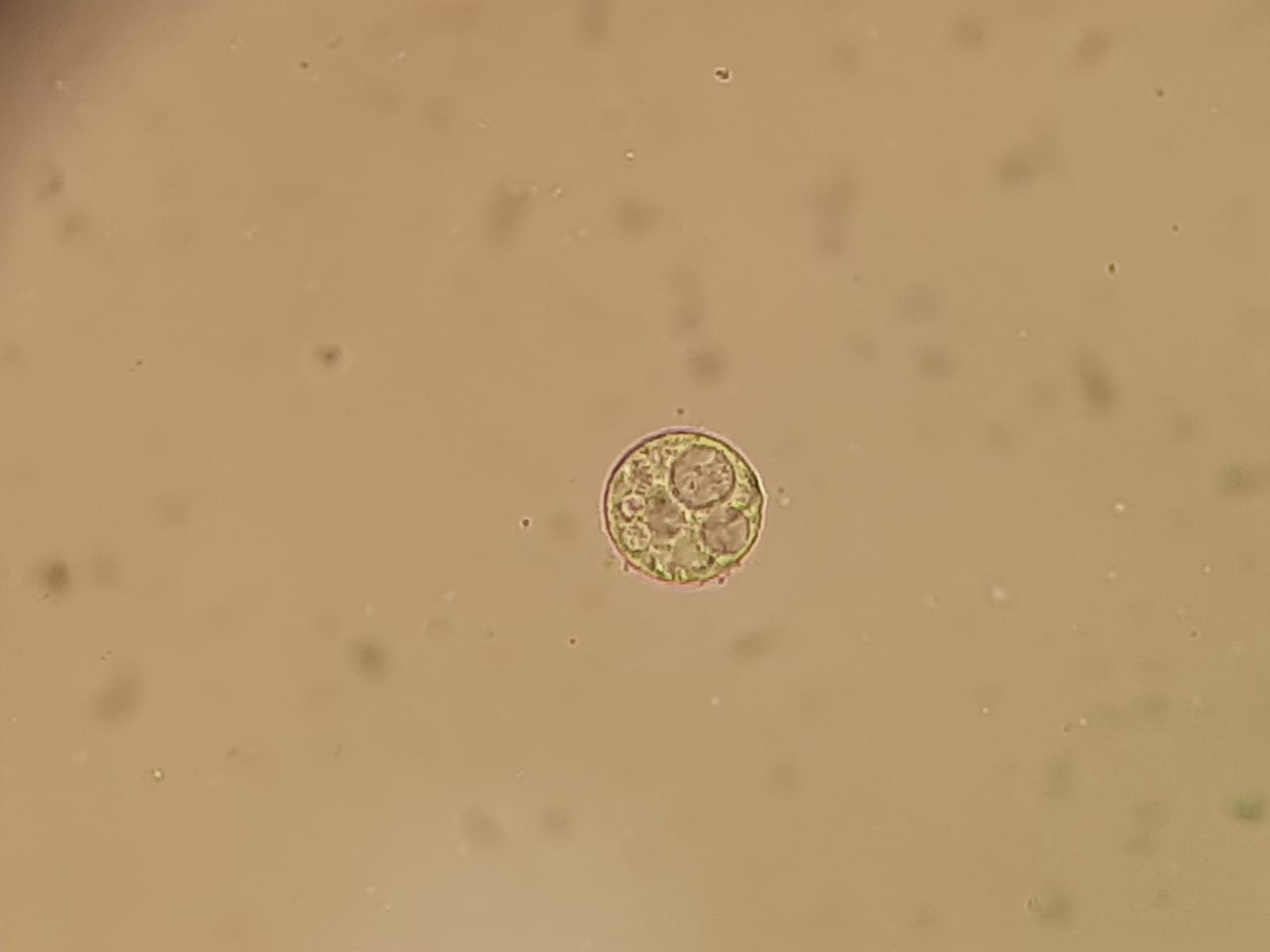
Parasite or Artifact? Identify.
Artifact- Fat Globules in stool

Parasite or artifact? Identify
Artifact- Starch cells
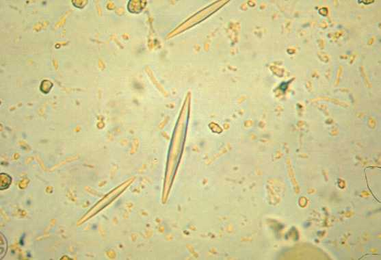
Parasite or Artifact? Identify.
Artifact- Charcot Leyden Crystals
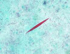
Parasite or Artifact? Identify.
Artifact- Charcot Leyden Crystals

Parasite or Artifact? Identify.
Artifact- Muscle Fibrils

Parasite or Artifact? Identify.
Artifact- Muscle Fibrils

Parasite or Artifact? Identify.
Artifacts- Air Bubbles

Parasite or Artifact? Identify.
Artifact- Yeast cells

Parasite or Artifact? Identify.
Artifact- Yeast cells

Parasite or Artifact? Identify.
Artifact- Vegetable spirals
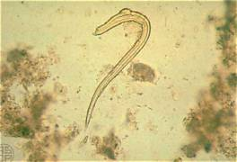
Parasite or Artifact? Identify.
Artifact- Plant Fibers

Parasite or Artifact? Identify.
Artifact- Plant Fibers

Parasite or Artifact? Identify.
Artifact- Pollen

Parasite or Artifact? Identify.
Artifact- Pollen
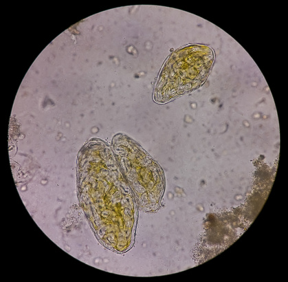
Parasite or Artifact? Identify.
Artifact- Vegetable Cells
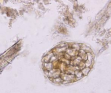
Parasite or Artifact? Identify.
Artifact- Vegetable cells
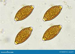
Parasite or Artifact? Identify.
Parasite- Helminth Eggs (Trichuris trichuria)
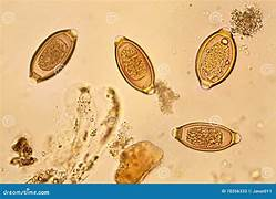
Parasite or Artifact? Identify.
Parasite- Helminth Eggs (Trichuris trichuria)

Parasite or Artifact?
Parasite (Enterobius vermicularis)

Parasite or artifact
Parasite (Taenia saginata egg)

Parasite or artifact
Parasite (Ascaris lumbricoides egg)

Parasite or artifact
Parasite (Ascaris lumbricoides)

Parasite or Artifact
Parasite (Hymenolepis diminuta formerly nana)

Parasite or Artifact
Parasite (Strongyloides stercoralis)
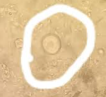
Parasite or Artifact
Parasite (E. histolytica)

Parasite or Artifact
Parasite (Giradia duodenalis cyst)

Parasite or Artifact? Identify.
Parasite. Cryptosporidium oocyst

Parasite or Artifact? Identify.
Parasite (Blastocystis hominis)
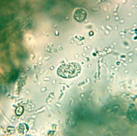
Parasite or Artifact? Identify.
Parasite (Endolimax nana)

Parasite or Artifact? Identify.
Parasite (Entamoeba coli)