Week 2 - Neuromuscular
 Call Kai
 Learn
Learn Practice Test
 Spaced Repetition
 Match
Match Flashcards
 Knowt Play
1/150

There's no tags or description
Looks like no tags are added yet.
Name | Mastery | Learn | Test | Matching | Spaced | Call with Kai | Chat |
|---|
No analytics yet
Send a link to your students to track their progress
151 Terms
CNS - components (2)
brain and spinal chord
PNS - components
nerves connecting the central nervous system to the body
Includes neuronal clusters (ganglia) at specific locations along peripheral nerves
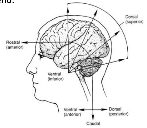
Naming of human head positions
Humans rostro-caudal = towards nose and tail bone -> back and front = dorsal and ventral
CNS of humans has a 90° bend because of cephalic flexure -> anything above the midline of the head has different names
cells of CNS - list (4)
can be loosely categorised into neurons (functional units) or glial cells
Astrocyte -> important components of synaptic transmission and in blood brain barrier
Microglia -> type of immune cell that plays surveillance role for dyeing cells or infections
Oligodendrocyte -> form myelin that insulates axons so action potentials can travel faster
Neuron -> functional cells
major regions of CNS - list (4)
Cerebrum
Diencephalon -> hypothalamus, thalamus, pineal gland (part of epithalamus)
Brainstem -> mid brain, pons, medulla, oblongata
Cerebellum
major folds of brain - list (4)
Longitudinal fissure separates left and right hemisphere
Central sulcus essentially divides brain into front and back bit (somewhat midline) -> can be followed from longitudinal fissure to down laterally
Parietal-occipital sulcus = separates parietal and occipital lobes
Transverse fissure defines location of temporal lobe
major folds of brain - pre and post central sulcus
Postcentral gyrus = after central sulcus -> location of primary sensory cortex
All sensory info ends up in postcentral gyrus
Precentral gyrus = before central sulcus -> location of primary motor cortex
Ability to move muscles comes from neurons in precentral gyrus
folds of cerebellum
undulations called folia instead of gyri like in rest of brain → looks like patterns on leaves
role of insular cortex
important part of limbic nervous system → emotions
role of corpus callosum
composed of axons that allow for communication between left and right hemispheres
Grey and white matter (2 grey 1 white)
Grey matter cortex = location of cell bodies of neurons -> not myelinated
Grey matter nuclei (basal ganglia or basal ganglia) -> pockets of grey matter deeper in the brain
Location of where electrodes would be placed to treat someone with Parkinsons
White matter = composed of myelinated axons
Oligodendrocytes (glial cells) provide insulation to nerve axons and gives them a white appearance
layers protecting the brain - list (4)
Skin of scalp
Periosteum -> lines bone
Bone of skill
Meninges and cerebral spinal fluid
meninges and spaces in-between- list (5)
dura mater
subdural space
arachnoid mater
subarachnoid space
pia mater
meninges and spaces in-between - dura mater (3)
Directly under skull
Very thick, tough, fibrous structure
Different folds of the dura mater sit within fissures of the brain
meninges and spaces in-between - folds of dura mater (3)
Falx cerebri = dural fold that sits in longitudinal fissure -> separates hemispheres and provides protection
Tentorium cerebelli = dural fold that sits in space between cerebellum and cerebrum (transverse sinus)
Falx cerebelli = dural fold that sits between hemispheres of cerebellum
meninges and spaces in-between - importance of subdural space
Dura mater is associated with lots of blood vessels -> if one were to rupture, blood would spill into the subdural space (subdural bleed)
Because dura mater does not expand, pressure forms on brain
meninges and spaces in-between - subarachnoid space
location of CSF
meninges and spaces in-between - pia mater
Very think and fine membrane that sits on surface of brain → hard to separate from brain
superior saggital sinus - summary
large blood vessel within dura mater
Arachnoid villus protrudes from arachnoid mater into superior sagittal sinus → important for reabsorption of CSF into vasculature
production and re-absorption of CSF - steps (4)
Blood flows into choroid plexus -> specialised capillary bed in vascular tissue
When capillary bed fills with blood, some of the plasma can be filtered via gaps between endothelial cells into the ventricles to form CSF
As ventricles fill, CSF eventually finds its way out into the subarachnoid space to bathe the brain
As CSF enters into arachnoid villi (granulations) and pressure increases, CSF is forced back into blood circulation
CSF - def
fluid filtrate from blood that bathes the brain and protects it by providing cushioning
Production occurs within ventricular system of central nervous system
consequence of CSF not being reabsorbed in adults
increase in pressure in subarachnoid space -> pressure exerted onto neural tissue -> widespread neurological problems
consequence of CSF not being reabsorbed in children - born without arachnoid granulations (4)
Results in condition called hydrocephalus (water on brain)
In children, sutures of the skull are not fused yet -> skull can expand resulting in an enlarged head
Treated by catheter inserted into lateral ventricles that drains CSF from ventricles into another vascular bed
Drainage tube usually introduced into peritoneal cavity with extra length to allow for growth of child
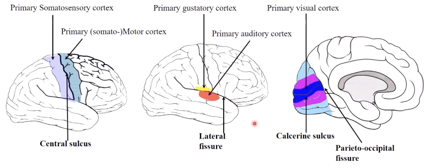
organisation of cerebral cortex
most primary cortices are located around deepest sulci/ fissures
Central sulcus -> surrounded by primary somatomotor and somatosensory cortices
Lateral fissure -> surrounded by primary auditory and gustatory cortices
Calcarine sulcus -> surrounded by primary visual cortex
brainstem - location
between diencephalon and spinal chord
brainstem - major divisions (3)
Midbrain -> mesencephalon
Pons -> "the bridge" between the rest of the brain stem and the cerebrum and cerebellum
Medulla oblonga -> myelencephalon = bulbus
protection of spinal chord
Meninges and CSF in sub-arachnoid space protect the spinal chord as well as the brain
spinal chord segments - list (5)
cervical
thoracic
lumbar
sacral
cocygeal
enlargement of spinal chord segments (4)
Cervical and lumbar portions are enlarged compared to thoracic portion → have nerves that extend into limbs
more cells in that area = increased diameter of chord
Cervical = nerves to upper limbs
lumbar = nerves to lower limbs
end of spinal chord
ends at L2
structure called conus medullaris
end of spinal chord - cauda equina (3)
Nerves extend down within the vertebral column and exit at each vertebral level until one single fibre is left (filum terminale) -> attaches to coccyx
Filum terminale is not a nerve but an extension of the pia mater
Bundle of nerves that extend down = cauda equina
spinal cord development - process (3)
First trimester: spinal chord fills enter vertebral column
later in embryonic development: chord starts to creep up vertical colum until it gets to around L2 → muscoskeletal growth occurs at faster rate than elongation of spinal cord
nerves that give rise to peripheral nerves that go into tissues stretch to accomodate and elong with vertebral coumn → forms cauda equina
spinal cord - lumbar puncture (2)
collection of CSF from thecal sac that surrounds spinal chord
Because spinal chord ends around L2, safer to collect CSF from subarachnoid space just below level of Conus medullaris -> between L3 and L4 without damaging spinal chord
grey and white matter of spinal chord
Grey matter found deeper within white matter of spinal chord -> arranged in butterfly shapes
grey and white matter of spinal chord - grey matter components (3)
Posterior (dorsal) horn = horn on posterior surface
Anterior horn = horn on ventral surface
Lateral horn = sticks out on lateral aspects
grey and white matter of spinal chord - white matter components (3)
Posterior funiculus
Lateral funiculus
Anterior funiculus
grey and white matter of spinal chord - white matter arrangement (3)
Efferent pathways = descending
Afferent pathways = ascending

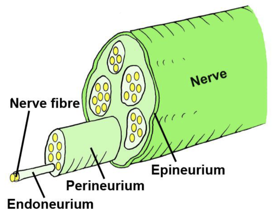
nerves - def and structure (3)
collections of nerve fibres (axons) within external connective tissue sheath (epineurium)
Axons grouped into fascicles within nerves and surrounded by perineurial sheath → can be either sensory or motor axons
Each nerve fibre surrounded by endoneurium
types of nerves (2)
Spinal nerves = nerves that arise from the spinal cord
Cranial nerves = nerves that arise from the brain or brain stem
spinal cord - location of neuron cell body
cell body of motor neurons = found within boundaries of CNS
cell body of sensory neurons = just outside boundary
PNS - types of neurons (2)
Sensory (afferent) neurons = periphery neurons that carry signals to the CNS
Motor (efferent) neurons = peripheral neurons that carry signals from the CNS
connection of neurons to CNS (3)
Separate spinal roots connect efferent and afferent neurons to the CNS
Ventral surface of spinal chord = axons of motor nerves -> ventral root
Dorsal surface of spinal chord = axons of sensory nerves -> dorsal root
Ventral horn contains ventral root -> dorsal horn contains dorsal root
Dorsal root ganglia = cell body of sensory neurons and their axons -> extend into the periphery and into dorsal horn of grey matter in spinal chord
spinal reflex - steps (5)
Sensory receptor
Sensory neuron
Interneuron
Motor neuron
Effector organ
pairings of spinal nerves - list (31)
8 cervical
12 thoracic
5 lumbar
5 sacral
1 coccygeal
number of vertebral bones to spinal nerve pairings
30 bones → 31 spinal nerve pairs
Cervical nerve 1 arises above vertebral bone C1 etc → cervical nerve 8 arises below vertebral bone C7
T1 is below the T1 vertebra etc
branching of dorsal and ventral roots
form spinal nerves → branch into dorsal or ventrla ramus
rami - def
branches of mixed spinal nerve -> contain both sensory and motor axons in contrast to dorsal and ventral root
dorsal and ventral rami
Dorsal ramus wraps around back of body to innovate all muscle and skin on posterior side of anatomy
Ventral ramus wraps around front of body to innovate all anterior structures
segmental innervation - def
concept that each spinal segment have a spinal nerve associated with it will have a distinct dermatome and a distinct myotome
Each spinal nerve will innovate a very defined region of the body -> each of the nerves that is part of the cord will have a very define pattern of innovation
clinical importance of segmental innervation
To identify levels of functional deficits due to nerve damage
segmental innervation - dermatome meaning and face
area of skin that is mainly supplied by a single spinal nerve
Face doesn’t have spinal dermatome -> nerves supplied by cranial nerves
No C1 dermatome as it innovates the meninges and other structures excluding the skin
segmental innervation - myotome
group of muscles that a single spinal nerve innervates
cranial nerves - mnemonic
Oh, Oh, Oh, To Touch And Feel Very Glossy Velvet, Ah
cranial nerves - cerebral hemisphere (2)
Olfactory
Primary sensory nerve for smell
Optic
Primary sensory nerve for vision
cranial nerves - midbrain (2)
Oculomotor
Eye movement (not vision)
Trochlear
Eye movement
cranial nerves - pons (4)
Trigeminal
Major sensory nerve for face (cranial nerve dermatome)
Abducens
Eye movement
Facial
Facial muscle control -> facial expression
Vestibulo-cochlear -> part of both pons and medulla
Nerve goes to inner ear for balance and hearing
cranial nerves - medulla (5)
Vestibulo-cochlear -> part of both pons and medulla
Glossopharyngeal
Sensory innovation to tongue for taste and to pharynx for other sensations like pain
Vagus
Major parasympathetic nerve
Innovates all internal organs and parts of gastrointestinal systems
Accessory
Innovates things in the neck like muscles
Hypoglossal
Movement of tongue
cranial nerves - sensory, motor or both
Not all nerves are mixed -> some are purely motor whilst others purely sensory
“Some Say Marry Money, But My Brother Says Big Brains Matter More”
somatic nervous system - def
controls voluntary movement of muscles and innovates skin in terms of sensory component
autonomic nervous system - def
effector system in CNS and PNS mediating unconscious homeostatic control of organ and body physiology
Can be divided into parasympathetic and sympathetic
somatic vs autonomic nervous system
somatic = voluntary
autonomic = involuntary
both contain efferent and afferent neurons
somatic motor neurons - location of cell body summary (1)
cell bodies in ventral horn of spinal cord give rise to axons that project to muscle
somatic motor pathway (2)
Pre-ganglionic neurons arise from anterior root with cell bodies in anterior column
Axons extend down peripheral or anterior ramus
autonomic neural pathway - summary
often has two neurons in the pathway
Neuron that resides in spinal chord (pre-ganglionic neuron) projects out
pre-ganglionic neuron synapses to a second neuron (post-ganglion neuron) in the chain before controlling tissue of interest
true for parasympathetic and mostly true for sympathetic (sympathetic nervous system also uses hormones like those from the adrenal gland)
autonomic neural pathway - ganglion
Connection between the neurons happens in the ganglion -> collections of cell bodies of the autonomic nervous system
autonomic system - axon length of parasympathetic vs sympathetic
Parasympathetic: pre-ganglion neuron has really long axon because ganglion is located in or very near target tissue
Sympathetic: pre-ganglion neuron has short axon because ganglion sits right next to spinal chord -> post-ganglion neuron has very long axon to reach tissue
autonomic nervous system - thoracolumbar outflow and craniosacral outflow
Sympathetic autonomic nervous system: pre-ganglionic neurons arise from either the thoracic or lumbar parts of the spinal chord
Parasympathetic autonomic nervous system: pre-ganglionic neurons arise form cranial nerves or sacral nerves within spinal chord
division of autonomic nervous system based on origin from spinal chord
thoracolumbar outflow or craniosacral outflow depending on where they arise from
sympathetic autonomic motor pathway - steps (5)
Pre-ganglionic neurons arise from lateral horn rather than ventral horn
Pre-ganglionic neuron exits spinal chord via ventral roots into mixed spinal nerve
Pre-ganglionic neuron enters sympathetic ganglion via white ramus rather than continuing like somatic motor pathway
Pre-ganglionic neuron synapses with post-ganglionic neuron in ganglion
Post-ganglionic neuron leaves via grey ramus to re-enter into mixed spinal nerve and heads towards target tissue
coordination of sympathetic responses
can be integrated up and down ganglia for coordinated effect
Eg. allows heart rate, gastrointestinal activity and blood pressure to change all at once
pre- and post-ganglionic neurons - white or grey
white ramus because pre-ganglionic neurons are myelinated
grey ramus because post-ganglionic neurons are unmyelinated
parasympathetic autonomic motor pathway - steps (5)
Pre-ganglionic neurons arise from lateral horn rather than ventral horn -> cell body in lateral horn
Pre-ganglionic neuron exits spinal chord via ventral roots into mixed spinal nerve
Pre-ganglionic neuron extends to the target tissue
At target tissue, pre-ganglionic neuron synapses with post-ganglionic neuron
Post-ganglionic neuron extends to target tissue -> axon is considerably shorter than pre-ganglionic neuron
parasympathetic cranial nerves - list (4)
Oculomotor nerve -> narrows pupil and focuses lens
Facial nerve -> tear, nasal, salivary gland
Glossopharyngeal nerve -> parotid salivary gland
Vagus nerve -> viscera as far as proximal half of colon, cardiac, pulmonary and oesophageal plexus
gated ion channel - def
transmembrane protein that can open and close in response to stimulus
Voltage gated ion channel - def
a channel whose open or closed state depends on the value (polarity) of membrane potential
voltage gated ion channel - cause of conformational change (3)
has positively charged amino acids on voltage sensor → attracted to negative charge of intracellular membrane and closed
when cell accumulates positive charged inside cell → protein sensor repelled causing conformational change to open configuration
allows ion flux and change in cell activity
voltage gated ion channels on action potentials (3)
Action potentials depend on presence of voltage gated ion channels in cell membrane -> open and close in response to changes in membrane potential
When membrane potential achieves threshold potential -> voltage gated channels open and close
At fixed membrane potentials (below threshold potential) -> voltage gated channels are open
restriction of voltage-gated ion channels on neurons
restricted to:
Axon hillock
Axon
Axon terminals
Action potentials can only occur if voltage gated ion channels are present
voltage gated ion channels responsible for action potentials (2)
Na+ and K+
Ca2+ = responsible for neurotransmitter release
voltage gated sodium ion channels during action potential - summary phases (3)
Resting state: voltage sensing amino acids attracted to negative charge on intracellular side of membrane -> closed
Depolarisation (threshold potential): voltage sensing amino acids repelled by accumulating positive charge in intracellular space -> change in conformation to open configuration
Refractory period: channel-inactivating segment blocks further influx of Na+
conductance states of voltage gated Na+ channels (3)
Closed at resting potential -> capable of opening, low conductance
Open from threshold to peak potential -> rapid opening, high conductance
Inactive from peak to resting potential -> slow closing triggered tat threshold, not capable of opening due to inactivation gate blocking channel
refractory period of voltage gated Na+ channels
when channel is unable to open again -> must close before they reopen
conductance states of voltage gated K+ channels (2)
Closed at resting potential to peak potential -> opening triggered at threshold but opening is slow
Open from peak potential through to after hyperpolarisation -> slow closing
type of feedback cycle for action potential
positive
Opening of some voltage-gated Na+ channels triggers influx of Na+ which further decreases membrane potential -> triggers more channels to open
Once membrane reaches threshold potential, action potential automatically generated
action potential - self-regenerating steps (4)
Initiated in one segment
Depolarisation travel to next segment of axon
Threshold in adjacent segment achieved
New action potential in adjacent segment initiated
factors affected membrane potential - list (3)
Opening of ligand gated ion channel on binding of an excitatory neurotransmitter
Opening of ligand-gated ion channel on binding of an inhibitory neurotransmitter
Activation of a stretch/ mechanoreceptor
features of action potentials - list (8)
All or nothing -> no summation
Driven by voltage-gated ion channels
Self-renewing
Travel the entire length of the axon without diminishment
High fidelity
Hyperpolarising the membrane will decrease the probability that threshold will be achieved
Depolarising the membrane will increase the probability that threshold will be achieved
Under normal physiological conditions action potential only initiate at the trigger zone
action potential steps - summary (5)
rest
depolarisation
repolarisation
hyperpolarization
refractory period
action potential steps - rest (2)
Cell membrane at rest is x75 times more permeable to K+ than Na+ -> due to presence of K+ leak channels on cell membrane
Voltage gated sodium and potassium ion channels closed
action potential steps - depolarisation (2)
Voltage gated Na+ channels open -> membrane very permeable to Na+ and Na+ enters cell, increasing intracellular membrane potential
Voltage gated K+ channels remain closed
action potential steps - repolarisation (2)
Voltage gated Na+ channels become inactive (close) -> Na+ can no longer enter cell
Rapid fall in Na+ conductance
Voltage gated K+ channels open and membrane becomes very permeable to K+ -> K+ leaves cell and membrane potential falls
Slow rise in K= conductance
action potential steps - hyperpolarisation (3)
Voltage gated Na+ channels remain closed
Voltage gated K+ channels remain open -> membrane even more permeable to K+ that it would be at rest
Fall in membrane potential overshoots and membrane potential becomes even more negative than RMP
action potential steps - refractory period summary (2)
During and immediately after an action potential -> periods when it is hard to generate new action potentials
Voltage gated ion channels unable to generate another action potential
types of refractory period - list (2)
absolute
relative
absolute refractory period
corresponds to time when voltage-gated sodium channels open or inactive -> needs to close before opening again
relative refractory period
another action potential can be generated because sodium channels have closed but open state of potassium channels makes it relatively harder to reach threshold potential
requires stronger stimulus
graded potentials - def
any change in membrane potential below the threshold potential
occur because gated ion channels are opened or close
occur only at position along emmbrane where gated ion channel was opened
postsynaptic potential - def
graded potential that occurs as a result of synaptic activity
is movement of graded potential along a membrane passive or active
passive
amplitude of change to membrane potential decreases as it travels along axon
excitatory post-synaptic potential (EPSP) - def (3)
type of graded potential
Movement of ions across cell membrane at synapse that depolarises membrane
membrane potential moves closer to threshold