e) f) g) The Cardiac Cycle
1/32
There's no tags or description
Looks like no tags are added yet.
Name | Mastery | Learn | Test | Matching | Spaced | Call with Kai |
|---|
No analytics yet
Send a link to your students to track their progress
33 Terms
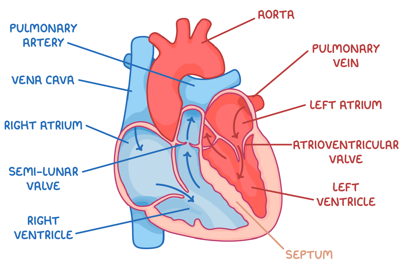
Structure of the Internal Heart
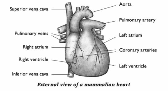
Structure of the External Heart
What is the cardiac cycle?
The sequence of contraction and relaxation of the heart chambers during 1 heartbeat.
Diastole means…
relaxes
Systole means…
contracts
What happens during diastole?
The heart is relaxed
The atria, then the ventricles fill with blood.
The volume and pressure of the blood in the heart build as the heart fills.
The pressure in the arteries is at a minimum.
What happens during systole?
The atria contract, closely followed by the ventricles.
The pressure inside the heart increases dramatically and blood is forced out of the right side of the heart to the lungs and from the left side to the main body circulation.
The volume and pressure of the blood in the heart are low at the end of systole, and the blood pressure in the arteries is at a maximum.
When volume decreases...
pressure increases
When volume increases...
pressure decreases
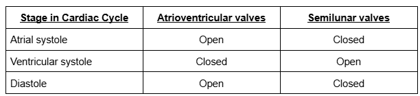
When do valves open?
When the pressure of blood behind them is greater than the pressure in front of them
When do valves close?
When the pressure of blood in front of them is greater than the pressure behind them
Why are valves important mechanisms?
They stop the backwards blood flow
Atrial Systole Process
1) The walls of the atria contract
2) The pressure in the atria rises above that in the ventricles, forcing the atrioventricular valves to open
3) Blood is forced into the ventricles
4) The ventricles are relaxed at this point
What coincides with atrial systole?
Ventricular diastole
When atria contract...
Atrial volume decreases
Atrial pressure increases
Ventricular systole Process
1) The walls of the ventricles contract
2) The pressure in the ventricles rises above that in the atria
3) The pressure in the ventricles rises above that in the aorta and pulmonary artery
4) During this period, the atria are relaxing. Atrial diastole coincides with ventricular systole
Diastole Process
1) The ventricles and atria are both relaxed
2) The pressure in the ventricles drops below that in the aorta and pulmonary artery, forcing the SL valves to close
3) The atria continue to fill with blood
4) Blood returns to the heart via the vena cava and pulmonary vein
5) Pressure in the atria rises above that in the ventricles, forcing the AV valves open
6) Blood flows passively into the ventricles without need of atrial systole
7) The cycle then begins again with atrial systole
Valves during the Cardiac Cycle
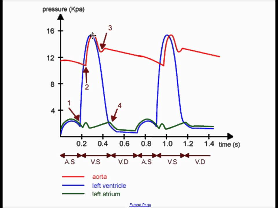
How to calculate heart rate from the cardiac cycle graph?
Step 1: Work out the length of one heart beat
Step 2: Calculate how many heart beats occur per second
Step 3: Calculate how many heart beats occur per minute

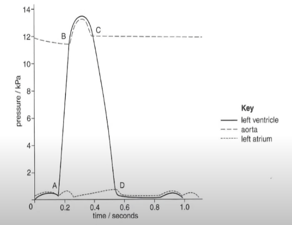
Explain what is happening at point A, B, C, and D.
At A, the ventricular pressure is higher than the atrial pressure. The atrioventricular valves close to prevent backflow.
At B, the ventricular pressure is higher than the aortic pressure, so the semi lunar valves open for blood to leave the ventricles into the aorta and out the heart.
At C, the aortic pressure is higher than the ventricular pressure, so the semilunar valves close to prevent backflow.
At D, the atrial pressure is higher than the ventricular pressure, so the atrioventricular valve open to allow blood into the ventricles.
What is cardiac output?
The term used to describe the volume of blood that is pumped by the heart (the left and right ventricle) per unit of time
Why do fitter people have stronger cardiac outputs?
Due to having thicker and stronger ventricular muscles in their hearts
Why does cardiac output increase when an individual is exercising?
So that the blood supply can match the increased metabolic demands of the cells
What is heart rate?
The number of times a heart beats per minute
What is stroke volume?
The volume of blood pumped out of the left ventricle during one cardiac cycle
Calculating cardiac output
Heart rate x Stroke volume
Units for cardiac output
cm3 min-1
Calculating Heart rate
cardiac output ÷ stroke volume
Calculating Stroke Volume
cardiac output ÷ heart rate
What does myogenic mean?
The heart will beat without any external stimulus
What is the SAN?
Sinoatrial node
Cells in the right atrium that act as pacemakers.
These cells initiate a nerve impulse without stimulation from the nervous system.
It is myogenic.

What is the AVN?
Atrioventricular node
It is the region of conducting tissue between atria and ventricles
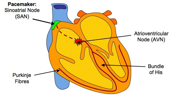
How heart action is initiated and coordinated:
The SAN initiates a wave of excitation, causing the atria to simultaneously contract. This is atrial systole.
However, a layer of non-conducting tissue prevents the excitation passing directly into the ventricles.
Therefore, the wave of excitation is instead picked up by the atrioventricular node (AVN)
The AVN introduces a short delay so that the ventricles contract after the atria.
After the short delay, the AVN sends the wave of excitation to the ventricles along the Bundle of His (conducting tissue made up of Purkyne fibres)
The Bundle of His splits into 2 branches and the Purkyne fibres conducts the wave of excitation down the septum of the heart to the apex.
The wave of excitation is then carried upwards in the walls of the ventricles. This triggers ventricular systole (contraction).
During ventricular systole, the blood pushes up from its base and is pushed upwards and outwards. Contraction starting from the apex allows for more efficient emptying of the ventricles.