Plant stems
1/9
There's no tags or description
Looks like no tags are added yet.
Name | Mastery | Learn | Test | Matching | Spaced | Call with Kai |
|---|
No analytics yet
Send a link to your students to track their progress
10 Terms
what are the different parts of the stem?
vascular bundles containing the xylem and the phloem
sclerenchyma fibres
structure of the xylem vessel?
very long tubes
made from dead cells joined end to end
hollow lumen - no cytoplasm
no end walls - uninterrupted tube allows water and mineral ions to pass
walls thickened with lignin to support the plant
pits in the cell walls where there is no lignin - water and mineral ions can transport across
function of the xylem vessel
transports water and mineral ions up the plant
provides support
structure of sclerenchyma fibres
made of bundles of dead cells
hollow lumen
have end walls
thickened cell walls with lignin
no pits
more cellulose than other cells
function of sclerenchyma fibres
to provide support
nothing to do with transport
structure of phloem tissue
contains sieve tube elements and companion cells
sieve tube elements are living cells joined end to end and forms sieve tubes
sieves have lots of holes to allow solute to pass
sieve tube elements have no nucleus, very thin layer of cytoplasm joined to adjacent cells through the holes in the sieve cells, few organelles
they need companion cells as they have no nucleus and can’t survive on their own
companion cells carry out living functions for both themselves and the sieve cells (energy for active transport)
function of phloem tissue
transport organic solutes from where they are produced to where they are needed (translocation)
nothing to do with support/structure
active transport
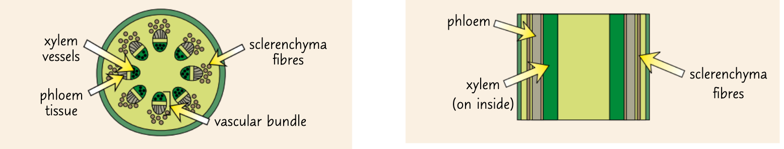
the position of the plant fibres in the stem
xylem and phloem cell in vascular bundles with phloem on the outside
sclerenchyma cells associated with vascular bundles on the outside of them

transverse vs longitudinal cross section
transverse = sections cut through each structure at a right angle to its length
longitudinal = section taken along the length of a structure
how to dissect plant stems
use scalpel and cut thin cross section (transverse or longitudinal)
use tweezers to place section in water to prevent them from drying out
transfer each section to a dish of toluidine blue O stain for 1 minute
this stains the lignin blue green and then rest of the tissue pink purple
rinse off excess stain with water
place sections on slide
place under microscope and adjust till you get a clear image
make a labelled drawing