Chapter 6: Integumentary system
 Call Kai
 Learn
Learn Practice Test
 Spaced Repetition
 Match
Match Flashcards
 Knowt Play
1/64

There's no tags or description
Looks like no tags are added yet.
Name | Mastery | Learn | Test | Matching | Spaced | Call with Kai | Chat |
|---|
No analytics yet
Send a link to your students to track their progress
65 Terms
dermatology
scientific study of the skin
the integumentary system
skin, hair, nails, and associated glands
integument
the skin. largest and heaviest organ
the skin composes ___ % of body weight
8%
epidermis
stratified squamous epithelium, top layer of the skin
dermis
deeper connective tissue layer of the skin
hypodermis
connective tissue layer (subcutaneous fat) beneath the dermis. not technically part of the skin
normal skin thickness ranges from __ and __
0.5 to 6 mm
skin thickness is based on relative thickness of the…
epidermis (stratum corneum)
thick skin
epidermis is 0.5 mm thick, and stratum corneum is thickest layer. covers palms, soles, fingers, and toes. Sweat glands but no hair follicles or oil glands
thin skin
covers the rest of the body. epidermis is 0.1mm thick and the stratum spinosum is the thickest layer usually
functions of the skin
resistance to trauma and infection
barrier functions (uv radiation, example)
vitamin d synthesis
sensation
thermoregulation
non-verbal communication
keratin
tough protein in epidermal cells. gives physical protection
dermicidin and defensins
antimicrobial peptides. helps skin resist infection
acid mantle
acidic film that protects against pathogens
vitamin D is used for…
bone development and maintenance
features of the epidermis
keratinized squamous epithelium
avascular
sparse nerve endings (most sensations due to nerve endings in the dermis)
keratinocytes
synthesize keratin. makes up majority of epidermal cells
stem cells of epidermis
undifferentiated cells that give rise to keratinocytes
found in the stratum basale (deepest layer)
melanocytes
synthesize melanin
branching process of melanocytes
branches spread among keratinocytes to distribute melanin via melanosomes
tactile cells
touch receptors associated with dermal nerve fibers
tactile disc
tactile cell and associated nerve fiber
dendritic cells
phagocytic immune cells found in stratum spinosum and stratum granulosum
5 cells of the epidermis
keratinocytes, stem cells, melanocytes, tactile cells, and dendritic cells
list the layers of epidermis from deep to superficial
stratum basale ➡ stratum spinosum ➡ stratum granulosum ➡ stratum lucidum ➡ stratum corneum
stratum basale
contains single layer of stem cells and keratinocytes on the basement membrane. Also contains melanocytes and tactile cells.
stratum spinosum
several layers of keratinocytes joined by desmosomes and tight junctions. also contains dendritic cells
stratum granulosum
three to five lanes of flat keratinocytes. contains keratohyalin granules
keratohylain granules
dark staining cells
stratum lucidum
thin layer only found in thick skin. keratinocytes packed with protein eleidin. cells have no nucleus or other organelles.
stratum corneum
up to 30 layers of dead keratinized cells
resists abrasion, penetration, and water loss
explain the history of the keratinocyte
produced in the stratum basale
mitosis stops when cells travel away from the dermis
new keratinocytes push old ones to the surface
keratinocytes flatten over time and produce lamellar granules
in 30 to 40 days the keratinocyte reaches the skin surface and exfoliates
calluses/corns
thick accumulations of dead keratinocytes on hands or feet
epidermal water barrier formation
formation occurs in the stratum granulosum
keratinocytes release filaggrin
cells produce tough envelope proteins beneath their membranes
membrane-coating vesicles release lipid mixture that spread out. over cell surface and waterproofs it
keratinocyte organelles degenerate and the cells die leaving waterproof face enclosing keratin bundles
epidermal water barrier
important for water retention and prevents dehydration.does not prevent absorption at the stratum corneum. tight junctions between keratinocytes and formed in stratum granulosum.
characteristics of the dermis
0.2 mm to 4 mm thick
contains mainly collagen but also elastic and reticular
vascular
contains nerve endings
hair follicles and nail roots are connected to the dermis
dermal papillae
upward, finger-like extensions of dermis

epidermal ridges
downward waves of epidermis
two zones of the dermis
papillary layer and reticular layer
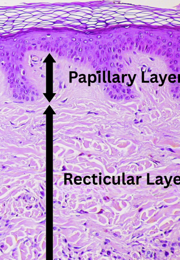
papillary layer
thin areolar tissue in and near dermal papillae
allows mobility of leukocytes and other defense cells
rich in small blood vessels
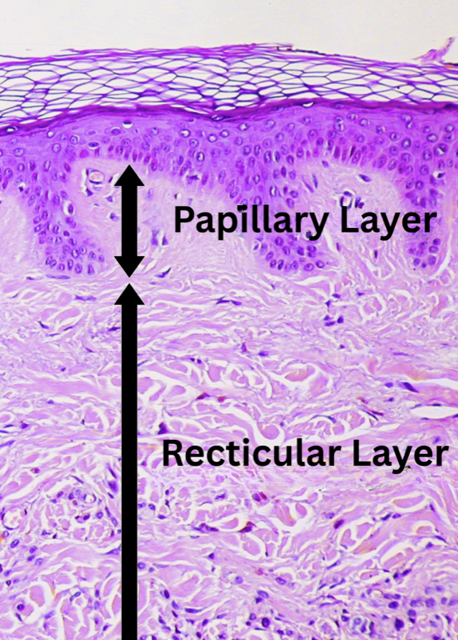
reticular layer
deeper and thicker layer
contains dense irregular connective tissue
striae
collagen fiber tearing in reticular layer
blister
serous fluid seep out of vessels, separating the epidermis from dermis
hypodermis characteristics
structure: more areolar and adipose tissue
function: pads the body and binds skin to the underlying tissue
subcutaneous fat
subcutaneous tissue that is comprised of predominately adipose tissue. function is energy reservoir and thermal insulation. thicker in women and thinner in elderly and infants
autograft
tissue from another location of same body
allograft
tissue form an unrelated person
eumelanin
dark melanin
Brown and black eumelanin
responsible for tan, brown, and black skin
Pheomelanin:
lighter, reddish brown
other factors that affect skin color
dermal blood vessels and white collagen
carotene
UV radiation
what causes variation in skin color?
same number of melanocytes but melanin is produced and accumulated differently
dark skin
greater amount of melanin produced by melanocytes
slower breakdown of melanin
melanin granules spread out in keratinocytes
Melanized cells seen throughout the epidermis
light skin
melanin clumped near keratinocytes nucleus
little melanin seen beyond stratum basale
cyanosis
blue skin due to oxygen deficiency
erythema
redness due to increased blood flow
albinism
genetic lack of melanin
pallor
pale due to decreased blood flow
jaundice
yellowing of skin due to high levels of bilirubin, maybe from poor liver function
hematoma
bruise (mass of clotted blood showing through the skin)
eccrine glands
sweat glands, dense on palms, soles, and forehead
sebaceous glands
produce oily sebum
ceruminous glands
apocrine glands found in external ear canal
apocrine glands
found in groin, anal region, areola, beard area
type of sweat gland
doesn’t use. apocrine method usually