B) Cardiac Shunts
 Call Kai
 Learn
Learn Practice Test
 Spaced Repetition
 Match
Match Flashcards
 Knowt Play
1/35

There's no tags or description
Looks like no tags are added yet.
Name | Mastery | Learn | Test | Matching | Spaced | Call with Kai | Chat |
|---|
No analytics yet
Send a link to your students to track their progress
36 Terms
patent ductus arteriosus
ventricular septal defect
atrial septal defect
tetralogy of fallot
persistent right aortic arch
PVATP
List all the cardiac shunts
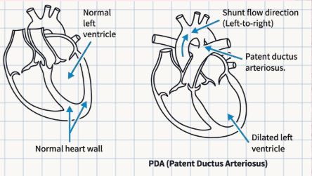
Patent Ductus Arteriosus (PDA)
Caused by the failure of the ductus arteriosus to close at birth.
ductus arteriosus
a vessel that connects the pulmonary artery and aorta in the fetus
Left-to-right shunting of blood from the aorta to the pulmonary artery.
Patent Ductus Arteriosus (PDA)
PDA causes blood to shunt in which direction, and between which two major great vessels?
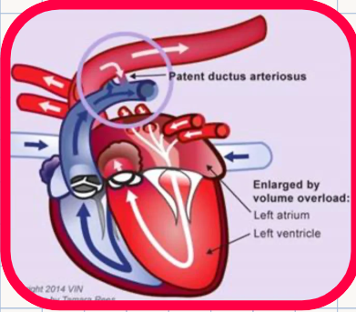
1. pulmonary overcirculation
2. left-sided volume overload
3. cardiac enlargement (dilatation)
4. left-sided congestive heart failure with pulmonary edema
Patent Ductus Arteriosus (PDA)
List the 4 sequential cardiopulmonary consequences that develop directly due to the left-to-right shunt in PDA:
dog and cat
Patent Ductus Arteriosus (PDA)
In which two domestic companion animal species is PDA commonly diagnosed?
Ventricular Septal Defect (VSD)
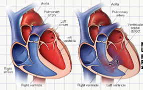
A pathological problem where there is a hole in the wall between the left and right ventricles that enables abnormal passage of blood between these two chambers.
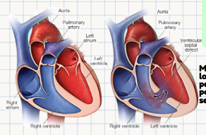
perimembranous portion of the septum.
Ventricular Septal Defect (VSD)
Within which specific micro-anatomical segment of the interventricular septum is a VSD most commonly located?
Shunting of blood from the left ventricle into the right ventricle or right ventricular outflow tract (moving along the high-to-low pressure gradient).
Ventricular Septal Defect (VSD)
In a typical VSD, blood initially shunts from which specific chamber into which target structure/outflow tract?
1. defect size
2. the pulmonary to systemic vascular resistance ratio
3. ventricular compliance
Ventricular Septal Defect (VSD)
What 3 specific physical and physiological factors determine the exact volume and impact of the blood shunted through a VSD?
Blood shunted to the right ventricle immediately circulates through the pulmonary vessels and left heart chambers, causing their volume overload and dilatation.
Ventricular Septal Defect (VSD)
Trace how a classic left-to-right VSD paradoxically causes Left-sided CHF chamber changes:
Eisenmenger Syndrome
Ventricular Septal Defect (VSD)
The development of a right-to-left shunt flow through a septal defect as a consequence of severe, chronic pulmonary hypertension.
1. cyanosis
2. fatigue
3. exercise intolerance
Ventricular Septal Defect (VSD)
List the 3 classic clinical signs that indicate a VSD patient has transitioned into Eisenmenger syndrome:
dog, cat, and cows
Ventricular Septal Defect (VSD)
In what 3 domestic animal species is a VSD most commonly diagnosed?
Atrial Septal Defect (ASD)
A pathological problem where there is an opening or hole within the interatrial septum, with shunting occurring in the direction of the pressure gradient between the two atria.
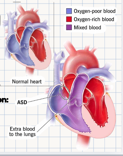
A midseptal location, specifically classified as a secundum-type defect.
Atrial Septal Defect (ASD)
What is the most common anatomical location and specific classification type for an ASD?
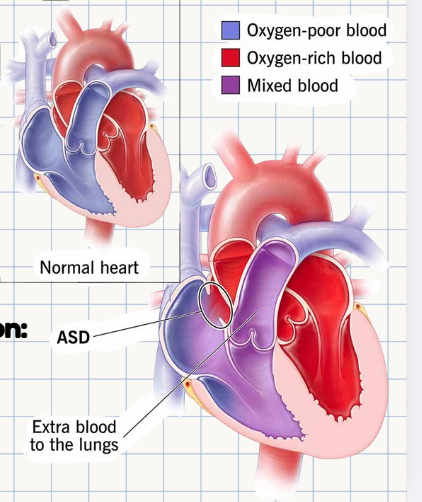
Atrial Septal Defect (ASD)
This causes volume overload of the right atrium and right ventricle, leading to dilation, hypertrophy, increased pulmonary blood flow, pulmonary hypertension, and possible right-sided CHF.
Atrial Septal Defect (ASD)
This causes volume overload of the right atrium and right ventricle, leading to ______, hypertrophy, ______ pulmonary blood flow, pulmonary hypertension, and possible ____-sided CHF.
In a VSD, shunted blood from the left ventricle goes into the right ventricular outflow tract and immediately heads straight to the lungs, returning to volume-overload the left atrium and left ventricle.
In an ASD, blood shunts from the left atrium back into the right atrium, meaning it must physically travel through and volume-overload the entire right side of the heart (RA and RV) before it can even reach the pulmonary circulation.
*addtl for diff
Contrast why a standard VSD chronically overloads and dilates the left heart chambers, whereas an ASD chronically overloads and dilates the right heart chambers:
1. Pulmonary Stenosis
2. Right Ventricular Hypertrophy
3. Overriding Aorta
4. Ventricular Septal Defect (VSD)
PROV
List the 4 distinct anatomical anomalies that define the complex congenital defect Tetralogy of Fallot (ToF):
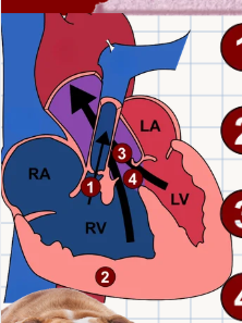
Pulmonary Stenosis
Tetralogy of Fallot
Narrowing of the exit from the right ventricle.
Right Ventricular Hypertrophy
Tetralogy of Fallot
Thickening of the right ventricular muscle (secondary to pumping against the stenotic valve).
Overriding Aorta
Tetralogy of Fallot
Where the aorta expands/shifts to allow blood from both ventricles to enter.
Ventricular Septal Defect
Tetralogy of Fallot
A hole allowing blood to flow between two ventricles
1. Reduced pulmonary blood flow
Generalized cyanosis
Tetralogy of Fallot
What are the two primary downstream consequences of ToF?
Reduced pulmonary blood flow
Tetralogy of Fallot
Results in fatigue and shortness of breath.
Generalized cyanosis
Tetralogy of Fallot
Leads to polycythemia and weakness driven by the mixing of oxygenated and deoxygenated blood in the ventricles entering the systemic circulation.
polycythemia
(increased red blood cell production due to chronic tissue hypoxia
Tetralogy of Fallot is a complex congenital defect vary based on the severity of pulmonic stenosis and the extent of right-to-left shunting.
Tetralogy of Fallot is a complex ______ defect vary based on the severity of pulmonic ______ and the extent of right-to-left ______.
Dog Breeds: Keeshonds and English Bulldogs
Also cats
Tetralogy of Fallot
Name the two dog breeds explicitly noted as predisposed to ToF, and what other companion animal species can develop it?
PERSISTENT RIGHT AORTIC ARCH (PRAA)
What specific vascular malformation forms a classic "vascular ring" around the mediastinal structures, trapping them at the base of the heart?
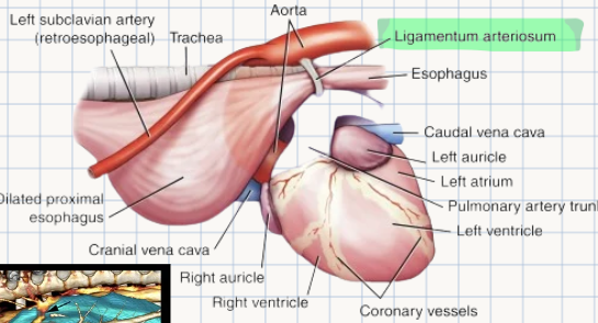
esophagus and trachea
PERSISTENT RIGHT AORTIC ARCH (PRAA)
Persistent right aortic arch leads to the entrapment and compression of which two non-vascular anatomical structures?
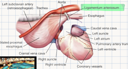
1. persistent right arch (Aorta)
2. left ligamentum arteriosum (stretching dorsally across)
3. ventral heart base
PERSISTENT RIGHT AORTIC ARCH (PRAA)
List the 3 specific structural boundaries that completely encircle the trachea and esophagus in a PRAA anomaly:
Esophageal compression causes severe dilation above the heart base (proximal megaesophagus).
PERSISTENT RIGHT AORTIC ARCH (PRAA)
What mechanical consequence happens to the esophagus due to the vascular ring compression, and exactly where does this change occur?
At weaning; presenting as regurgitation.
PERSISTENT RIGHT AORTIC ARCH (PRAA)
At what specific dietary milestone or life stage do clinical signs of PRAA classically manifest, and what is the primary clinical sign?
Aspiration pneumonia
PERSISTENT RIGHT AORTIC ARCH (PRAA)
What secondary, highly life-threatening respiratory condition frequently occurs in animals affected by PRAA due to chronic regurgitation?
dog: German Shepherd
cattle
cat
horse
PERSISTENT RIGHT AORTIC ARCH (PRAA)
Which dog breed is explicitly noted as being highly predisposed to PRAA, and what other 3 domestic animal species can develop it?