chp. 1 intro
1/34
There's no tags or description
Looks like no tags are added yet.
Name | Mastery | Learn | Test | Matching | Spaced | Call with Kai |
|---|
No analytics yet
Send a link to your students to track their progress
35 Terms
Comparative Vertebrate Anatomy
the study of vertebrate structure (or morphology) and the functional aspects of these structures.
a) It is a study of vertebrate morphology from an evolutionary perspective
ontogenesis
the development of an individual organism or anatomical or behavioral feature from the earliest stage to maturity.
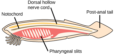
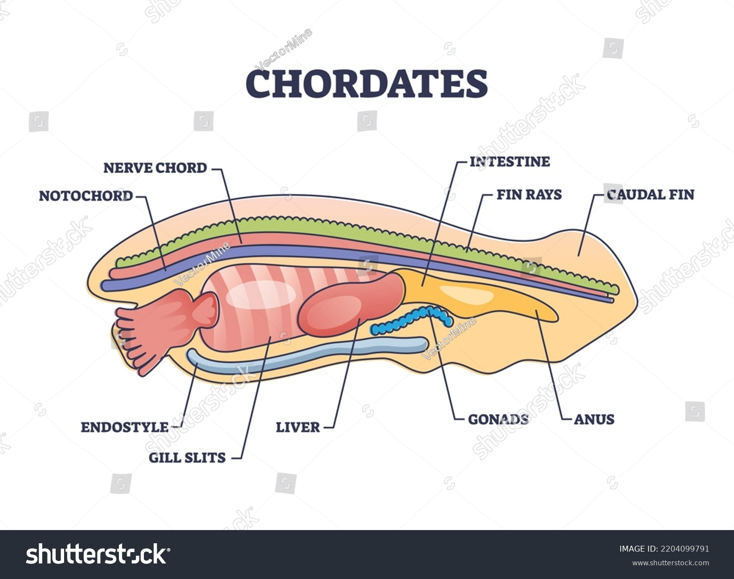
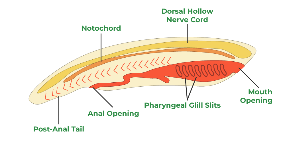
chordates
phylum chordata is made up of a group of animals all possessing four common features:
-notochord
-dorsal hollow nerve cord
-postanal tail
-endostyle

notochord
a rigid cartilaginous rod defining the longitudinal axis in the embryo
-the first skeletal feature to appear in the embryo
-In some vertebrates, such as agnathans, it will be maintained throughout the animal’s life span. In others it is replaced by the vertebral centra to varying degrees.
-In reptiles, birds, and mammals, the notochord is mostly replaced by the vertebrae and becomes very small or disappears. In mammals, the only remnants of the notochord are found between vertebrae, where they help form part of the intervertebral discs.

dorsal hollow nerve cord
the spinal cord and brain
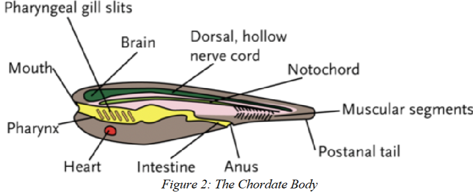
postanal tail
The postanal tail is a body extension that continues past the anus or cloaca. It may disappear in adults but is always present at some stage of development.
endostyle
a glandular groove on the floor of the pharynx.
-In non-vertebrate chordates they are paired grooves possessing cilia and are used in filter feeding.
-In vertebrates the endostyle will develop into the thyroid, an organ of the endocrine system.
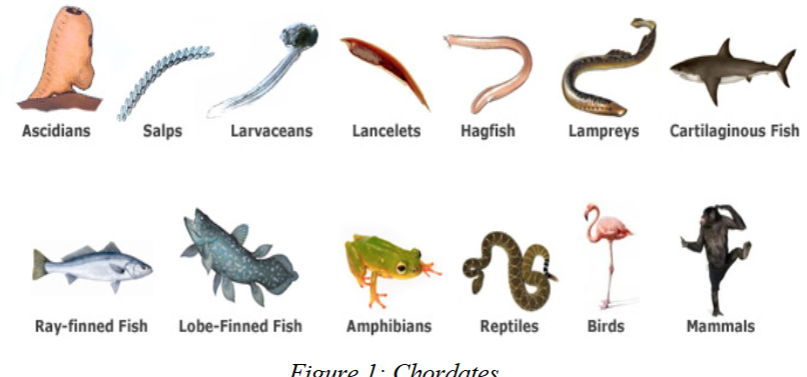
3 subphyla of chordata
1) Urochordata, the tunicates and sea squirts
2) Cephalochordata, amphioxus (lancelets)
3) Vertebrata/Craniata, chordates having a vertebral column of bone or cartilage
Cranialization
development of the head
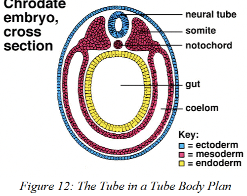
coelom
a fluid-filled body cavity between the outer body wall and the digestive tube that houses internal organs and allows them to grow and move independently.

Gnathostomata
jawed vertebrates
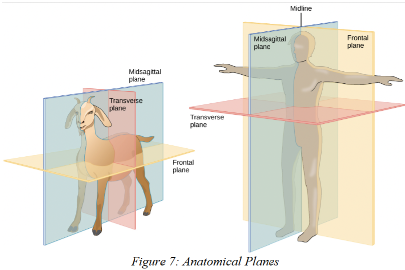
transverse plane
(aka; horizontal plane, cross section) –
separates cranial and caudal structures along the trunk and proximal from distal structures on the limb in most vertebrates.

Sagittal Plane
a vertical plane dividing the body into left and right portions.

dorsal plane
divides the body into ventral and dorsal
portions.
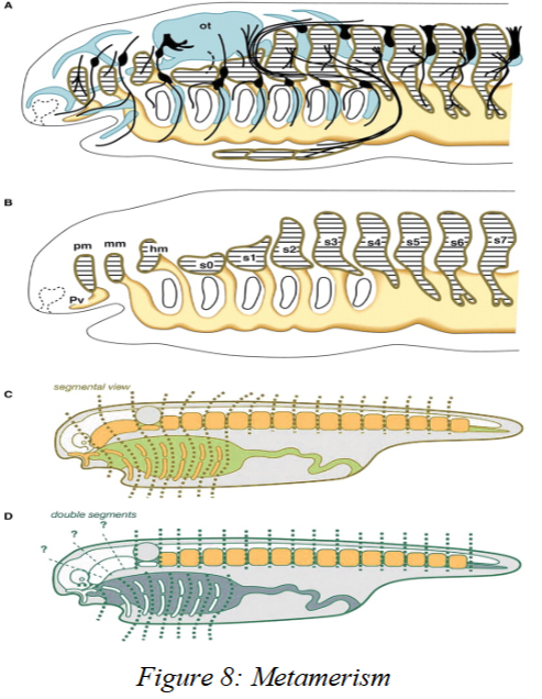
Metamerism
the serial repetition of structures along the long axis of the body.
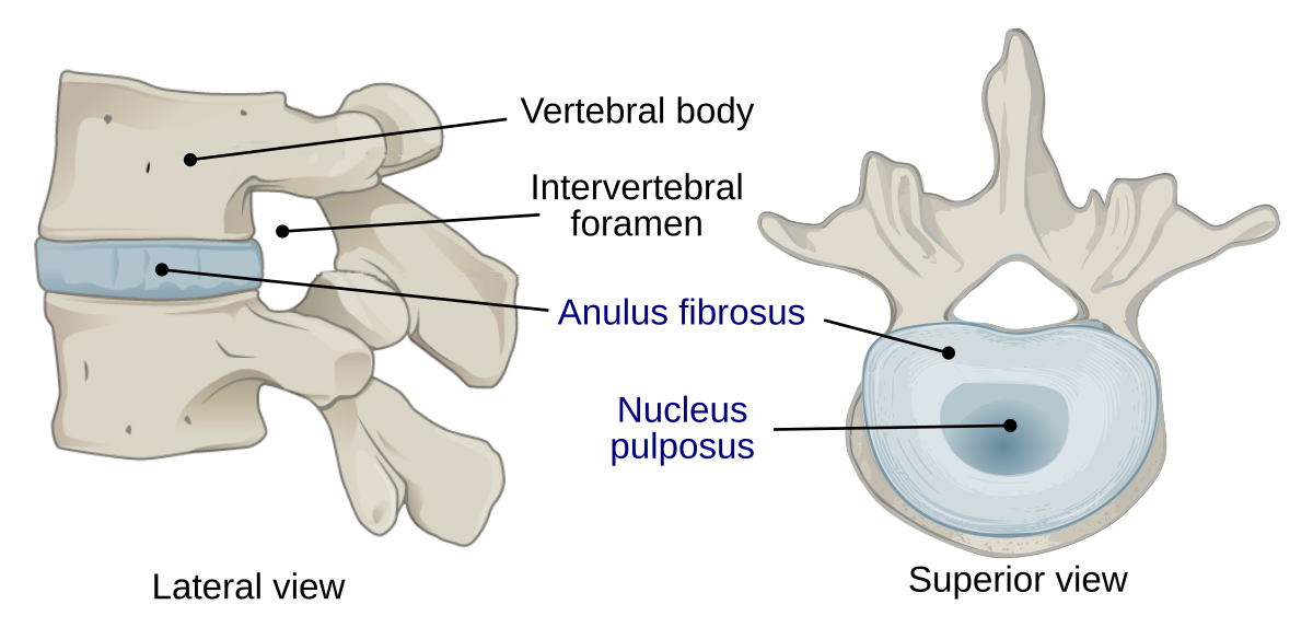
Nucleus Pulposus
In mammals remnants of the notochord will remain as portions of the intervertebral discs
-absorbs shock and provides flexibility to the spine
agnathans
jawless vertebrates
-notochord grows and is maintained throughout life
- It will even develop extensions called Lateral Neural Cartilages that will be found lateral to the spinal cord
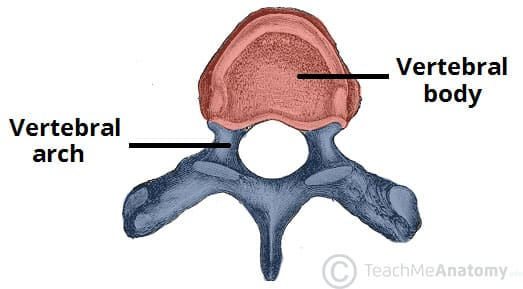
Vertebral/ Neural Arch
will extend dorsally from the centra to surround and protect the spinal cord.
-A number of processes will radiate from the arch.
- In some vertebrates a second arch, the Hemal Arch, will extend from the centrum ventrally in caudal vertebrae
Neurocoel
The hollow center of the dorsal hollow nerve cord.
-The neurocoel includes the central canal of the spinal cord
Neuralation
The process in which the dorsal hollow nerve cord originates. typically occurs along the longitudinal axis of the embryo dorsal to the notochord.
-During neurulation, ectoderm cells fold (neural groove) inward to form the neural tube, which develops into the dorsal, hollow nerve cord.
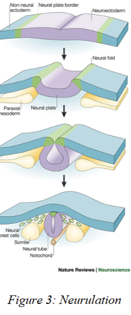
neural tube
becomes the dorsal hollow nerve cord after neuralation.
-develops from the deepening of the neural groove.

neural groove
part of neuralation to form dorsal hollow nerve cord.
During neurulation, cells of the ectoderm (the outer embryonic layer, above the notochord) divide rapidly. This growth creates a neural groove
- the neural groove will sink into the embryonic body and close off to form the Neural Tube

neuralation (in Agnathans and Neopterygians (the gars, bowfins, and teleosts)
Instead of forming a neural groove, they form a neural keel (a wedge of ectoderm above the notochord). The keel hollows out to become the nerve cord, which expands at the front into the brain. Cranial and spinal nerves then grow from the cord, forming the PNS, which connects the CNS to the rest of the body
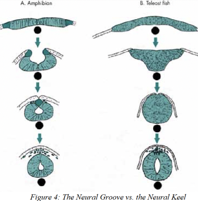
neural keel
a solid, wedge-shaped block of cells (from the ectoderm) in some fishes that later hollows out to form the neural tube, which becomes the dorsal, hollow nerve cord. IN NEURALATION OF JAWLESS FISH

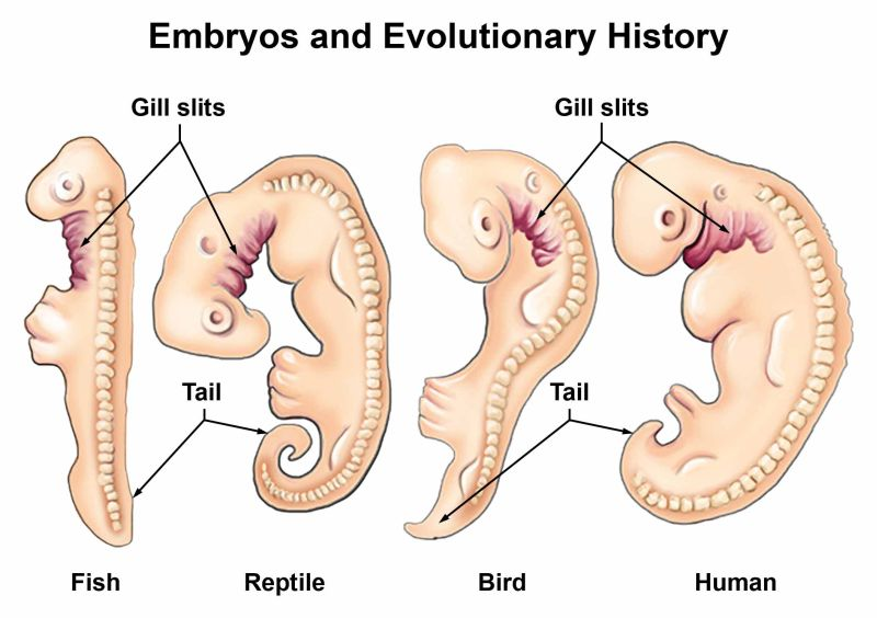
pharynx
a tube-like region behind the mouth and nose that connects to both the digestive system (esophagus/stomach) and the respiratory system (lungs or gills).
- It shows the relationship between vertebrates and other chordates.It produces a number of structures:
1] gills in fish
2] lungs in tetrapods
3] jaw skeleton and musculature
4] some endocrine glands
5] the middle ear in tetrapods
6] and serves a source of stem immune cells in the human fetus
![<p>a tube-like region behind the mouth and nose that connects to both the digestive system (esophagus/stomach) and the respiratory system (lungs or gills).<br>- It shows the relationship between vertebrates and other chordates.It produces a number of structures:</p><p>1] gills in fish</p><p>2] lungs in tetrapods</p><p>3] jaw skeleton and musculature</p><p>4] some endocrine glands</p><p>5] the middle ear in tetrapods</p><p>6] and serves a source of stem immune cells in the human fetus</p>](https://knowt-user-attachments.s3.amazonaws.com/80143067-35eb-47d7-89a1-110e1501bfd6.png)
pharyngeal slits
actual openings to the outside, which only form if a pouch connects with an ectodermal groove and the thin tissue (branchial plate) between them ruptures.
-In fishes pharyngeal slits are maintained throughout life for gills.
- In tetrapods pharyngeal slits are only temporary structures
—mammals:
1–2 pouches rupture, then reseal.
1st pouch → Eustachian tube
2nd pouch → palatine tonsils
Posterior pouches → some endocrine glands
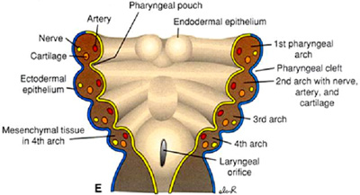
pharyngeal pouches
outpocketings of gut endoderm (grow outward).
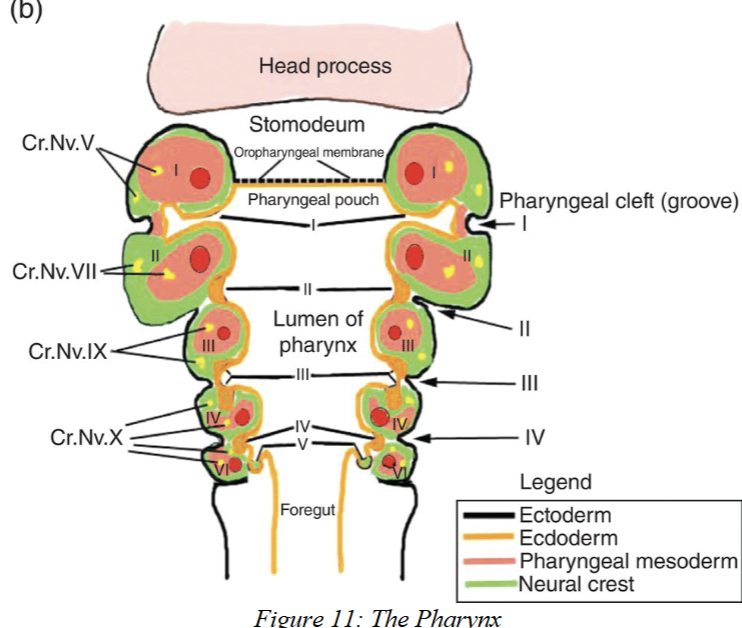
ectodermal grooves
inpocketings of ectoderm (grow inward and towards developing pouches)

branchial plate
thin tissue layer that separates where pharyngeal pouches and ectodermal grooves grow close together. If branchial plate ruptures, pharyngeal slit is formed.

pharyngeal arch
Pharyngeal arches are paired tissue structures in the embryo that give rise to parts of the skeleton, muscles, nerves, and blood vessels of the head and neck. In fishes, they form gills; in jawed vertebrates, the first two arches become the jaws and hyoid.
-found between adjacent pharyngeal pouches/slits
-Usually 7 pairs
-Each Arch Has 4 Components
Skeletal element → forms the pharyngeal/visceral skeleton (cartilage or bone).
Muscle → branchiomeric muscles (skeletal muscle for moving the arches).
Cranial nerve branch → motor + sensory innervation. (Cranial nerves V, VII, IX, X).
Blood vessel (aortic arch) → links dorsal and ventral aorta.
4 components of each pharyngeal arch
Skeletal element (pharyngeal/visceral skeleton)
Can be cartilage or bone.
Provides structure to the arch.
In fishes → forms gill supports.
In jawed vertebrates → forms jaws, hyoid, and some other head bones.
Branchiomeric muscles
Skeletal muscles attached to the arch.
Move the arch (e.g., opening/closing jaws or gills).
Cranial nerves (V, VII, IX, X)
Provide motor (movement) and sensory (feeling) input.
Each arch is innervated by a specific cranial nerve.
Aortic arch (blood vessel)
Connects dorsal and ventral aorta, helping blood circulate through the gills in fishes.
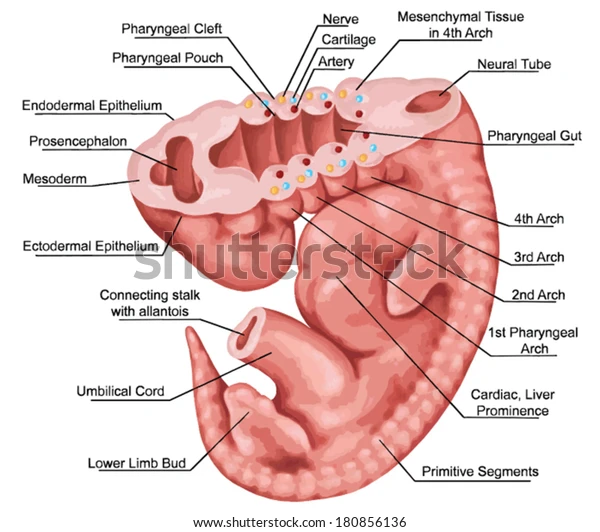
mandibular arch
the first pharyngeal arch is typically referred to as the Mandibular Arch since it contains the upper and lower jaws and related structures
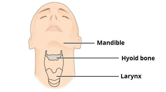
hyoid arch
2nd pharyngeal arch, supports jaw (hyoid bone)
branchial arches
The remaining pharyngeal arches are numbered 3 through 7 since they resemble unmodified gill arches.
coelum organization
Fishes, amphibians, reptiles:
Pericardial cavity → houses the heart
Pleuroperitoneal cavity → houses the rest of the viscera (including lungs in tetrapods)
Partition between these → transverse septum (fibrous connective tissue)
Birds and mammals:
Coelom is divided by diaphragm (mammals) or oblique septum (birds) into:
Thoracic cavity → heart (pericardial cavity) + lungs (pleural cavities)
Abdominopelvic cavity → abdominal cavity + pelvic cavity
All coelom regions are lined with serous membranes, named after the organs they surround (e.g., pleural membranes around lungs).