Biological Psychology Test 1
1/120
There's no tags or description
Looks like no tags are added yet.
Name | Mastery | Learn | Test | Matching | Spaced | Call with Kai |
|---|
No analytics yet
Send a link to your students to track their progress
121 Terms
Describe significant contributions to neuroscience in ancient Egypt and Greece,
Edwin Smith Surgical Papyrus (text)
-Dated to 1700 BC, Egyptian text
-First recorded writing about the brain
-Egyptians thought the brain was less important than other organs.
-Greeks started seeing the importance of the brain
Renaissance
-Artists like Leonardo da Vinci and Michelangelo began depicting the brain and nervous system in art.
-Andreas Vesalius (1514-1564) created detailed anatomy books
Age of Enlightenment
-René Descartes (1596-1650)
-The nervous system worked like a machine
-dualism: The mind and brain are separate
-monism: The mind is what the brain does. They are one.
1800s-1900s
1800s-1900s
-Time of great advancements in research methods, theories, and staining techniques
-Charles Darwin
-Wilhelm Wundt - father of modern psychology
-Santiago Ramón y Cajal - father of modern neuroscience
Describe the historical connection between artists and neuroanatomy by providing some examples.
-Artists like Leonardo da Vinci and Michelangelo began depicting the brain and nervous system in art.
-Andreas Vesalius (1514-1564)created detailed anatomy books
Explain the difference between dualism and monism. Which of these philosophies is the way we address neuroscience today, and why?
Most researchers today are in favor of monism since it's accepted that the mind is a function of the brain's processes
Dualism
The mind and Brain are 2 separate Entities.
Monism
The mind is what the brain does, they are one in the same
an applied approach to neuroscience.
-focus on creating solutions or remedies for problems
-Creating psychoactive drugs
-Clinical psychology
What is the Basic Approach to neuroscience
Discovery
-Theory building
-Understanding fundamental principles
Diagram and label a typical neuron (standard multipolar neuron)
Dendrite, cell body, axon, axon terminal
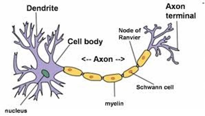
Dendrites
structure of neuron- receive information from other neurons and send electrical information to the cell body
cell body (soma)
structure of neuron- the region where inputs are integrated
Axon (nerve fiber)
structure of neuron- conducts output information away from the cell body as an electrical impulse. and action potentials are propagated along the length of axons and are the electrical signal that carries information from one place to another in the nervous system
axon hillock
structure of neuron- place where electrical signal is initiated
Axon terminals
structure of neuron- at the end of the axon, they release chemicals to communicate with other cells
synapse
structure of neuron- place where one neuron communicates with another
Cell membrane and ion channels
structure of neuron- Ion channels are large proteins in the cell membrane of neurons that selectively permit the flow of specific ions into and out of a neuron
Diagram types of neurons and how they differ from each other.
multipolar, bipolar, unipolar
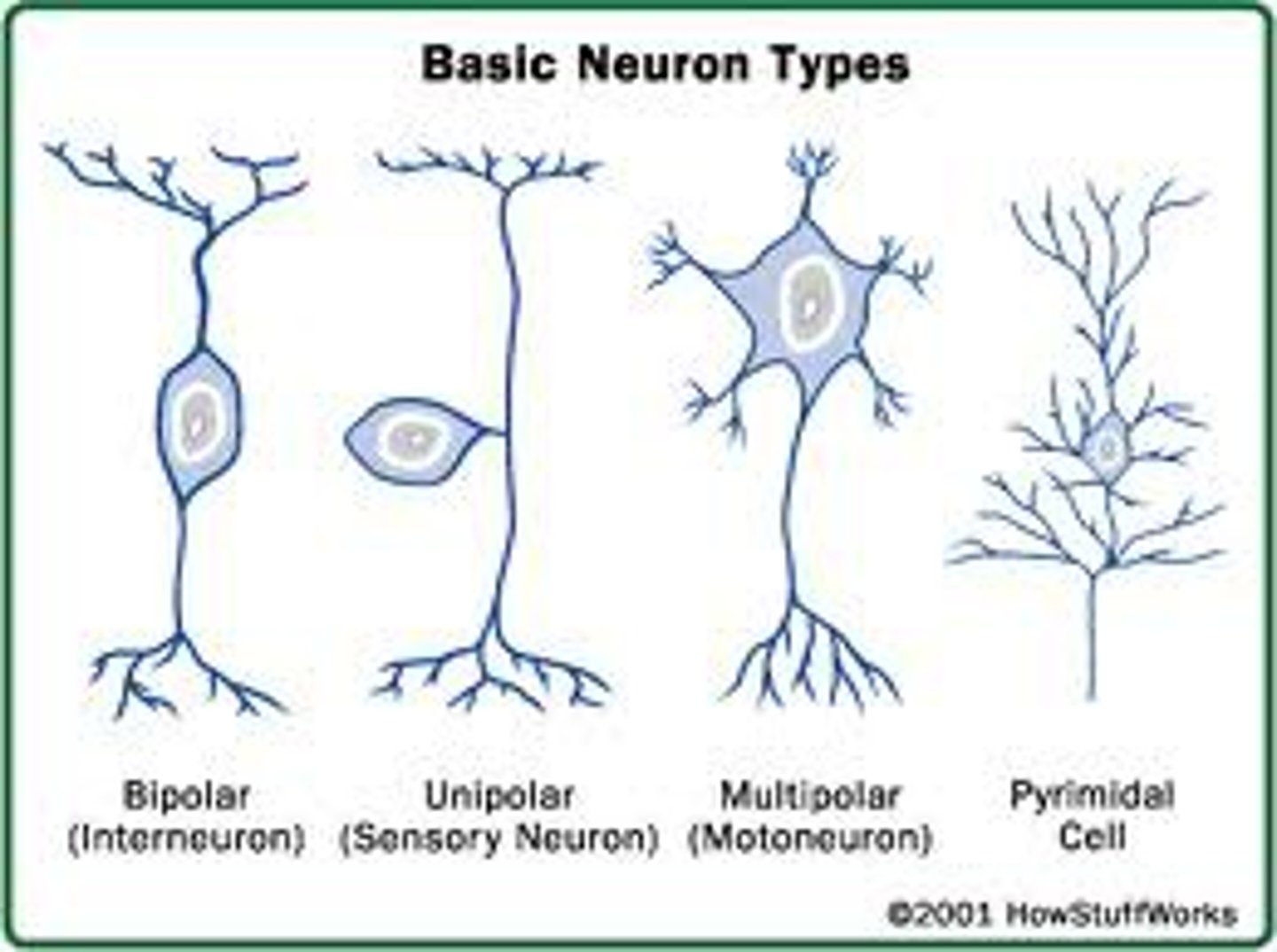
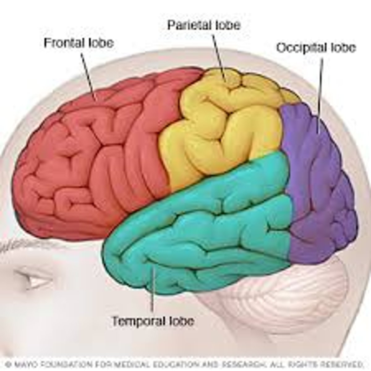
Diagram the different lobes of the cerebral cortex
frontal lobe, parietal lobe, temporal lobe and occipital lobe.
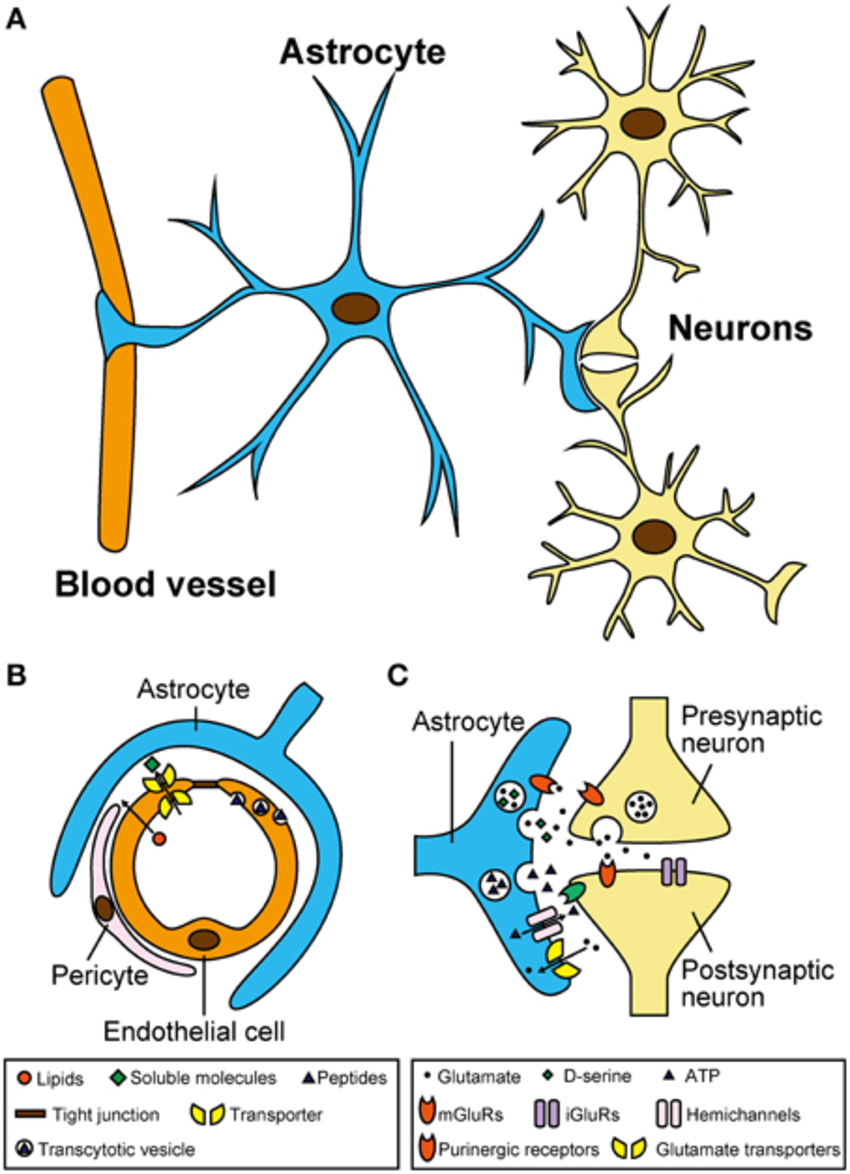
Glial Cells
non- neuronal cells in the central nervous system and the peripheral nervous system that do not produce electrical impulses.
Astrocytes
glial cell in the CNS
nutrients
Repair
help in signals
protect the blood-brain barrier
Microglia
glial cell in the CNS
immune function
removal of dead neurons
Oligodendrocytes
glial cell
CNS
creates myelin for axons
aids in development
Schwann cells
glial cells in the peripheral nervous system
creates myelin for neurons in PNS
creates pathway for regrowing axons after injury
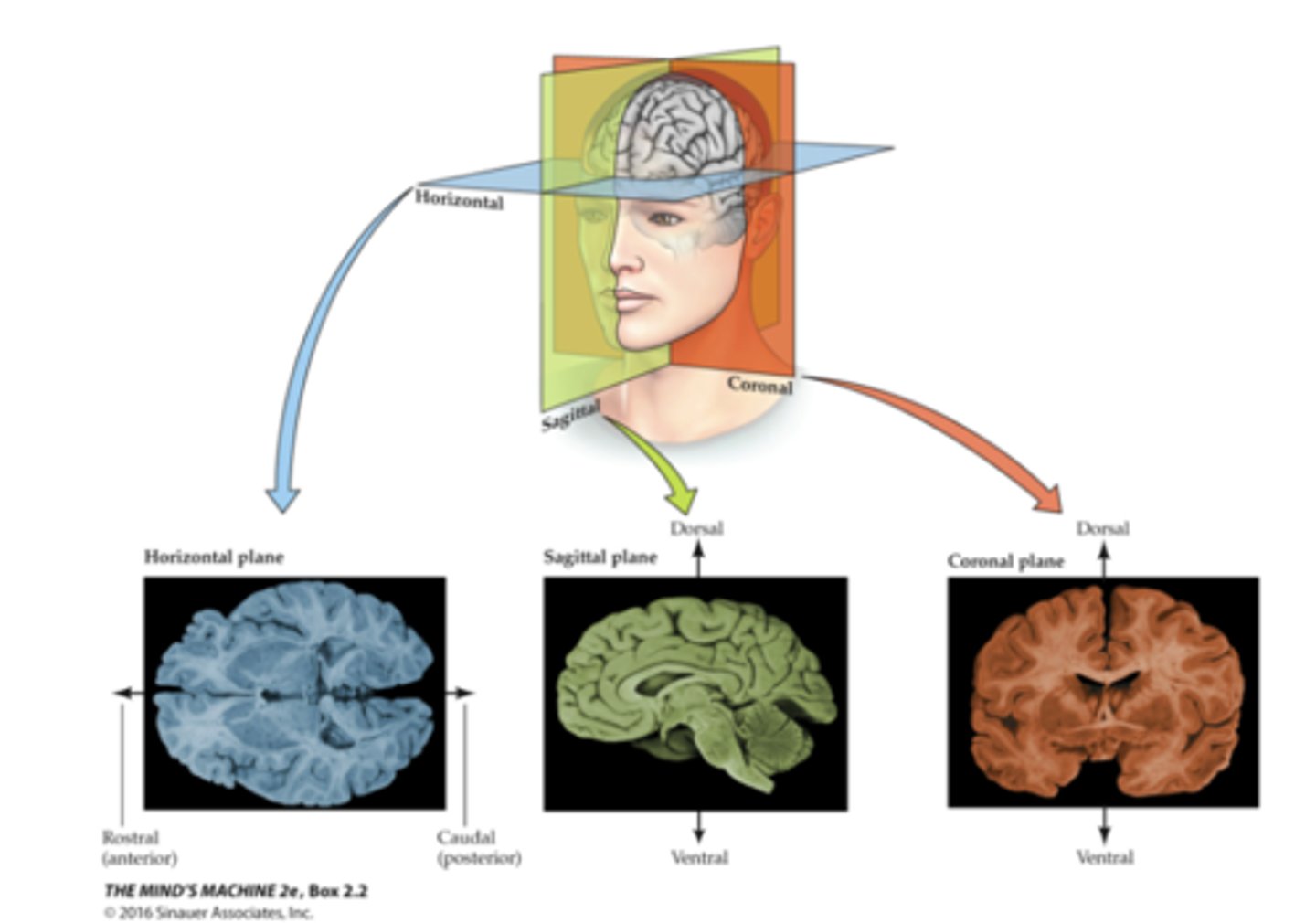
Coronal Plane
divides the brain into front (anterior) and back (posterior) regions

Medial
going inwards to the center of the brain
Lateral
going out towards your ears
Sagittal plane
Divides the brain into right and left halves
Horizontal Plane
Divides brain horizontally into a top part and a bottom part
Dorsal
towards the dome of the head
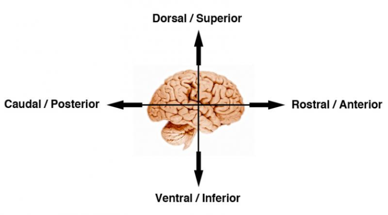
Ventral
towards v
anterior, and posterior.
The front of the brain toward your forehead is anterior
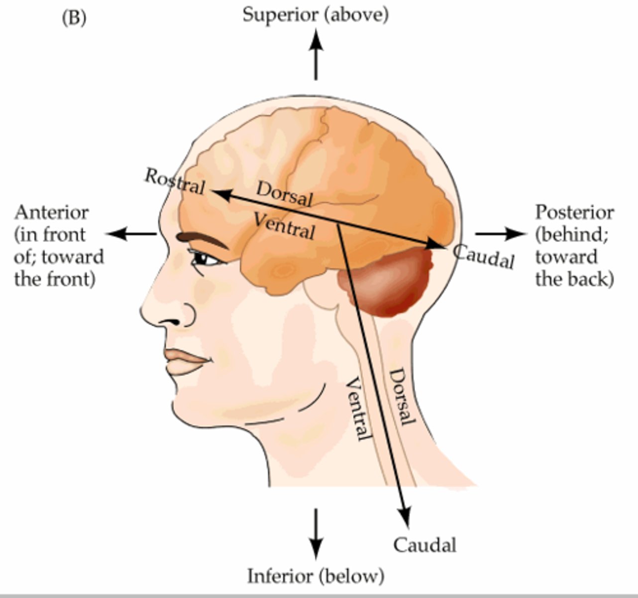
Posterior
Towards the back of your head
Be able to identify the three different planes (cuts) of the brain.
Horizontal, Sagittal, Coronal
Describe the basic divisions of the nervous system.
-central nervous system (CNS):
Brain and spinal cord
-Peripheral nervous system (PNS): Nerves outside the CNS throughout the body
PNS has 2 divisions: Somatic and Autonomic
Peripheral nervous system (PNS): Somatic Division
Spinal nerves (31 pairs) connect to the spinal cord
• The nerves of the somatic nervous system form two anatomical groups:
• Cranial nerves (12) innervate the head, neck, and visceral organs directly from the brain
Peripheral nervous system (PNS) : Autonomic Division
the main system for controlling the body's organs.
The ANS has two divisions:
Sympathetic & Parasympathetic
The sympathetic and parasympathetic nervous systems have different effects on organs due to different neurotransmitters.
Parasympathetic Nervous System
helps the body relax and recuperate. ("rest & digest")
Sympathetic Nervous System
prepares the body for action—the"fight-or-flight" response.
Diagram and label the basic anatomy of the spinal cord.
The nerves of the somatic nervous system form two anatomical groups:
• Cranial nerves (12) innervate the head, neck, and visceral organs directly from the brain
Be able to identify the different ventricles in the brain and the function of the cerebral spinal fluid.
Cerebral spinal fluid (CSF): clear liquid made by the choroid plexus
Found in ventricles
Removes waste
Also found in the spinal cord and around the brain
-Ventricles
Spaces within the brain
Two lateral ventricles
Third ventricle
Fourth ventricle
Describe the five major embryological divisions of the brain
Telencephalon-
--limbic system emotions, learning, motivation, sexual behavior
--basal ganglia
coordinated smooth muscle movement motivation
--cerebral cortex (see other flashcard)
Diencephalon-
Mesencephalon-
Cerebellum (Metencephalon)-
Means "little brain"Motor control, balance, coordination, learning, and memory
Pons (Metencephalon)-
Sleep, arousal, breathing, eye movements, and facial expressions
Medulla (Myelencephalon)-
Breathing, heart rate, blood pressure, vomiting
Telencephalon parts
(know functions, see other flashcards)
limbic system, basal ganglia, and cerebral cortex
Be able to identify the different layers of the meninges.
-dura mater: thick outer meninges layer against the skull
-pia mater: thin membrane following the contours of the cerebral cortex
-arachnoid:
web-like membrane between the dura and pia mater
contains CSF (cerebrospinal fluid) and blood vessels
Limbic system functions and what it is a part of
Part of Telencephalon
emotions, learning, motivation, sexual behavior,
amygdala, hippocampus, cingulate cortex, fornix, septumhalamus• relay for sensory information
Basal ganglia functions and what it is a part of
Part of Telencephalon
primarily concerned with coordinating smooth muscle movement
Has influence in motivation
Structures:
striatum
globus pallidus
nucleus accumbens
Striatum
Made up of Caudate and Putamen
Important for smooth muscle movement
Globus Pallidus
Works with other structures in the basal ganglia to keep movement smooth and deliberate
Nucleus Accumbens
Motivated behaviors including addictions
Cerebral cortex functions and what it is a part of
Part of Telencephalon
frontal lobe: decision making, motivation, attention, higher cognitive reasoning
parietal lobe: integration of sensory
information, specifically touch
occipital lobe: basic visual processing
temporal lobe: language, detailed visual and auditory perception
Diencephalon functions and parts
thalamus- relay for sensory information
hypothalamus
mammillary bodies: connect to
thalamus and plays a role in memory
Hypothalamus
Regulates body temperature, hunger, thirst, and controls the release of hormones from the pituitary gland.
Mesencephalon parts
Tectum
Tegmentum
Substantia nigra
ventral tegmentum or ventral tegmental area (VTA)
Tectum
superior colliculus
-eye tracking and visual reflexes
Inferior colliculus
-auditory reflexes
Tegmentum
periaqueductal gray (PAG)
Receives information from amygdala about fear
Affects pain perception
Substantia nigra
Sends tracts to striatum for smooth muscle movement
Uses the neurotransmitter dopamine
ventral tegmentum or ventral tegmental area (VTA)
Sends projections to cortex and nucleus accumbens
Important in motivation and addiction
Cerebellum functions and what it is a part of
Part of Metencephalon
Means "little brain"
Motor control, balance, coordination, learning, and memory
Medulla functions and what it is a part of
part of Myelencephalon
Breathing, heart rate, blood pressure, vomiting
Label the four lobes of the cerebral cortex and be able to discuss each lobe's basic function.
-frontal lobe: decision making, motivation, attention, higher cognitive reasoning
-parietal lobe: integration of sensory information
-occipital lobe: visual processing
-temporal lobe: language, visual and auditory perception
Describe the functions of the basal ganglia and identify its major structures.
coordinated smooth muscle movement, motivation
-striatum:
caudate
putamen
globus pallidus
nucleus accumbens
Pons functions and what it is a part of
Part of Metencephalon
Sleep, arousal, breathing, eye movements, and facial expressions
List the five stages of the scientific method and give examples of what happens at each stage.
1. empiricism
All knowledge comes from our sensesof the natural world.
Begins with observation
2. hypothesis
Statement of prediction of howsomething in nature works
Comes from empirical observation
Is testable
3. experimentation
Independent variable (the manipulation)• Dependent variable (the measurement)• Control groups• Quantification and statistics
4. theory• Use the results to create or modify theories• Good theories create new questions
5. replication• Repeat experiment• Conducts meta-analysis of similar research
reticular formation functions and what it is a part of
Myelencephalon
Part of Arousal, sleep, and attention, respiration, heart rate
Describe the difference between a hypothesis and a theory.
hypothesis•
Statement of prediction of how something in nature works
• Comes from empirical observation
• Is testable
theory•
Use the results of a study to create or modify theories• Good theories create new questions
Provide reasons why animals are used in research in psychology and neuroscience.
Fundamental principles of genetics, physiology, learning, and neuroscience are similar across species of animals
• The underlying mechanisms of behavior are similar across species and often easier to study in nonhuman species
• We are interested in animals for their own sake.
• What we learn about animals sheds light on human evolution.
• Some experiments cannot use humans because of legal or ethical reasons.
List the types of animals most often used in research
Rodents and pigeons most often used in psychology
• Rodents and in rare circumstances, monkeys, are most often used in neuroscience
Bees, sea slugs, flies, flat worms because of simple nervous system
Nissl Staining
Staining purple areas of protein synthesis in the cell body• Stains areas of dense cell bodies• Image shows density of neurons in the cortex.
neurohistology
Study of the tissue of the nervous system
Golgi staining method
Method of neurohistology
Potassium dichromate and silver nitrate
Taken up randomly by neurons turning them black (entire neuron is stained- but NOT all neurons are stained
green fluorescent proteins (GFP)
Method of neurohistology
Gene isolated from a bioluminescent jellyfish or coral
• The GFP can be spliced into markers found in neurons, making them glow green.
Differentiate immunostaining from other types of neuron staining methods.
Method of neurohistology
Antibodies identify and attach to specific antigens (proteins).• Antibodies can be used to attach fluorescent dyes or radio active elements to specific proteins.• Good for identifying different types of neurons
Describe the difference between anterograde and retrograde tracing and techniques used for neuron tracing.
Tracing is determining the location of where axons project to and were they come from
anterograde tracing
Injecting tracer molecules at the cell body
Molecules are transported to the terminal ends where they are found.
retrograde tracing
Injecting at the terminal ends and searching for the tracers in the cell body
Example: Horseradish peroxidase
Dendrites
Branch-like structures on a neuron that receive signals from other neurons and transmit them towards the cell body.
Soma (Cell Body)
The main part of a neuron that contains the nucleus and other cellular organelles. It integrates incoming signals from dendrites and generates electrical impulses to transmit information along the axon.
Axon
Long fiber that transmits electrical impulses away from the cell body of a neuron.
Synapse
A junction where two neurons communicate, allowing the transmission of electrical or chemical signals between them.
Types of neurons : Anaxonic
A neuron where no axons are able to be differentiated from the dendrites
Most Critical Ions in Neuron Communication
Sodium (Na+)Potassium (K+)Chloride (Cl-)Calcium (Ca+)
Ions outside the Cell at Rest
Chloride (Cl-) and Sodium(Na+) more positive outside the cell at rest
Resting Membrane Potential RMP
-70 mV
Ions mostly inside the cell at rest
Potassium (K+) and Negatively charged Anions
TO Forces that contribute to RMP
Diffusion (Osmotic Pressure)andElectrostatic Pressure
Osmotic Pressure (Diffusion)
Causes ions to spread TOWARDS A UNIFORM CONCENTRATION(The same) along a concentration gradienthigh to low
Electrostatic Pressure
Causes Ions to flow towards oppositely charged areas.
Threshold Potential
A specific membrane polarity that opens voltage-gated ion channels.
Action Potential
-Disproportionally large depolarization
Threshold of Excitement
a level above which any stimulation produces a massive depolarization
All or None Property
all action potentials are all or none
Graded Potentials
not an all-or-none polarity change like an action potential but has levels of strength that can ultimately influence the production of an action potential.
Where and How is Action Potential Created at
Axon Hillock or Axon Initial Segment (AIS) This is an area that contains a high density of ion channels
Depolarization
The Neuron membrane becomes less polarized (more positive) -Caused by Na+ ions entering inside the neuron
Hyperpolarization
Neuron becomes more polarized (more negative than at RMP)
Absolute Refractory Period
1-2 ms during action potential when no other action potentials can be produced
Relative Refractory period
short time where the neuron membrane is hyperpolarized (more negative than at resting potential) Difficult but not impossible to create another action potential (but will need powerful stimulation)
Sodium Potassium Pump
After an Action Potential Na+ and K+ must be pumped against the gradient back to their original locationUses ATPPumps out 3 Na+ for every 2 K+ it pulls in (pulling against the gradient)
Saltatory Conduction
jumping of action potentials from node to nodeincreasing the speed of the action potential propagating down the axon.
Synapse
a site of functional contact between two neurons where an electrical impulse is transmitted from one to another neuron.
Synaptic Cleft
the space between neurons at the nerve synapse across which a nerve impulse is transmitted by neurotransmitters.