Cellular organisation of the CNS
1/44
There's no tags or description
Looks like no tags are added yet.
Name | Mastery | Learn | Test | Matching | Spaced | Call with Kai |
|---|
No analytics yet
Send a link to your students to track their progress
45 Terms
what are the 2 main types of cells in the brain?
neurones
glia
what is the vasculature separated by?
goes through the brain but not apart of it - separated by blood brain barrier
what is the Nissl stain?
cresyl violet- nucleic acid mostly identifies ER
what is the golgi stain?
silver staining technique; identifies entire cells
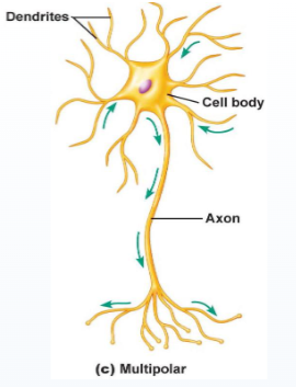
what is the basic CNS neuronal structure?
Multipolar neurones (unipolar and bipolar rare in CNS)
Cell soma with multiple processes (axon, dendrites)
Synapses formed between axon (outgoing) and dendrites (incoming)
what is a projection neurone?
neurones with long axons that project to other parts of the CNS
What are local/relay neurones?
neurones with short axons that contact other cells in the local brain area
what are interneuones?
type of relay neurone
usually GABAergic
Occasionally cholinergic, dopaminergic
what are GABAergic interneurons?
Inhibitory local neurones; vast majority of interneurones
Prevent overactivity (q.v. epilepsy)
Refinement of signals to other neurones; e.g. visual cortex for acuity
Co-ordinate activity across multiple neurones; e.g. oscillations
how can neurones be classified by their electrical properties?
Tonic / regular firing: constantly fire APs
Phasic / bursting: fire APs in short bursts of several APs
Fast spiking: fire APs at high rates
what is the soma of the neurone?
Main body of the cell; contains all normal cell organelles
Nucleus, Golgi apparatus, lysosomes
Only source of protein synthesis & degradation in neurones
what is the neuronal cytoskeleton?
Similar to cytoskeleton of other eukaryotic cells
Highly dynamic; heavily involved in cell structure, growth, transport
what are the three main components of the neuronal cytoskeleton?
Microtubules
Neurofilaments
Microfilaments
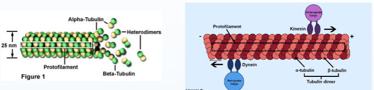
what are microtubules?
Tubulin polymers particularly associated with cellular transport
Helical structure of 13 protofilaments
Numerous neurological disorders connected to dysfunction of proteins associated with microtubules
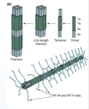
what are neurofilaments?
Equivalent to non-neuronal intermediate filaments, ~10nm diameter
Particularly common in axons
Important for development and radial growth of axons, maintenance of axon calibre and action potential conduction
Also heavily associated with neurological disorders accumulation observed in amyotrophic lateral sclerosis, Charcot-Marie- Tooth disease, Alzheimer’s, etc.
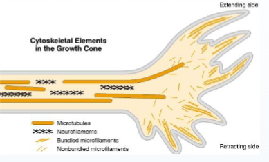
what are microfilaments?
Formed of actin, ~4-6nm diameter
Highly present in dendritic spines, axon terminals, growth cones
Important for:
Membrane integrity
Distribution of membrane proteins
Cell morphology
Interactions with extracellular environment
what is fast and slow transport?
Fast (~400 mm/day, < 1 μm/s) or slow (8mm, <0.1 μm/s)
Along microtubules; anterograde transport mediated by kinesin motors, retrograde by dynein
Fast anterograde e.g. synaptic vesicles
Fast bidirectional e.g. mitochondria, mRNA
Fast retrograde e.g. endosomes
Slow anterograde e.g. neurofilaments, tubulin
Transport directionally balanced to prevent accumulation
what are examples of neurological and neurodevelopmental disorders?
Tau protein (MAP): Alzheimer’s (tau tangles)
Neurofilaments: Charcot-Marie-Tooth disease
Dynactin: Motor Neurone Disease
Huntingtin (?): Huntington’s Disease
Dynein & kinesin: Forms of intellectual disability
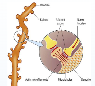
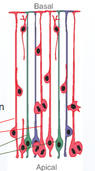
what are dendrites?
“Proximal” dendrites start as perikaryal extensions; wide
Become progressively smaller diameter towards “distal” dendrites, multiple branching
Main receptive field of neurones
Irregular outline: dendritic spines
Some organelles (e.g. mitochondria)

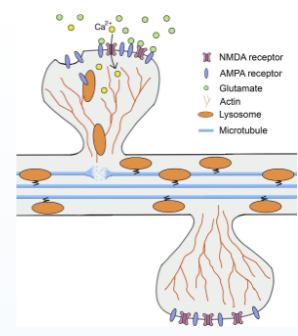
what are dendritic spines?
Small membranous extension from the dendrites which form postsynaptic area of the synapse
Structure and contents key to proper synaptic function
Compartmentalisation: spine “neck” restricts movement between spine and dendrite (e.g. Ca2+)
Contain ribosomes & some ER
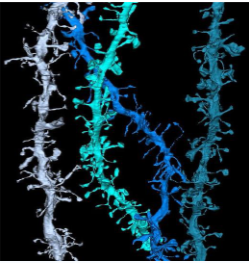
what is the structure of dendritic spine?
Rich in microfilaments; microtubules and intermediate filaments absent
Contain ribosomes & some ER
“Thin”, “stubby”, “mushroom”, etc
what is dendritic transport?
Similar in process to axonal transport; motor proteins along microtubules
Dendrites rich in microtubules
Delivery of key dendritic proteins (e.g. receptors), lysosomes, etc. anterograde and retrograde
Dendritic vesicles distinct from axonal vesicles; sorting mechanism unknown
what is glia?
Support cells for the CNS: astrocytes, oligodendrocytes (Schwann cells in periphery), microglia, ependymal cells, radial cells, etc.
Interact heavily with neurones
Number of glial cells highly uncertain
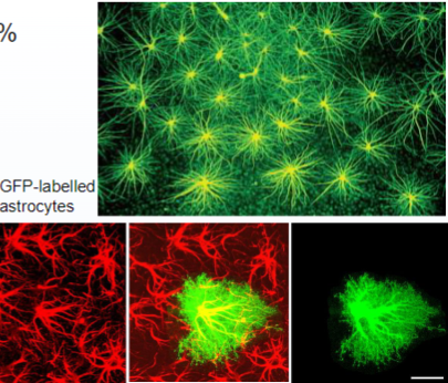
what are astrocytes?
Most numerous glial cell? (20-40% brain cells?)
Structural & metabolic support, transmitter uptake, ion balance
Protection
Brain-vascular interaction
what is the metabolic support of the glia?
Regulation of ion homeostasis (particularly K+)
Transmitter reuptake and metabolism (Glu, GABA, DA, etc.); release NT precursors back into extracellular space
Glycogenolysis & gluconeogenesis; release of lactate to neurones during high energy demand
what is neuroprotection?
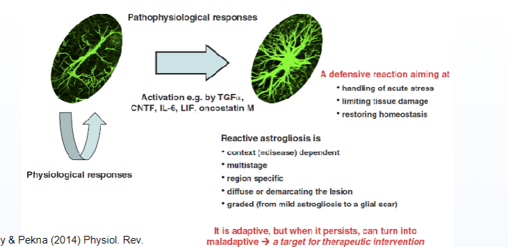
Astrogliosis (aka astrocytosis, reactive astrocytosis)
Proliferation & symptomatic alteration in astrocytic function due to CNS damage
Associated with trauma, stroke, infection, neurodegeneration, epilepsy, etc.
“Glial scar”
May be pathological if overactive
what is functional hyperemia?
Brain has high energy requirements and low energy storage
Mechanism to increase blood flow to areas of high activity; change in blood flow ~ 1-3s after activity
Signalling from neurones (via NO) and astrocytes (PLA2 / AA)
Impaired functional hyperemia may be associated with pathological conditions (e.g. Alzheimer’s, diabetic retinopathy)
what are astrocytes functions?
Have receptors for major NTs (mostly metabotropic)
Show Ca2+-evoked release of transmitters (ATP, glutamate, d- serine; “gliotransmission”) that excite neurones
Are not electrically excitable, but are excitable: Ca2+ activity in response to neuronal transmission
Can respond differently depending on neurone and neuro- transmitter released
what is astrocyte signalling?
Astrocytes connected by connexin hemichannels
Syncytium; although not in truest sense as astrocytes more capable of discrete, independent activity.
Show Ca2+ waves (IP3 receptors most heavily implicated)
Ca2+ waves associated with gliotransmitter release (and subsequent neuronal activation)
Activated by NT release
what is the relation between astrocytes & synapse?
The concept of the astrocyte’s anatomical and function integration with pre-and postsynaptic neurones.
Exchange of information between the astrocyte and neurones
Astrocytic involvement in synaptic activity, synaptic plasticity, neurological disorders
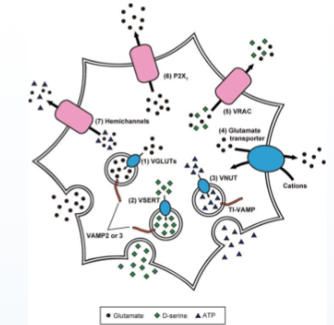
what is gilotransmission?
Astrocytes release transmitters which activate neurones – termed gliotransmitter
Main gliotransmitters:
Glutamate
D-serine
ATP (& adenosine)
ATP may generate astrocytic Ca2+ waves
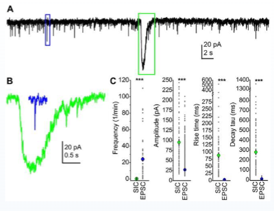
what is extra synaptic signalling?
“Slow inward currents” (SICs) & “slow outward currents” (SOCs)
Poorly understood
Astrocyte-derived Glu/GABA release activating neurones
Capable of synchronising neuronal activity
what is the CNS immune system?
CNS has a “privileged” immune system; separate from periphery
No lymphatic system
Separated from blood by BBB
CNS & peripheral immune systems recognise each other as foreign
what is microglia?
First line defence, innate & adaptive immune systems express immune proteins (e.g. MHCs, TLRs, etc.) can mediate recruitment of peripheral immune system
what is the function of microglia?
5-20% of glial cells
Normally exist in ramified form monitoring CNS; small soma and processes
Activation for immune response converts to reactive microglia, with amoeboid form, and proliferation
what are the two microglia stages?
Non-phagocytic stage: soma enlarges, proliferation, processes shrink
Phagocytic stage: Become fully amoeboid
what is microglial immune signalling?
Activated by infection, injury, neurodegenerative diseases
Although different cells for CNS immune system, similar signalling molecules involved
Recognition of pathology-associated molecules e.g. LPS, amyloid beta (Aβ), thrombin, IFN-γ, proinflammatory cytokines
Release of inflammatory mediators e.g. IL-1, IL-6, TNFα
what are pathological immune responses?
Chronic immune responses in the brain become pathological
Associated with neurodegenerative disorders, epilepsy, etc.
Release of cytotoxic chemicals e.g. ROS, glutamate → enhanced neuronal damage
May be exacerbated by recruitment of peripheral immune system
what are oligodendrocytes?
Form myelin in the CNS
Pathology: multiple sclerosis
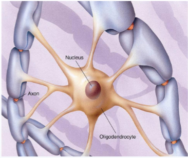
what is oligodendrocyte myelination?
Selectively myelinate axons with diameter >0.2 μm; recognition mechanism unknown
Myelination stimulated by electrical activity; inhibition of AP generation inhibits myelin formation
Myelination stimulated by Leukaemia Inducing Factor (LIF) induced by axonal ATP release
Efficient myelination also affected by various axonal membrane proteins
what are oligodendrocyte vulnerable to?
oxidative stress
High metabolic demand of myelination
Toxic byproducts
High intracellular iron concentration
Low antioxidant concentrations
Expression of pro-excitotoxic receptors
Often seriously degraded as byproduct of neuronal / astrocytic death
Myelin sheaths vulnerable to autoimmune attack
what is multiple sclerosis?
Immune-mediated CNS disorder
Symptoms: muscle weakness, decreased co-ordination, visual & sensory deficits, autonomic deficits, cognitive deficits (rarely).
CNS lesions & inflammation
Death of oligodendrocytes and demyelination
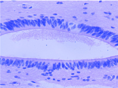
what are ependymal cells?
Line walls of ventricles
Rod-shaped & ciliated
Role in production and regulation of CSF
what are radial cells?
Important in neural development
Progenitor cells for neurones and glia
Also guide migration of cells
what are other glial cells?
Bergmann glia: Cerebellar radial cells that persist into adulthood where they function similarly to astrocytes
Muller glia: Retinal radial cells; function similarly to astrocytes
Schwann cells: Oligodendrocytes in the periphery
Satellite glia: Perform a similar function to astrocytes at autonomic ganglia