Parathyroid & Other Neck Pathology
 Call Kai
 Learn
Learn Practice Test
 Spaced Repetition
 Match
Match Flashcards
 Knowt Play
1/24

There's no tags or description
Looks like no tags are added yet.
Name | Mastery | Learn | Test | Matching | Spaced | Call with Kai | Chat |
|---|
No analytics yet
Send a link to your students to track their progress
25 Terms
the most common cause of enlargement of a parathyroid gland
parathyroid adenoma
if a patient has elevated serum calcium (hypercalcemia), analyze the neck carefully for signs of a
parathyroid adenoma
clinical findings of a parathyroid adenoma
elevated serum calcium
elevated PTH
sonographic findings of a parathyroid adenoma
hypoechoic mass adjacent to the thyroid
enlargement of the cervical lymph nodes, or —
lymphadenopathy
lymphadenopathy usually measure less than —
1 cm
clinical findings of abnormal lymph nodes
palpable neck mass (possibly)
enlarged nodes may be painful
sonographic findings of abnormal lymph nodes
enlargement of the node >1 cm
rounded shape
loss of the echogenic hilum
calcifications
may be hyperemic or demonstrate abnormal vascular patterns with color doppler
thyroglossal duct cysts are
benign congenital cysts located within the midline of the neck superior to the thyroid gland and are typically located below the level the hyoid bone
brachial cleft cysts are
congenital neck cysts as well
brachial cleft cysts are most often found near the
angle of the mandible
fibromatosis colli is a rare,
pediatric fibrous tumor located within the sternocleidomastoid muscle
the likely cause of fibromatosis colli
malposition in utero or a traumatic birth that leads to the development of fibrosis (scar tissue) within the muscle
congenital muscular torticollis
the mass within the sternocleidomastoid muscle causes the muscle to shorten, resulting in teh twisting of the infancts chin toward the nonaffected side
clinical findings of a thyroglossal duct cyst
palpable mass within the midline of the neck superior to the thyroid gland
infected cysts may be painful
sonographic findingd of a thyroiglossal duct cyst
anechoic, well-defined, and unilocular cyst with posterior enhancement
may have internal components
clinical findings of a branchial cleft cyst
palpable neck mass located near the angle of the mandible
infected cysts may be painful
sonographic findings of branchial cleft cyst
anechoic mass near the angle of the mandible
clinical findings of fibromatosis colli
pediatric palpable neck mass
torticollis (twisted neck with the chin angled to the nonaffected side
sonographic findings of fibromatosis colli
fusiform-shaped mass within the sternocleidomastoid muscle that is hypoechoic, hyperechoic, or even isoechoic to the adjacent tissue
may also contain calcifications that shadow
may yield a hyperemic pattern with color doppler
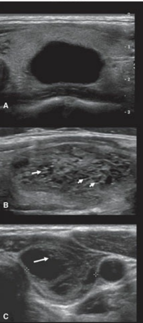
low risk thyroid cancer
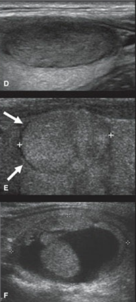
intermediate risk thyroid cancer
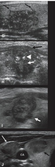
high risk thyroid cancer
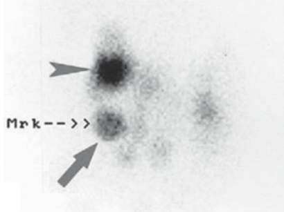
Nuclear medicine images of thyroid nodules. A radioactive marker (Mrk) was placed over a 2-cm palplable nodule (arrow) in the right thyroid lobe. This nodule represents a “hot” nodule, which is most likely benign.
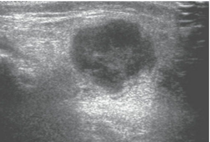
Abnormal lymph node. This lymph node is enlarged, hypoechoic, and has lost its echogenic hilum.