Electrophysiology of cardiac muscle
1/22
Earn XP
Description and Tags
lecture 5
Name | Mastery | Learn | Test | Matching | Spaced | Call with Kai |
|---|
No study sessions yet.
23 Terms
How does electrical excitation propagate through cardiac muscle?
cardiac cells form an electrical syncytium
electrical signal propagates through gap junctions
structure of cardiac myocytes in the SAN
fewer myofibres and mitochondria
smaller sarcoplasmic reticulum compared to atrial or ventricular cardiac myocytes
primary function generation and conduction of a pacemaker potential
Why is conduction through the AVN slower than the SAN?
AVN surrounded by a fibrous AV ring
Why is the conduction delay between the SAN and AVN important?
ensures sequence of atrial and ventricular contractions is well time
Where does the electrical signal propagate after the AVN?
bundle of His and Purkinje fibres
propagation fast because their main function is to activate contraction in the ventricle
purkinjie fibres split into 2 branches into the 2 ventricle walls
ventricles contract from the apex to the base
Why do ventricles contract from the apex to the base?
ensures max blood leaves the ventricles purely through the mechanical squeezing and pressure gradient.
What happens with damage to the SA node?
cells in the AVN and Purkinje fibres can also function as pacemakers
intrinsic pacemaker rate is slower than the SAN
fastest pace and therefore normal heart rate determined by the SAN
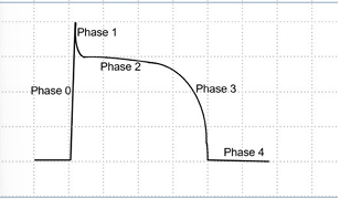
What is phase 0 of action potential generation in cardiac myocytes?
rapid depolarisation phase
driven by voltage-gated Na+ activation and slow opening of Ca2+ channels
more or less fast depending on the type of the cell

What is phase 1 of action potential generation in cardiac myocytes?
the initial brief rapid repolarisation
driven by opening of K+ channels
Na+ channels close to inactive state

What is phase 2 of action potential generation in cardiac myocytes?
the plateau
Ca2+ influx balances K+ efflux
Ca2+ channels are L-type voltage-gated channels (dihydropyridine receptors)

What is phase 3 of action potential generation in cardiac myocytes?
terminal repolarisation
delayed activation of different K+ channels
restores the membrane potential to the resting level

What is phase 4 of action potential generation in cardiac myocytes?
electrical diastole
returns to resting potential
heart waiting for another electrical trigger to restart the cycle
What is a consequence of this prolonged action potential?
results in long refractory period
prevents summation and tetanus from occuring
How is the SAN action potential different?
does not display rapid phase 1 depolarisation
plateau phase not very prominent
SAN cells do not have a true resting potential - membrane potential always changing
do not depolarise as much as other muscle cells
funny current
spontaneous slow increase in current during phase 4 depolarisation in SAN cells
due to non-specific cation channel (HCN channel)
HCN channel has mixed permeability for both Na+ and K+
called funny because HCN channel opens on hyperpolarisation, not depolarisation
When does the HCN channel open?
end of phase 3 depolarisation
leads to an inwards cation current
drives slow membrane depolarisation in phase 4
What does the slope of the phase 4 slow depolarisation determine?
heart rate
When potential reaches threshold (~40mV) in SAN?
Ca2+ channels open
rapid depolarisation but still slower than in ventricles as no Na+
at peak Ca2+ channels close & K+ open leading to repolarisation
effect of noradrenaline and adrenaline on funny current
increase funny current
reduce phase 3 depolarisation
increased heart rate
shorter but higher plateau in ventricular cells
stronger contraction
Calcium induced calcium release
ryanodine receptors activated through the extracellular Ca2+ that enters through dihydropyridine receptors
Consequences of calcium induced calcium release
risk of calcium overload therefore a regulatory mechanism needed to balance Ca2+ levels with each action potential
extracellular Ca2+ influences how much tension is being produced
How is Ca2+ concentration regulated during CICR?
high activity of Na+/Ca2+ exchanger in the sarcolemma
re-sequestered in SR between action potentials, controlled by SERCA pump
SERCA pump inhibition and stimulation
inhibited: Phospholamban (PLN), relieved upon phosphorylation of PLN
stimulation: adrenaline. PKA acts downstream of adrenaline & phosphorylates PLN. During fight-or-flight response, adrenaline leads to activation of SERCA pump