Kidneys and Suprarenals
1/23
There's no tags or description
Looks like no tags are added yet.
Name | Mastery | Learn | Test | Matching | Spaced |
|---|
No study sessions yet.
24 Terms
What are the two functions of the urinary system?
Filtration of blood & formation of urine
Waste excretion
fluid & electrolytes balance -> body fluid volume & blood pressure
Acid-base balance
Formation & release of hormones
Renin > blood pressure
Erythropoietin > red blood cell production
Calcitrol (vit D) > calcium balance
Describe the location of the kidneys, ureter, and suprarenal
Kidneys:
retroperitoneal in the posterior abdominal region, lying in the extra- peritoneal connective tissue lateral to the vertebral column.
extend from approximately Tv12 –Lv3
Right Kidney = Lower then Left Kidney
Ureter:
run inferiorly from the kidneys , passing over the pelvic brim at the bifurcation of common iliac arteries.
suprarenals are associated with the superior pole of each kidney
Describe the Hilum of the Kidneys
Which Border?
Transmits?
Continious w/?
On medial Border
Transmits
renal vein,
renal artery,
ureter
Lymphatics
Nerve Plexus
continuous with renal sinus which contains the upper expanded end of ureter (renal pelvis)
Describe the Relationships to other structures:
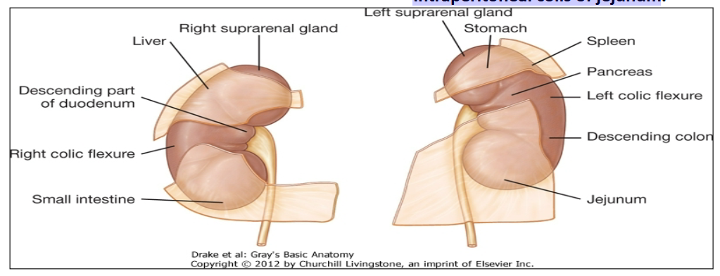
Anterior Surface Right Kidney
Anterior Surface Left Kidney
posterior surface of the Right and Left
Kidneys
Anterior surface of Right Kidney:
right suprarenal,
liver,
descending part of duodenum,
right colic flexure,
small intraperitoneal part of the small intestine.
Anterior surface of Left Kidney:
left suprarenal and intraperitoneal part of stomach
and spleen,retroperitoneal pancreas,
left colic flexure,
descending colon,
intraperitoneal coils of jejunum
posterior surface of the Right and Left Kidneys
diaphragm superiorly, psoas major, quadratus lumborum, and transversus abdominis inferiorly.
subcostal nerves and vessels, as well as the iliohypogastric and Ilioinguinal nerves, also pass posterior to both kidneys.
Left kidney is anterior to 11th & 12th ribs
The right kidney is anterior to the 12th rib only.
The pleural sacs and the Costodiaphragmatic recesses lie posterior to the kidneys.
Describe the three coverings of the kidneys
Perirenal or Perinephric fat: covers the fibrous capsule and adrenal glands.
Fibrous capsule: Surrounds the kidney , closely applied to its outer surface
Renal fascia (of Gerota )
condensation of extra peritoneal fibrous connective tissue enclosing the perirenal fat.
encloses the kidneys & suprarenal glands, the two being separated by a thin septum.
is incised during any surgical approach to the region
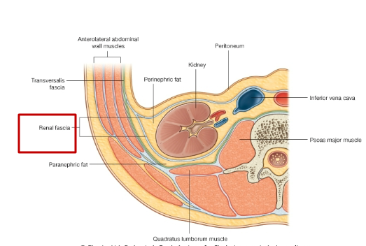
Describe the Coverings of the kidneys:
Superior
Lateral
Medial
Inferior
Pararenal fat or paranephric fat
Superiorly
renal fascia is continuous with the diaphragm
Laterally
renal fascia is continuous with the fascia transversalis
Medially
anterior layer is continuous with the sheath of renal vessels, the aorta, and the Inferior vena cava,
posterior layer blends with the fascia of psoas major.
Inferiorly
renal fascia encloses the ureter and directs the perinephric infection inferiorly into the pelvis.
Pararenal fat or paranephric fat:
external to renal fascia; often in large quantity; part of retroperitoneal fat.
Describe the Kidney Internal Structure
Each kidney consists of an outer cortex and an inner medulla.
Extensions of renal cortex (the renal columns) project in the
medulla and divide it into discontinuous aggregates (renal pyramids).The bases of these pyramids are directed towards the cortex, while
the apices are inwards to the renal sinus. The apical projections (renal papilla) are surrounded by a minor calyx.In the renal sinus, several minor calyces unite to form a major calyx, and 2-3 major calyces unite to form the renal pelvis, which is the funnel-shaped superior end of the ureter.
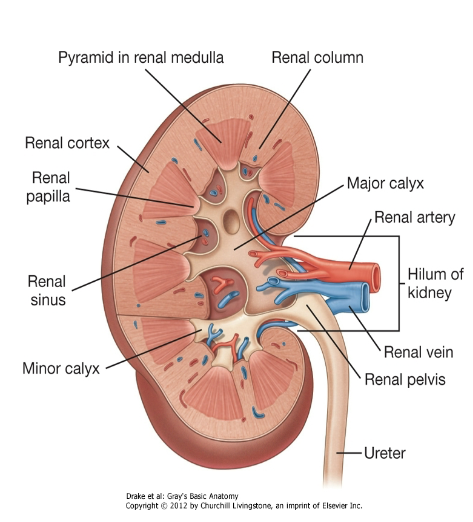
What does the Cortext and Medulla Contain?
Cortex
renal corpuscles ( glomerulus & Bowman's capsule)
proximal parts of proximal and distal convoluted tubules and collecting ducts .
Medulla
thick and thin limbs of loops of Henle
distal parts of proximal and distal convoluted tubules and distal collecting ducts
Describe the Renal Vasculature:
Renal Artery?
Renal Veins?
Renal artery:
Branch of the abdominal aorta.
arises just below the origin of the superior mesenteric artery, between Lv1 and Lv2.
The right renal artery is longer and passes behind the IVC.
Accessory renal arteries are common, may enter through the hilum or at any other point
extra hilar arteries.
Renal Veins
Multiple renal veins unite to form the Left & Right Renal Veins, both of which lie anterior to the arteries at the hilum and drain into the IVC.
Left renal vein is longer, crosses midline between the aorta and the SMA, and can be compressed by an aneurysm in either of these two vessels.
Describe the branching of the renal artery into the kidneys
Each renal artery divides into an anterior and a posterior branch, which further subdivide into 5 segmental end arteries (do not anastomose )
Five segments in each kidney, supplied by segmental arteries are: superior, inferior, posterior, anterosuperior& anteroinferior.
Lobar a.>>interlobar a. >>> arcuate a. >>> interlobular a.
>>> afferent arterioles >>> glomerulus >>> efferent
arteriole >>> cortical peritubular capillaries (cortex) or vasa rectae spuriae (medulla)
Describe the descending portion of the vasa recta and the ascending venous side
The descending portion of the vasa recta is arterial and
is composed of small- diameter vessels with continuous endothelium.
The ascending venous side is larger-diameter vessels with
thin , fenestrated walls .The ascending venous limb drains into interlobular vein >>> arcuate vein >>> interlobar vein >>>> finally exiting each kidney via the renal vein
Describe the Clinical Correlates:
What is clinically important about Segmental renal arteries
What is renal vein entrapment syndrome?
Segmental renal arteries:
end arteries with no effective collateral circulation
If a segmental artery is occluded (thromboembolus from
the left atrium or an atheromatous lesion) → infarction; Infarction =:clinically silent or cause pain with costovertebral angle tenderness and hematuria
lack of collateral circulation also means that a renal segment can be surgically resected, leaving the adjacent segments functioning
renal vein entrapment syndrome:
left renal vein crosses toward the inferior vena cava through the angle between the superior mesenteric artery and the aorta.
Vein Compression = renal vein entrapment syndrome (nutcracker syndrome)
left flank pain and hematuria.
Male patients may develop a varicocele and left testicular pain.
NOTE:
renal vein entrapment syndrome differs from the superior mesenteric artery syndrome, in which the third part of the duodenum is compressed at the same angle.
Describe the location of the Ureter
Upper half in the abdomen (retroperitoneum) on the
medial aspect of the psoas major muscle.At the pelvic brim, the ureter crosses the common iliac
artery or the beginning of the external iliac artery >> before
it enters the urinary bladder in the pelvic cavity.Adheres to the peritoneum. Mobilization of the peritoneum puts the ureter at risk.
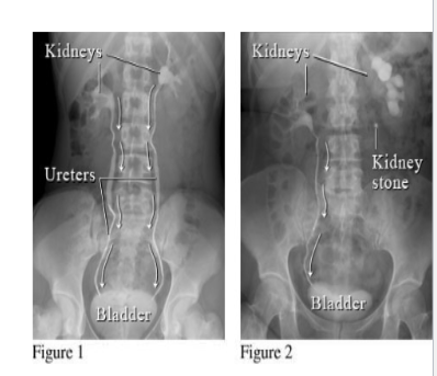
Where are normal spots in which renal calculi (stones) may get impacted
ureteropelvic junction: where the renal pelvis joins the ureter.
as the ureter crosses the pelvic brim: at the bifurcation of the common iliac artery.
ureterovesical junction: where the ureter pierces Obliquely through the wall of the urinary bladder.
Describe Urinary Tract Stones (renal calculi )
Etiology
Patient presentation
Treatment?
Etiology:
More common in men ( 20-60 years) and associated with sedentary lifestyle.
Patient Presentation:
patient complains of severe intermittent pain ( ureteric colic) which may be felt from loin to groin , proximal anterior aspect of thigh or external genitalia . (T11- L2).
May also have blood in the urine ( hematuria )
Treatment:
Lithotripsy sends a shock wave that breaks the stone into
fragments which then pass in the urine.
Describe Staghorn Calculus
also sometimes called coral calculi, obtain their characteristic
shape by forming a cast of the renal pelvis and calyces, thus
resembling the horns of a stag.
Describe the Blood Supply to Ureter
Upper End
Middle
Pelvic Cavity
Arterial
Upper end:
branches of the renal artery
Middle:
branches from the testicular / ovarian arteries , the abdominal aorta, and the common iliac arteries .
In the pelvic cavity:
branches of the internal iliac arteries.
Arteries divide into ascending and descending branches, which form a longitudinal anastomosis.
Veins:
Upper end: renal v.
Middle: gonadal v.
In the pelvic cavity: branches of the internal iliac veins.
Describe the Lymphatic Drainage of the Kidneys and Ureters
Kidneys: paraaortic (lateral aortic or lumbar) nodes, around the origin of renal artery
Ureters:
upper: paraaortic (lateral aortic) nodes
middle: common iliac nodes
inferior: external, or internal iliac nodes
Describe the Nerve Supply of kidneys & Ureters/ Referred Pain?
Sympathetic fibers:
Kidneys: T10-T12
Ureters: T11-L2
Parasympathetic fibers:
Kidneys: vagus
Ureters: vagus & S2-S4
Fibers pass through renal or hypogastric plexuses and are
distributed along branches of blood vessels
Ureteric pain is referred to the cutaneous areas supplied by the
T11-L2 spinal cord level .
posterior and lateral abdominal wall below the ribs and above the iliac crest, the pubic region, scrotum in males, and labia majora in females, and the s and proximal anterior part of the thigh. ( lion to groin)
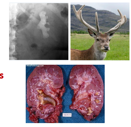
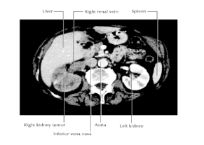
Describe Renal Cell Carcinoma:
Patient Presentation
Complication
Renal cell carcinomas:
Grow outward of the kidney into fat & fascia
Patient Presentation:
Most patients complain of blood in the urine (hematuria), pain in the infrascapular region (loin), and a mass.
Complication:
invade the renal vein Spread to the inferior vena cava, right atrium, and across the tricuspid valve into the pulmonary artery.
What is an ideal location for Renal Transplant?
Iliac fossa is an ideal location for the transplant because a
new space is created without compromising the other structures
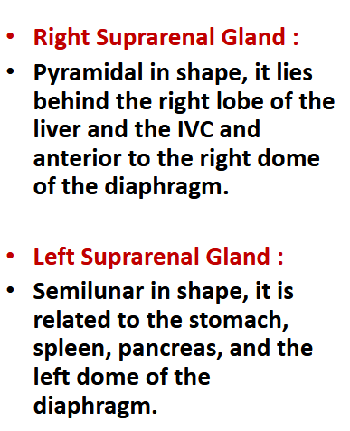
[REVIEW] shape of suprarenal glands
Describe the blood supplies to the Supra Renal Gland
A. Arteries
Superior suprarenal a. <<< inf. phrenic a.
Middle suprarenal a. <<< abdominal aorta
Inferior suprarenal a. <<< renal a.
B. Veins
Right suprarenal v. >>> IVC
Left suprarenal v. >>> left renal v.
Describe the Nerve Supply to Suprarenal Gland
Primarily Sympathetic:
Primarily sympathetic fibers via the greater, lesser, and least splanchnic nerves.
Preganglionic fibers end in the adrenal medulla
Postganglionic fibers supplying blood vessels arise from ganglia around the aorta.