Radiology Alternate Imaging Modalities 1 and 2
 Call Kai
 Learn
Learn Practice Test
 Spaced Repetition
 Match
Match Flashcards
 Knowt Play
1/46

There's no tags or description
Looks like no tags are added yet.
Name | Mastery | Learn | Test | Matching | Spaced | Call with Kai | Chat |
|---|
No analytics yet
Send a link to your students to track their progress
47 Terms
What if fluoroscopy?
an imaging technique commonly used to obtain real-time moving radiographic images of the internal structures
it is inverted = bone is black and gas is white
How does a CT scanner work?
an x-ray tube spins around the patient
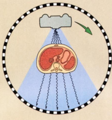
What is the difference between a single slice and a spiral?
Single slice involves the complete rotation of the tube then the table moves before the next rotation of the tube. Spiral involves continuous rotation of the tube linked to continuous movement of the table.

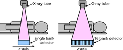
How does a multi-detector CT scanner work?
It creates a reconstruction of an object from multiple projections by utilizing multiple rows of detectors.
This allows them to capture multiple slices of the body in a single rotation, resulting in faster scans and higher resolution images.
what is the difference between a pixel and a voxel?
a pixel is a picture element that has only two dimensions (x,y). A voxel is a volume picture element and has three dimensions (x,y,z).
pixel = radiograph
voxel = MRI and CT
What do the different voxel values (x,y,z) represent?
X and Y are determined by the image matrix size. Z is based on the slice thickness.

What effect does slice thickness have on spatial resolution?
thicker slices cause a loss of spatial resolution, therefore an isotopic voxel should be used.
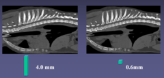
What is an isotopic voxel?
x, y, and z dimension are equal. This allows for multiplanar reconstructions and volume rendering without the loss of resolution.

What are Hounsfield units (HU)?
HU values are used to represent different tissue densities in CT, but there are limitations on how many of the values can be displayed.
The typical monitor can only display 256 shades of grey even though there are >4000 HU values.
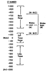
What is window?
the range of HU represented on a specific image (the picture shows a window of 1818 HU to -1818 HU.

What is level?
the central value of the HUs displayed (the image has a level of 0 HU)

A wide window (400-2000 HU) is best used for what?
Imaging tissue types that vary greatly (ex. bone and lung)
The goal is to see all the various tissues in one image
A narrow window (50-400 HU) is best used for what?
imaging tissue types with similar densities (ex. white and grey matter of the brain)
It provides greater density discrimination and contrast
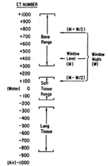
what does hyperattenuating mean?
WHITE- blocking x-ray from detector
Highest HU values (bone, metal)
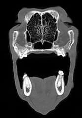
What does hypoattenuating mean?
BLACK
Lowest HU values (air)

What are the advantages of CT?
1. more detailed view of anatomy without superimposition
2. faster and less expensive than MRI
3. best for bone, lung, abdomen
What are the disadvantages of CT?
1. radiation exposure
2. more expensive than radiographs
3. poor soft tissue contrast (compared to MRI)
What is the basic principle behind how MRI scans work?
atoms with an odd number of protons possess a magnetic moment (hydrogen contains one proton and therefore acts like a tiny magnet). Normally, magnets are in random alignment which cancel out any net magnetic moment. With an MRI, the hydrogens will align with/against the magnetic moment of the machine
How is an image formed in an MRI?
some hydrogen atoms line up with the magnetic field and other hydrogen nuclei line up opposite to the direction of the magnetic field (it takes less energy to align with the field than against). Transiently, a radiofrequency coil applies energy which allows the hydrogen nuclei in line with the magnetic field to flip in the other direction. As the H returns to its normal orientation, there is energy released which can be detected.

What is weighting?
the use of certain parameters to optimize the differences in relaxation rates to different tissues in order to provide contrast in the image
What are the two main weights?
T1-weighted
T2-weighted
What is T1-weighted?
fluid is hypointense (BLACK) bc they have long relaxation times
fat is hyperintense (WHITE) bc it has short relaxation times
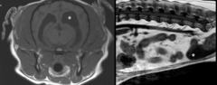
what is T2-weighted?
fluid is hyperintense (WHITE)
Fat is hyperintense (WHITE)
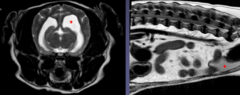
what does hypointense mean?
less bright than tissue you compare to
What does isointense mean?
same brightness as tissue you compare to
What does hyperintense mean?
brighter than the tissue you compare to
What are the advantages of MRI?
1. better soft tissue contrast and detail
2. noninvasive
What are the disadvantages of MRI?
1. long scan times (30 min-2 hours)
2. general anesthesia
3. cost
What are the main differences between CT and MRI images?
CT gives better bone detail and is good for the thorax and abdomen. MRI is better for soft tissue detail and good for neuroimaging and musculoskeletal
CT is not good for neuro
what are the main differences between CT and MRI image aquisition?
CT requires heavy sedation or general anesthesia. It has faster scan times (seconds to minutes)
MRI requires general anesthesia and has a longer scan time of 1-2 hours
What are the main differences in CT and MRI safety issues?
CT has ionizing radiation
MRI has projectiles, metal implants
What are some things that you should remember about the MRI?
1. the magnet is ALWAYS on
2. ferromagnetic objects = projectile
3. anything that can be demagnetized should be removed (credit cards, ID cards, etc.)
4. a screening form should be completed before entering the room
How do US machines create sound waves?
it transmits (1%) of the time- propagates sound waves into the tissues
it receives 99% of the time- measure the intensity and time of arrival of returning sound waves
What is sound?
a vibration that transmits energy by alternating regions of high pressure (compression) and low pressure (rarefaction)
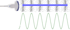
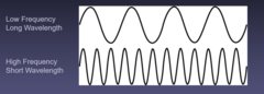
lower frequency transducers penetrate _________________
FARTHER- but they have lower resolution
good for deep or large patients
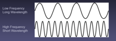
Higher frequency transducers penetrate ________________
NOT AS FAR- but they have higher resolution
How is the image formed for US?
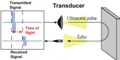
using the echoes returning from the tissues. the distance of the reflector from the transducer is based on the time of flight of sound wave
What speed does sound travel in tissues?
a relatively constant speed of 1540 m/s
How does US interact with matter?
differences in acoustic impedance (resistance to flow) in the various tissues will alter the direction of the sound wave
tissues can scatter waves (don't see good margins)

What does anechoic mean?
black = no echoes bouncing back (fluid; bile, urine)
What does hypoechoic mean?
darker (dark grey) than compared structure
What does isoechoic mean?
the same echogenicity to compared structure
What does hyperechoic mean?
brighter than compared structure (bone, gas, organ boundaries)
What is the echogenicity of body tissues and substances
Big, Ugly
Red
Moose
Really
Likes
Stealing
People's
Bananas and Grapes

What is the doppler effect?
the perceived change in frequency that occurs as a result of the motion between the sound source and the observer
Allows determination of velocity and direction of blood flow. Toward us = higher frequency

What is color flow mapping?
signals from the moving RBCs are displayed in color as a function of their motion towards or away from the transducer

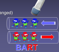
What is the BART method?
BLUE = away
RED = toward