Regional anatomy 7 -- the neck 1
1/27
There's no tags or description
Looks like no tags are added yet.
Name | Mastery | Learn | Test | Matching | Spaced |
|---|
No study sessions yet.
28 Terms
The neck –
A tube providing continuity from the head to the trunk
It extends –
Anteriorly from lower border of the mandible to the upper surface of the manubrium of sternum
Posteriorly from the superior nuchal line on the occipital one of the skull to the intervertebral disk between C7 & T1 vertebrae –
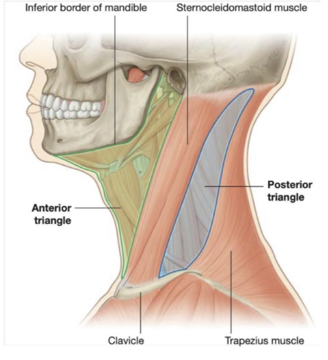
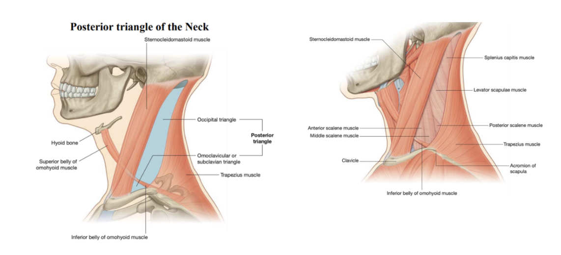
Triangles of the neck —
Divided into anterior & posterior triangles –
The boundaries of the anterior triangle are the anterior border of the sternocleidomastoid muscle, the inferior border of the mandible, and the midline of the neck
The boundaries of the posterior triangle are the posterior border of the sternocleidomastoid muscle, the anterior border of the trapezius muscle, and the middle one-third of the clavicle
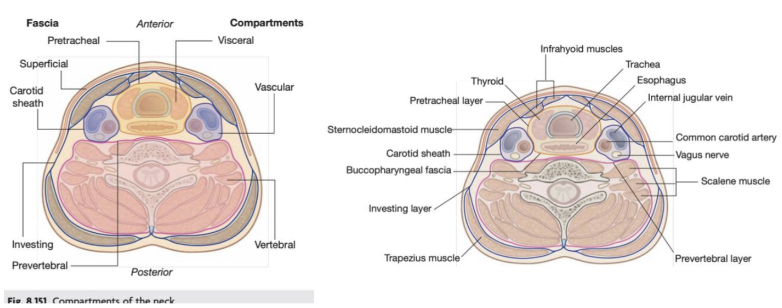
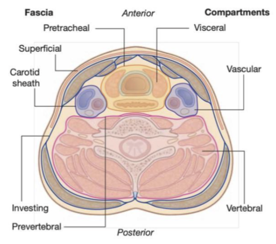
Four compartments in the tube provide longitudinal organization –
Visceral compartment –
Anterior and contain parts of the digestive & respiratory system, and several endocrine glands
Vertebral compartment –
Posterior and contains the cervical vertebrae, spinal cord, cervical nerves, and muscles associated with the vertebral
2 Vascular compartments —
One on each side – lateral & contain the major blood vessels (Carotid, jugular vein) & the vagus nerve (X)
Neck fascia –
The soft connective tissue of the neck gets denser & creates a generally vascular plane – used as surgical planes
2 types —
Superficial fascia
Deep cervical fascia

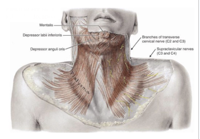
Superficial fascia
Contains a thin sheet of muscle (the platysma) –
Begins in the superficial fascia of the thorax & runs upwards to attach to the mandible & fuse with the mimic muscles of the face –
Innervated by the cervical branch of the facial nerve (VII), and is only found in this location
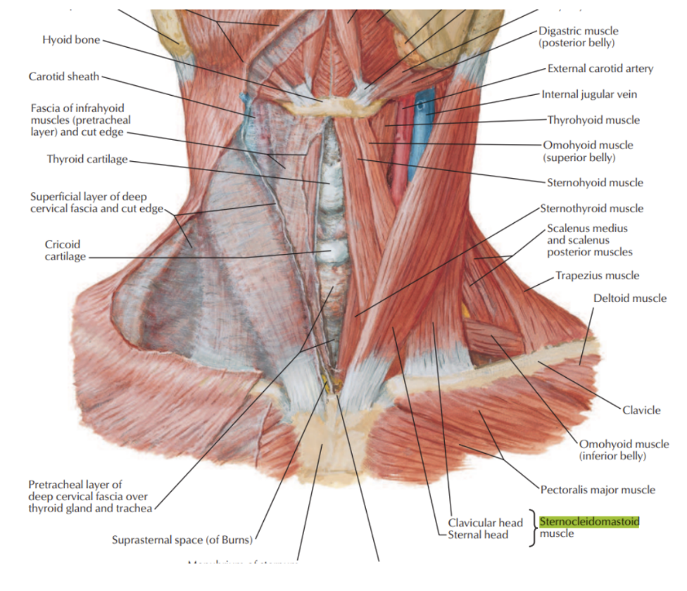
The deep cervical fascia –
Organized into several distinct layers, including –
An investing layer
Surrounding all structures in the neck
The prevertebral layer
Surrounds the vertebral column and the deep muscles associated with the back
The pretracheal layer
Encloses the viscera of the neck
The carotid sheaths –
Receive a contribution from the other three fascial layers and surround the 2 major neurovascular bundles on either side of the neck
The investing layer –
Completely surrounds the neck
Attaches posteriorly to the ligamentum nuchae and the spinous process of the CVII vertebrae,
Splits as it posses forward to enclose the trapezius muscle & reunites into a single layer as it forms the roof of the posterior triangle
Splits again to surround the sternocleidomastoid muscle, and reunites again to join its twin from the other side
Anteriorly, it surrounds the infrahyoid muscles
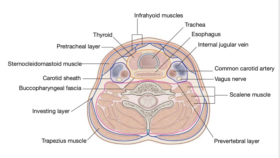
Prevertebral layer –
A cylindrical layer of fascia surrounding the vertebral column and muscles associated to it
These include –
The prevertebral muscles, the anterior, middle, & posterior scalene muscles, and the deep muscles of the back
Pretracheal layer –
Consists of a collection of fascias surrounding the trachea, esophagus, & thyroid gland
Anteriorly – consists of a pretracheal fascia crossing the neck just posteriorly to the infrahyoid muscles
Covers the trachea & thyroid gland
Begins superiorly at the hyoid bone & ends inferiorly in the upper thoracic cavity
Laterally, this fascia continues & covers the thyroid & the esophagus
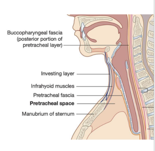
Posteriorly, is referred to as the buccopharyngeal fascia –
Separates the pharynx & esophagus from the prevertebral layer
Begins superiorly at the base of the skull & ends inferiorly in the thoracic cavity
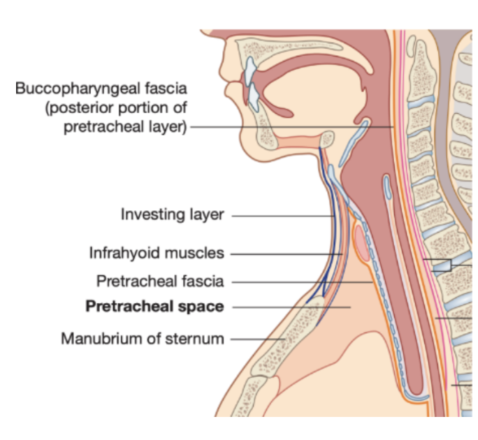
Fascial spaces
Betweeen the investing layer of cervical fascia covering the posterior surface of the infrahyoid muscles and the pretracheal fascia, which passes between the neck
Pretracheal space — covering the anterior surface of the trachea and the thyroid gland
Retropharyngeal space between the buccopharyngeal fascia and the prevertebral fascia, which extends from the base of the skull tothe upper part of the posterior mediastinum
Buccopharyngeal fascia — posterior surface of pharynx & esophagus
Prevertebral fascia — anterir surface of the transverse processes and bodies of the cervical vertebrae
Prevertebral space — In the vertebral layer covering the anterior surface of the transverse processes
Danger space —
Bounded at the top by the skull base, at front by alar fascia and behind by the prevertebral fascia
Ends at the level of the diaphragm
The retropharyngeal space —
Anterior to the danger space, between the alar fascia & buccopharyngeal fascia
There exists a midline raphe in this space so some infections of this space appear unilateral
Carotid sheath –
A column of fascia surrounding the common carotid artery the internal carotid artery, the internal jugular vein, and the vagus nerve as these structures pass through the neck
It receives contributions from the investing, prevertebral, & pretracheal layers, though the extend of each component’s contribution varies
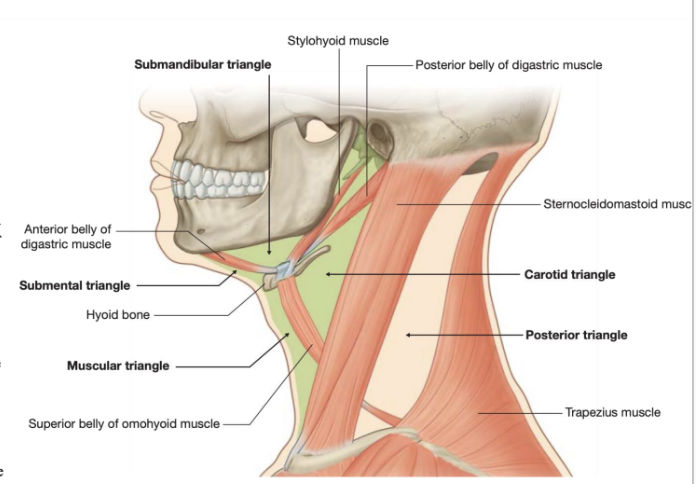
Anterior triangle of the neck subdivision –
Further subdivided into several smaller triangles as follows –
The submandibular triangle
Outlined by the inferior border of the mandible superiorly * anterior and posterior bellies of the digastric muscle inferiorly
The submental triangle –
Outlined by the hyoid bone inferiorly, the anterior belly of the digastric muscle laterally, and the midline
The muscular triangle
Outlined by the hyoid bone superiorly, the superior belly of the omohyoid muscle, & the anterior border of the sternocleidomastoid muscle laterally, and the midline
The carotid triangle –
Outlined by the superior belly of the omohyoid muscle anteroinferiorly, the stylohyoid muscle and posterior belly of the digastric superiorly, and the anterior border of the sternocleidomastoid muscle posteriorly
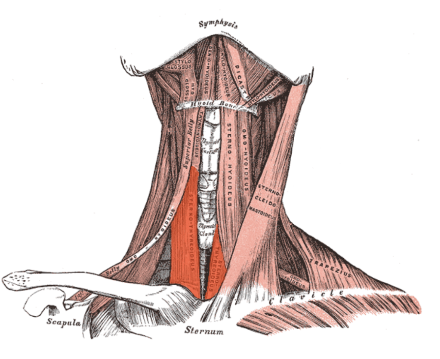
Suprahyoid vs infrahyoid muscles –
Muscles in the anterior triangle of the neck can be groups based on their location relative to the hyoid bone
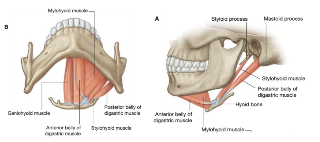
Suprahyoid muscles –
Stylohyoid, digastric, mylohyoid, and geniohyoid
Give mobility to the hyoid bone & form the floor of the oral cavity
Infrahyoid muscles
Omyhyoid, sternohyoid, thyrohyoid, and sternothyroid
Stylohyoid muscle
Suprahyoid muscle
Arises from the base of the styloid process and passes anteroinferiorly to attach to the lateral area of the body of the hyoid
During swallowing it pulls the hyoid bone posterosuperiorly and it is innervated by the facial nerve (VIII)
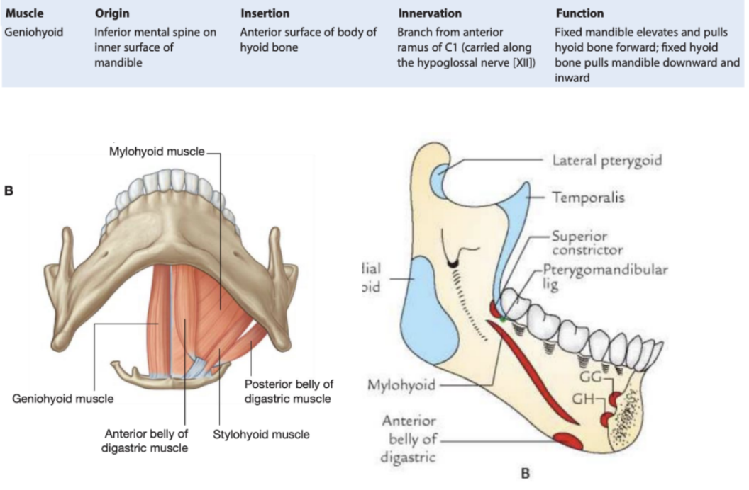
Geniohyoid muscle
Suprahyoid muscle
Parallel to the anterior belly of the digastric muscle
Superior to the floor of the oral cavity — not generally considered a muscle of the anterior trignalge of the neck, but regarded as a suprahyoid muscle
Narrow & superior to the medial part of each mylohyoid muscle
The muscles from each side are next to each other in the midline
The geniohyoid arises from the inferior mental spine of the mandible and passes backward and downward to insert on the body of the hyoid bone
2 functions depending on which bone is fixed —
Fixation of the mandible elevates and pulls the hyoid bone forward
Fixation of the hyoid bone pulls the mandible downward and inward
Innervated by CI
Digastric muscle —
Suprahyoid muscle
2 muscular bellies connected by an intermediate tendon & innervated by 2 different nerves — thus digastric
Anterior belly —
Mylohyoid nerve (branch of mandibular (trigeminus) inserting in the digastric fossa
Inferior apex of the roots of the anterior belly are superior to the mylohyoid line
Posterior belly —
Facial nerve — starting from tip of mastoid on the mastoid notch, and continues at the anterior belly of the muscle at the level of the intermediate tendon
Posterior roots finish posterior to the mylohyoid lines
Intermediate tendon —
Attaches to body of hyoid bone & point of insertion of both bellies
Due to this arrangement, multiple actions depending on whivch bone is fixed —
When mandible is fixed, functions to raise hyoid bone
When hyoid bone is fixed, opens mouth by lowering mandible
Roots —
Inferior apex of anterior belly roots — superior to mylohyoid, while posterior finish posteriorly —
Important due to infectons —
Infections from the posterior belly directly go into the neck — infection of the morals can thus directly spread to neck
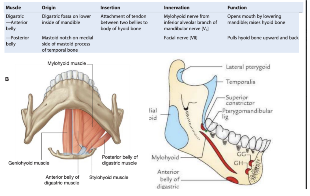
Mylohyoid muscle
Suprahyoid muscle
Origin —
Mylohyoid line of mandible
Insertion —
Mylohyoid raphe, body of hyoid bone
Action —
Forms floor of oral cavity
Elevates hyoid bone & floor of mouth
Depresses mandible
Innervated —
Nerve to mylohyoid (of inferior alveolar nerve (CN V3))
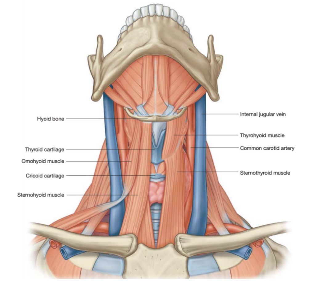
Sternohyoid
Infrahyoid muscle
Origin —
Posterior aspect of sternoclavicular joint & adjacent manubrium of sternum
Insertion —
Body of hyoid bone medial to attachment of omohyoid muscle
Innervation —
Anterior rami of C1 to C3 through the ansa cervicalis
Function —
Depresses hyoid bone after swallowing
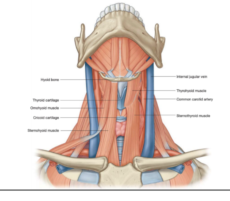
Omohyoid muscle
Infrahyoid muscle — also called the inferior digastrc muscle
Lateral to the sternohyoid muscle — consists of 2 bellies with an intermediate tendon in both the posteiror & anterior triangles of the neck —
Inferior belly —
Begins on superior border of scapula (medial to the suprascapular notch) and passes forward and upward across the posterior triangle ending at the tendon
Superior belly
Begins at intermediate tendon & ascends to attach to the body of the hyoid bone just lateral to the sternohyoid
Intermediate tendon —
Attached to the clavicle, near its medial end, by a fascial sling
Function —
Depresses & fixes the hyoid bone
Innervation —
The anterior rami of C1 to C3 through the ansa cervicalis

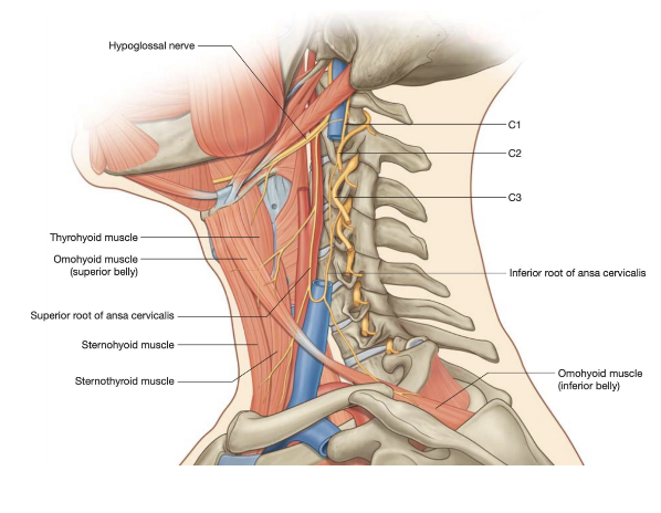
Ansa cervicalis –
Gives innervation to pre-laryngeal muscle
Descending branch of hypoglossal nerve (superior root of ansa cervicalis), and posteriorly connects to CI, CII, & CIII, (cervical roots) (sometimes even CIV) – thus creating an innervation to all superficial prelaryngeal muscles
Loop of nerves made from anterior branches of C1 connected to posterior branches of C3 (& everything in between) that supply infrahyoid muscles
Thyrohyoid muscle —
Deep to the superior parts of the omohyoid and sternohyoid
Origin — oblique line on the outer lamina of the thyroid cartilage
Passes upward to insert into greater horn & adjacent aspect of the body of the hyoid bone
Functions — depend on which bone is fixed
Generally depresses hyoid
When hyoid is fixed it raises the larynx (ex. when high notes are sung)
Innervation
Innervated by fibers from the anterior ramus of C1 that travel with the hypoglossal nerve (XII)
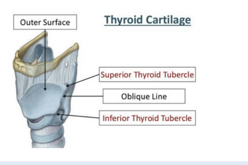
Sternothyroid muscle
Beneath the sternohyoid & in continuity with the thyrohyoid
Arises from posterior surface of the manubrium of the sternum & passes upward to attach to the oblique line on the lamina of the thyroid cartilage
Draws the larynx (thyroid cartilage) downward & is innervated by the anterior rami of C1 to C3 through the ansa cervicalis
Muscles overview

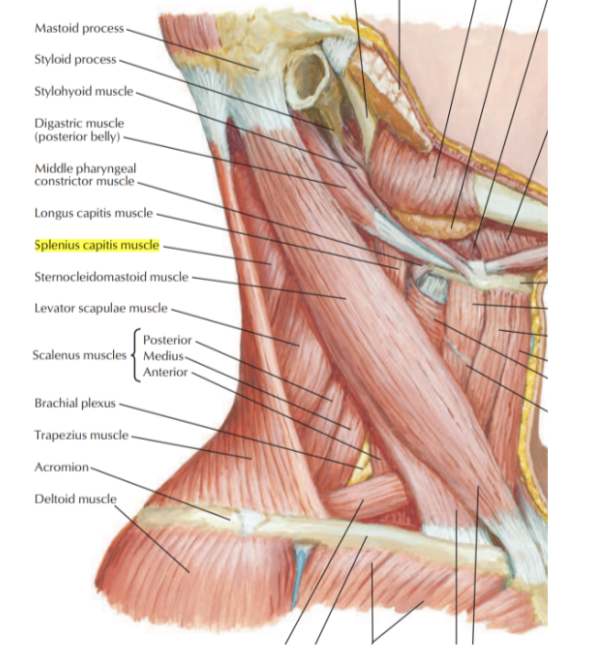
Muscles of the posterior triangle — image
Sternocleidomastoid muscle —
Origin —
Sternal head
Upper part of anterior surface of manubrium of sternum
Clavicular head
Superior surface of medial one-third of clavicle
Insertion —
Sternal head —
Lateral one-half of superior nuchal line
Clavicular head —
Lateral surface of mastoid process
Innervation —
Accessory nerve (XI) and branches from anterior rami
Trapezius muscle
Origin —
Superir nuchal line, external occipital protuberance, ligamentum nuchae, spinous processes of vertebrae CVII to TXII
Insertion —
Lateral one-third of clavicle, aronium, spine of scapula
Innervation —
Motor — accessory nerve (XI) proprioception — C3 and C4
Function —
Assists in rotating the scapula during abduction of humerus above horizontal —
Upper fibers — elevate
Middle fibers — adduct
Lower fibers — depress scapula
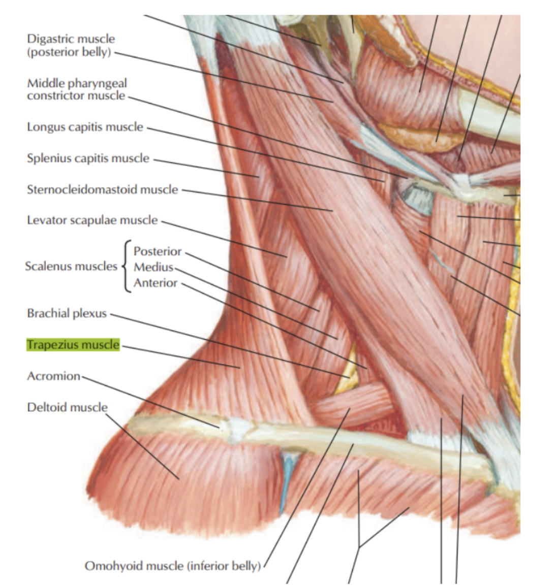
Splenius capitis muscle
Origin —
Lower half of ligamentum nuchae — spinous processes C3-T3
Insertion —
Mastoid process, skull below lateral one-third of superior nuchal line
Innervation —
Posterior rami of middle cervical nerves
Function —
Together, draw head backward
Individually, draw & rotate head to one side
(turn face to same side)