Case 13: Let Me Clear My Throat
 Call Kai
 Learn
Learn Practice Test
 Spaced Repetition
 Match
Match Flashcards
 Knowt Play
1/23

There's no tags or description
Looks like no tags are added yet.
Name | Mastery | Learn | Test | Matching | Spaced | Call with Kai | Chat |
|---|
No analytics yet
Send a link to your students to track their progress
24 Terms
False
The normal esophagus is readily identified in thoracic images.
TRUE OR FALSE
False
Justification: The esophagus is DORSAL to the trachea in the lateral views in a thoracic study.
The esophagus is ventral to the trachea on the lateral views in a thoracic study.
TRUE OR FALSE
A = trachea
B = carina
List the structures in this image, labeled A and B
c. Heart + Trachea + Esophagus + Blood Vessels + Nerves + Lymphatics
What structures live in the mediastinum of an adult dog?
a. Trachea + Esophagus + Blood Vessels + Nerves + Lymphatics
b. Heart + Trachea + Esophagus + Blood Vessels + Nerves
c. Heart + Trachea + Esophagus + Blood Vessels + Nerves + Lymphatics
d. Heart + Trachea + Esophagus
b. Cranial, Middle, Caudal
The mediastinum is divided into what anatomic regions?
a. Orad, Middle, Aborad
b. Cranial, Middle, Caudal
c. Frontal, Intermediate, Caudal
d. None Of The Above
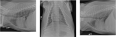
Right lateral, left lateral and VD thoracic projections are available for review.
Describe this study
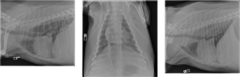
Patient positioning is adequate for interpretation.
Study technique is adequate for interpretation.
Describe patient positioning and technique
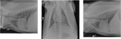
There is marked fluid and gas distension of the thoracic esophagus on all projections (primarily fluid filled on the right lateral projection). There is focal interstitial infiltrate within the ventral aspect of the right cranial lung lobe. More ill-defined increased soft tissue opacity causing vascular margin indistinction is identified within the left caudal lung and cranial lobes on the DV projection, with mild volume loss, that is not as apparent on the right lateral projection. There is widening of the cranial mediastinum likely secondary to fat accumulation given the patient's obese body condition. The cardiac silhouette and vasculature are within normal limits. There is marked hepatomegaly.
Describe findings in this study
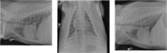
Megaesophagus with aspiration pneumonia, right cranial lung lobe. Atelectasis left lung with possible underlying interstitial infiltrate/aspiration. Hepatomegaly.
Describe the conclusions in this study
Investigate for possible definitive cause for megaesophagus. Institute appropriate therapy for management of megaesophagus.
Describe the next steps in this study
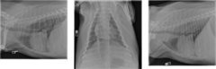
It increases scatter and decreases quality of the film
This dog is in significantly overweight condition. Why does this matter?
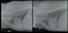
Tracheal stripe sign (superimposition of ventral esophageal wall with dorsal tracheal wall)
What is the name of this soft tissue band which is typically seen when an esophagus is distended with air?
Aspiration pneumonia
What is the common complication associated with megaesophagus?
d. All Of The Above
Megaesophagus can be divided into what categories?
a. Congenital or Acquired
b. Segmental or Generalized
c. Cervical or Thoracic
d. All Of The Above
Idiopathic
What is the most common cause for megaesophagus in the dog?
d. Both B and C
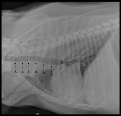
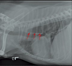
What are the arrows pointing to in this radiograph?
a. Longus Colli Muscle
b. Tracheal Stripe Sign
c. Superimposed Dorsal Tracheal Wall and Ventral Esophageal Wall
d. Both B and C
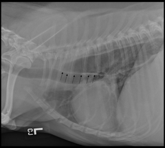
Esophagus
What structure is outlined by the arrows?
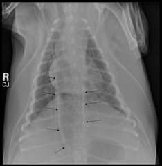
Aspiration pneumonia
What common sequela to megaesophagus is illustrated in this image?
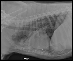
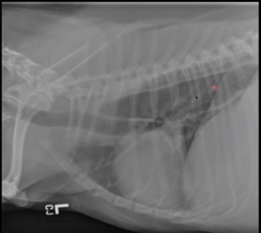
Esophagus enlarged (megaesophagus) and filled with soft tissue opacity
note: there is a line around where the caudal vena cava runs that you can see the differentiation/margin where the normal darker aerated lung is below the line, and the fluid opacity in the esophagus is above the line
Describe the esophagus in this image
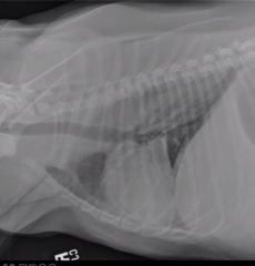
Esophagus enlarged and filled with air opacity + tracheal stripe sign (dorsal trachea overlining wall of esophagus = soft tissue line).
Slight aspiration pneumonia
Describe the esophagus in this image
Idiopathic or myasthenia gravis
What is the most common cause of megaesophagus?
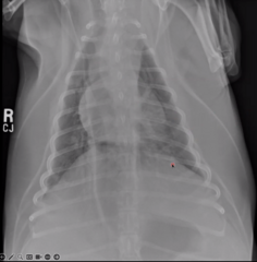
Enlarged esophagus, air filled
Widening of cranial mediastinum likely due to fat. Slight amount of aspiration pneumonia. Interstitial pattern in left caudal lung lobe (but likely due to atelectasis)
Describe the esophagus in this image
Cranioventral
What is the typical location of aspiration pneumonia?
Ribs and sound waves don't pass thru air (because the esophagus lives by the lungs)
What makes ultrasound of mid-esophagus difficult to truly determine megaesophagus?