Other Structural Elements of The Cell
1/36
There's no tags or description
Looks like no tags are added yet.
Name | Mastery | Learn | Test | Matching | Spaced | Call with Kai |
|---|
No analytics yet
Send a link to your students to track their progress
37 Terms
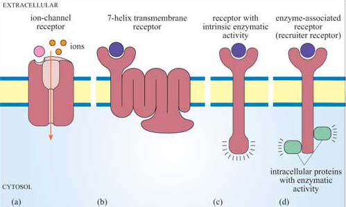
What is the function of cell-surface receptors?
They transmit signals from the extracellular space into the cytoplasm, triggering cellular responses.
What is a ligand?
A molecule, such as a hormone or neurotransmitter, that binds specifically to a receptor.
What is signal transduction?
The process by which binding of a ligand to a receptor triggers a response within the cell.
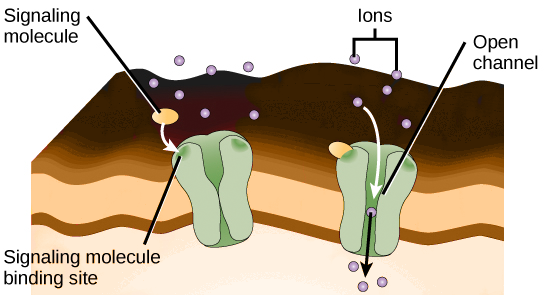
What happens when a ligand binds to a ligand-gated ion channel?
The ion channel opens, allowing ions to flow through the membrane.
Give an example of a ligand-gated ion channel and its function.
The sodium channel at the neuromuscular junction
Nicotinic Acetylcholine Receptor (nAChR): This channel opens when acetylcholine, a neurotransmitter, binds to it.
It allows sodium ions (Na⁺) to enter the cell and potassium ions (K⁺) to leave the cell, playing a crucial role in muscle contraction and nerve signal transmission.
What characterizes catalytic receptors?
Type of cell surface receptor that, when bound by a specific signal molecule (ligand), directly trigger an enzymatic activity inside the cell

What is an example of a catalytic receptor?
The insulin receptor is a catalytic receptor that, when bound by insulin, activates its tyrosine kinase activity to trigger a series of internal cellular processes that help the cell take in glucose and manage energy.

What is the role of a G-protein-linked receptor?
A G-protein-linked receptor is like a "signal receiver" on the surface of a cell.
When a specific molecule (like a hormone) binds to the receptor on the outside of the cell, it activates the receptor.
The activated receptor then turns on a G-protein inside the cell.
The G-protein acts like a "messenger" that carries the signal inside the cell.
This messenger activates other molecules inside the cell, called second messengers.
Explain step-by-step how epinephrine would activate a G-protein-linked receptor
Epinephrine attaches to receptor
It activates helpers inside the cell called G-proteins
G-proteins swap out old battery (GDP) for GTP to get energy
Energized G-proteins move through cell membrane to turn on adenylyl cyclase
Adenylyl cyclase takes building blocks of ATP and makes new helpers called cAMP
cAMP activates special workers called cAMP-dependent protein kinases (cAMP-dPK) inside the cell
cAMP-dPK add a tag (phosphate) to the enzymes of the cell
Some tools help break down stored energy (like glycogen), and these get turned on. Other tools help store energy, and these get turned off.
Epinephrine acts like a key that starts a chain reaction inside the cell, ultimately turning on and off tools that help the cell use or store energy. It + HR, + blood glucose, dilate airways, + blood flow, enhance alertness
What is the cytoskeleton and its main functions?
It provides structural support, facilitates cell movement, and aids in intracellular transport.

What are the three components of the cytoskeleton?
Microtubules, intermediate filaments, and microfilaments.

What are microtubules made of and what is their function?
Made of tubulin protein, they are involved in cell division and intracellular transport.
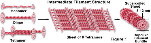
What is the role of intermediate filaments?
They provide mechanical support for the cell.
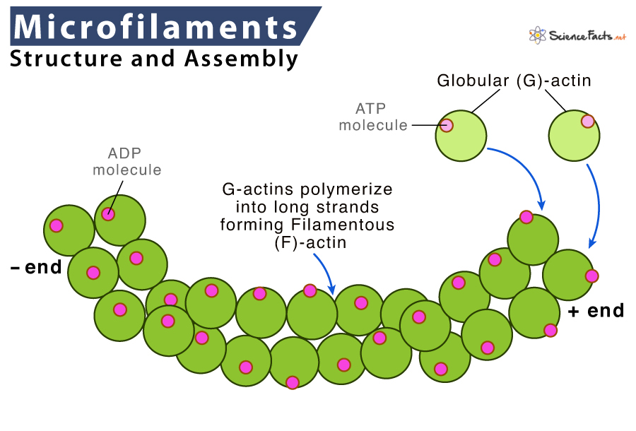
What are microfilaments composed of, and what is their function?
Composed of actin protein, they are involved in cell movement and shape changes.

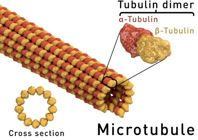
What are microtubules composed of?
Microtubules are composed of α-tubulin and β-tubulin dimers.
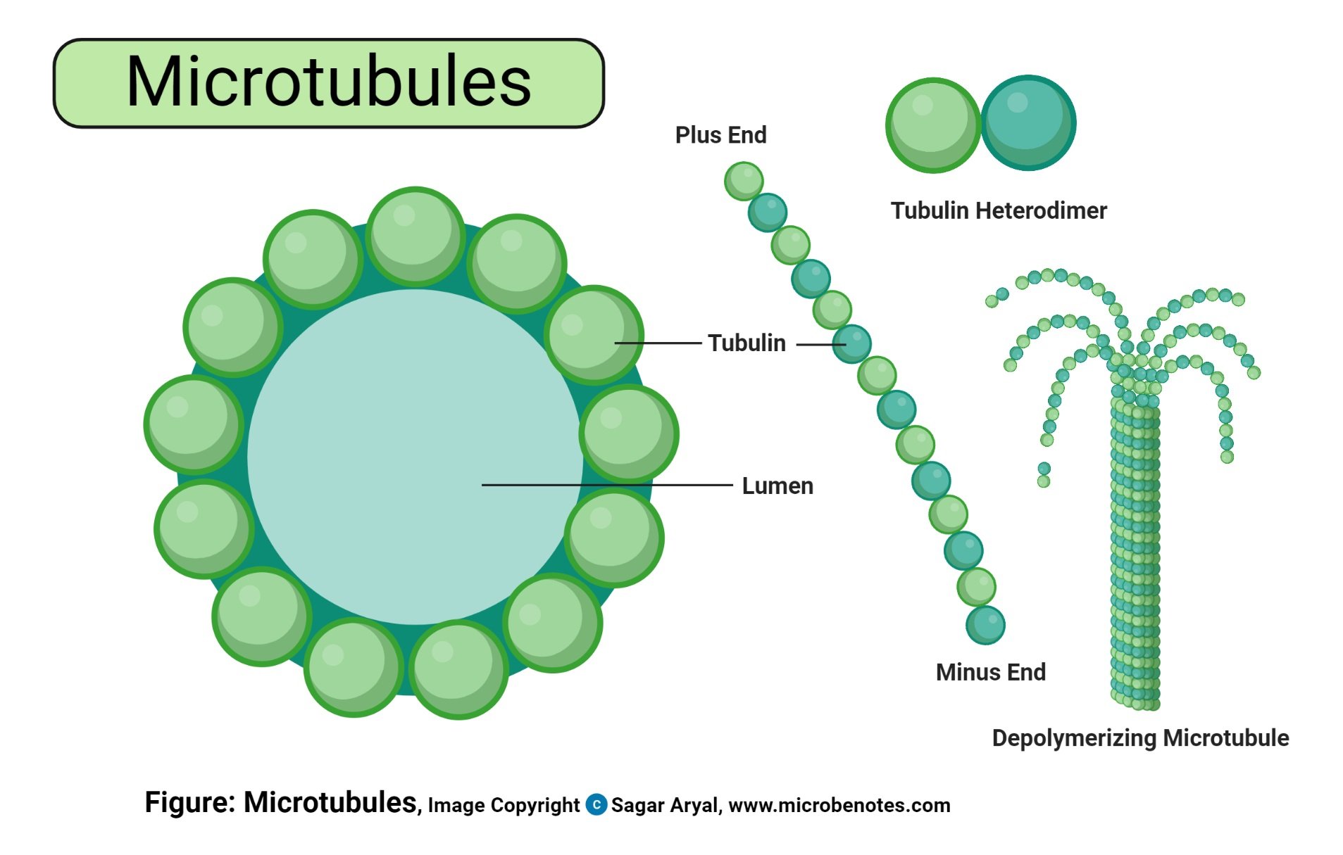
How do microtubules form?
α-tubulin and β-tubulin form dimers (molecule of 2 identical molecules linked together), which polymerize noncovalently (pairs stick together without strong bonds) into sheets that roll into tubes.
How do microtubules elongate?
By adding αβ-tubulin dimers to one end, while the other end is anchored/attached to the microtubule organizing center (MTOC).
What is the microtubule organizing center (MTOC)?
A structure near the nucleus that anchors one end of the microtubule and contains a pair of centrioles.
Centrosome are organizers of cell division

What role do microtubules play in cell division?
They form the mitotic spindle, which separates chromosomes during mitosis.

Why are centrioles not essential for mitosis?
Centrioles are not essential for mitosis because animal cells have other strategies to build a spindle
How do microtubules facilitate intracellular transport?
By serving as "railroads" for proteins that hydrolyze (add water) ATP and act as molecular motors.
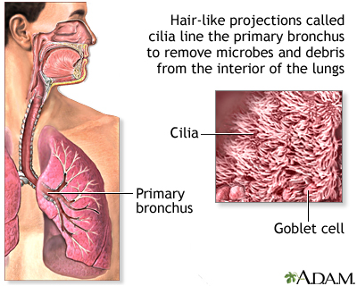
What are cilia, and what is their function?
Cilia are small "hairs" that move fluids past the cell surface, such as sweeping mucus in the respiratory tract.
What is a flagellum, and what is its function?
A flagellum is a large "tail" that propels cells by wiggling, such as the flagellum of sperm cells.
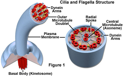
Describe the structure of cilia and flagella
Both have a "9 + 2" arrangement of microtubules, with nine pairs around two central microtubules.
What protein is involved in the movement of cilia and flagella, and how does it work?
Dynein causes movement by enabling microtubules to slide past each other.
How are cilia and flagella anchored to the plasma membrane?
They are anchored by a basal body, which has the same structure as a centriole (nine triplets of microtubules).
What protein forms microfilaments, and how do they polymerize?
Microfilaments are formed from actin, polymerizing from globular actin (G-actin) monomers.
Describe the structure of microfilaments.
Actin monomers form chains that wrap around each other to create actin filaments.
What is the function of microfilaments in cells?
Microfilaments are dynamic and involved in cell movement and division, such as amoeboid movement (locomotion) and cytokinesis (division of cytoplasm).
What is the role of intermediate filaments in cells?
Intermediate filaments provide mechanical strength to cells, particularly in tissues subject to mechanical stress. Found in muscle and skin cells
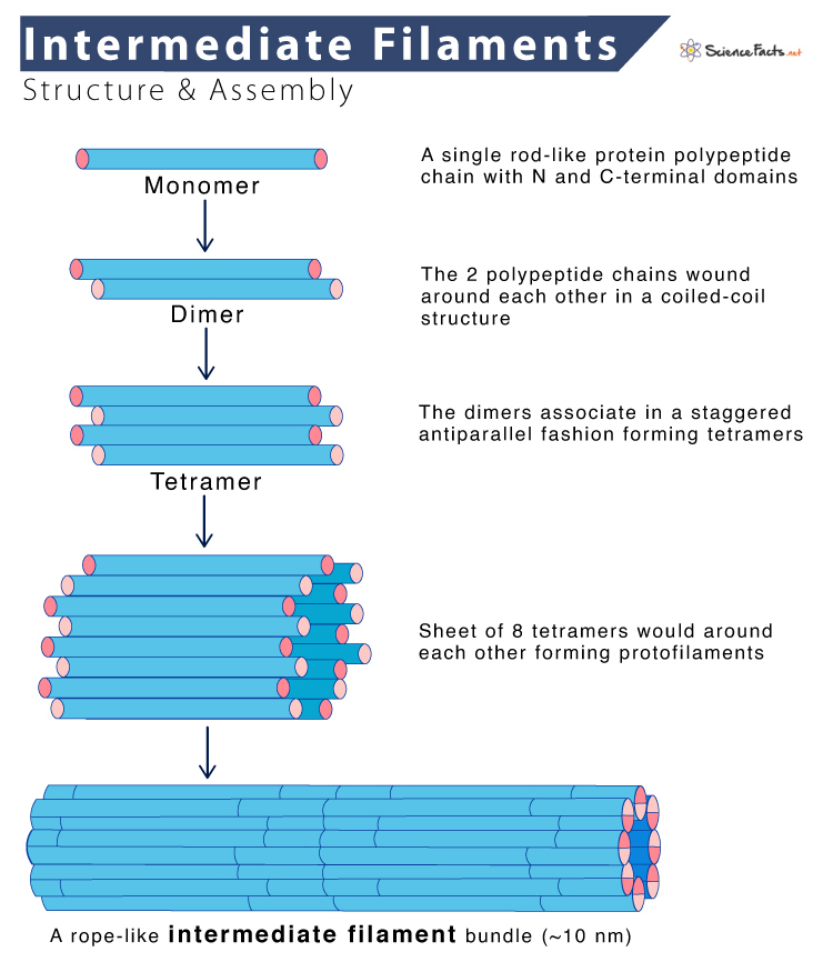
How do intermediate filaments differ from microfilaments and microtubules?
Intermediate filaments are more permanent structures composed of diverse polypeptides, unlike the dynamic nature of microfilaments and microtubules.
What are tight junctions, and where are they found in the body?
Tight junctions form seals between adjacent cells, blocking the movement of molecules across the cell layer; found in tissues like intestinal epithelium.

Describe the structure of desmosomes and their function.
Desmosomes are spot-like junctions that anchor cells together with fibers spanning plasma membranes; they provide mechanical stability to tissues.
Found in tissues that need to withstand stretching and pulling, like skin and heart muscle. They prevent cells from separating easily under mechanical stress.
What are gap junctions, and what is their function in tissues?
Gap junctions are pores between cells that allow ions and small molecules to pass directly between them; found in tissues like cardiac muscle for synchronized action potentials.
Which type of cell junction forms a seal between cells, preventing the mixing of molecules across the cell layer?
Tight junctions form seals between cells.
What type of junction anchors cells together but does not form a complete seal?
Desmosomes anchor cells together.
Where are gap junctions particularly important, and what do they allow to pass between cells?
Gap junctions are important in tissues like cardiac muscle, allowing ions and small molecules to pass between cells.