Module 7: Chapters 25 (Panoramic Imaging), 26 (Extraoral Imaging), & 27 (Three-Dimensional Digital Imaging)
1/29
There's no tags or description
Looks like no tags are added yet.
Name | Mastery | Learn | Test | Matching | Spaced |
|---|
No study sessions yet.
30 Terms
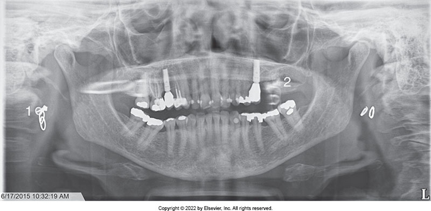
1.) You are examining your patient's panoramic image. You notice that the molars and premolars on the right side are larger than the teeth on the left side. What patient positioning error did you make?
a) The patient's head was turned to the right.
b) The patient's head was turned to the left.
c) The patient is slumped.
d) The patient's teeth are behind the focal trough.
a) The patient's head was turned to the right.
b) The patient's head was turned to the left.
c) The patient is slumped.
d) The patient's teeth are behind the focal trough.
2.) (1) The panoramic image is the preferred image for the detection of caries and periodontal disease. (2) The panoramic image is preferred for the evaluation of eruption patterns, growth, and development.
a) Both statements are true.
b) Both statements are false.
c) The first statement is true; the second statement is false.
d) The first statement is false; the second statement is true.
a) Both statements are true.
b) Both statements are false.
c) The first statement is true; the second statement is false.
d) The first statement is false; the second statement is true.
3.) In panoramic imaging, the ___________ is a theoretical concept used to determine where the dental arches must be positioned to achieve the clearest image.
a) waypoint
b) focal trough
c) neutral zone
d) home zone
a) waypoint
b) focal trough
c) neutral zone
d) home zone
4.) If the patient's lips are not closed on the bite-block during the exposure of a panoramic image, a _____________ shadow results that obscures the anterior teeth.
a) light radiopaque
b) dark radiopaque
c) light radiolucent
d) dark radiolucent
a) light radiopaque
b) dark radiopaque
c) light radiolucent
d) dark radiolucent
5.) A "reverse smile line" is seen on the image if the patient's
a) chin is tipped down.
b) chin is tipped up.
c) teeth are positioned too far back on the bite-block.
d) teeth are positioned too far forward on the bite-block.
a) chin is tipped down.
b) chin is tipped up.
c) teeth are positioned too far back on the bite-block.
d) teeth are positioned too far forward on the bite-block.
6.) The anterior teeth will appear "skinny" or "narrow" if the
a) chin is tipped down.
b) chin is tipped up.
c) teeth are positioned too far back on the bite-block.
d) teeth are positioned too far forward on the bite-block.
a) chin is tipped down.
b) chin is tipped up.
c) teeth are positioned too far back on the bite-block.
d) teeth are positioned too far forward on the bite-block.
7.) A disadvantage of panoramic dental imaging when contrasted with intraoral dental imaging is
a) panoramic imaging results in higher exposure to the patient than intraoral imaging.
b) fewer anatomic structures can be viewed on a panoramic image than on a complete intraoral imaging series.
c) the images seen on a panoramic image are not as sharp as those on intraoral images because of the intensifying screens.
d) the exposure of a panoramic image is readily accepted by the patient because there is no discomfort involved.
8.) An extraoral image receptor is placed __________ the mouth during x-ray exposure. Extraoral imaging is used to image _________ areas of the skull or jaws.
a) outside; small
b) outside; large
c) inside; small
d) inside; large
a) outside; small
b) outside; large
c) inside; small
d) inside; large
9.) Which of the following would you evaluate using an extraoral lateral cephalometric image?
a) Gutta percha placement in the root canal
b) Level of crestal bone
c) Recurrent decay
d) Growth and development
a) Gutta percha placement in the root canal
b) Level of crestal bone
c) Recurrent decay
d) Growth and development
10.) The lateral jaw projection
a) requires the use of a special x-ray unit.
b) provides for more diagnostic information than a panoramic image.
c) is used to examine the anterior portion of the mandible.
d) is valuable for patients with limited jaw opening because of a fracture or swelling.
a) requires the use of a special x-ray unit.
b) provides for more diagnostic information than a panoramic image.
c) is used to examine the anterior portion of the mandible.
d) is valuable for patients with limited jaw opening because of a fracture or swelling.
11.) For the _____________ projection, the patient's face is against the receptor with both the forehead and chin touching the receptor.
a) posteroanterior
b) lateral cephalometric
c) reverse Towne
d) Waters
a) posteroanterior
b) lateral cephalometric
c) reverse Towne
d) Waters
12.) (1) Three-dimensional imaging provides a more accurate image than traditional two-dimensional imaging. (2) Locations, distances, sizes, and shapes of pathology and anatomic landmarks, including eruption patterns, are more accurately represented with three-dimensional imaging.
a) Both statements are true.
b) Both statements are false.
c) The first statement is true; the second statement is false.
d) The first statement is false; the second statement is true.
a) Both statements are true.
b) Both statements are false.
c) The first statement is true; the second statement is false.
d) The first statement is false; the second statement is true.
13.) Common uses of three-dimensional imaging include which of the following?
1. Implant placement
2. Extraction or exposure of impacted teeth
3. Definition of anatomic structures
4. Airway and sinus analysis
5. Evaluation of temporomandibular joint disorders
a) 1, 2, 3, 4, 5
b) 2, 4, 5
c) 1, 2, 3
d) 3, 4
a) 1, 2, 3, 4, 5
b) 2, 4, 5
c) 1, 2, 3
d) 3, 4
14.) In panoramic imaging, the x-ray tube rotates around the patient's head in one direction while the receptor rotates __________________ direction.
a) horizontally in the same
b) horizontally in the opposite
c) vertically in the same
d) vertically in the opposite
a) horizontally in the same
b) horizontally in the opposite
c) vertically in the same
d) vertically in the opposite
15.) A ghost image appears ___________ than/of its actual counterpart.
a) on the same side and lower
b) on the opposite side and higher
c) more distinct
d) smaller
a) on the same side and lower
b) on the opposite side and higher
c) more distinct
d) smaller
Purposes and Uses of Panoramic Imaging
panoramic imaging: extraoral technique that is used to examine the maxilla AND the mandible on a wide view in a SINGLE PROJECTION
Used to evaluate:
impacted teeth
eruption patterns, growth, and development
detect diseases, lesions, and conditions of the jaws
examine the extend of large lesions
evaluate trauma
Panoramic Imaging Concepts
tomography: the process by which the movement of the receptor and the tubehead produce an image; an imaging technique that allows the imaging of one layer or section of the body while blurring images from structures in other planes
The receptor and tubehead move around the patient in OPPOSITE DIRECTIONS.
focal trough: a 3-D curved zone in which structures are clearly demonstrated on a panoramic image; structures IN the focal trough are well-defined, and structures OUTSIDE of the tough are blurred or indistinct
Images produced:
real image: results when a structure lies BETWEEN THE RECEPTOR AND MOVING ROTATION CENTER (aka “true” image)
double image: results when a structure that is located BEHIND THE MOVING ROTATION CENTER is penetrated TWICE by the beam (“mirror image”); ex: cervical spine, hyoid bone
ghost image: results when a structure is located OUTSIDE OF THE FOCAL PLANE AND CLOSE TO THE X-RAY SOURCE; objects are often near the midline (appears blurred and distorted, higher and on the opposite side of the true image); ex: earrings, ramus, hard palate
Panoramic X-Ray Unit Components: x-ray tubehead (with narrow vertical slit-shaped collimator), head positioner, exposure controls
Image receptor: may be direct digital, PSP, or film
Exposure factors: mA and kV can be adjusted to accommodate patients of different sizes; exposure time can be adjusted based on the type of image obtained; exposure time ranges from 10-30 secs
Procedures for Panoramic Imaging
Equipment Preparation
Patient Preparation: explain procedure, place lead apron (NO THYROID COLLAR, double-sided, secured on front), remove all metallic objects from head-and-neck area (glasses, jewelry, hearing aids, etc.)
Patient Positioning: pt must sit/stand as tall as possible, bite on the block in an end-to-end position and close lips, swallow and hold tongue at the palate, align patient’s planes:
Frankfort Plane: passes through the floor of the orbit and the EAM (tragus to ala)
Midsagittal Plane: midline of patient’s face
Evaluating the Diagnostic Quality of a Panoramic Image
Diagnostic images result when equipment prep, pt prep, and pt positioning are completed correctly.
free of all errors
should demonstrate accurate anatomic features
dentition: smile-like curve, crowns and apices visible
ramus & cervical spine: ramus should be the same width on each side; cervical spine may be present along edges but should not overlap ramus
nasal cavity & maxillary sinus: hard palate double image should appear ABOVE the apices of max teeth
body of mandible: inferior border is smooth and continuous
condyle: centered, on the same horizontal plane, and of equal size on each side
hyoid bone: hyoid bone double image may slightly overlap mandible
exhibit diagnostic density and contrast from proper exposure settings (smaller pts require less exposure, density shouldn’t be too dark or too light, contrast should allow for identification of the junction between the enamel and dentin of molars so that impacted/unerupted teeth can be seen)
Common Panoramic Errors: Equipment Preparation
overexposure/underexposure
Common Panoramic Errors: Patient Preparation
Ghost Images: REMOVE jewelry/dentures/partials/retainers/metallic objects
Lead apron artifact: use an apron WITHOUT a thyroid collar and place it LOW around the patient’s neck
Positioning Errors: lips/tongue, chin tipped up/down, teeth anterior/posterior to focal trough, turned head, slumped posture
Common Panoramic Errors: Positioning Errors
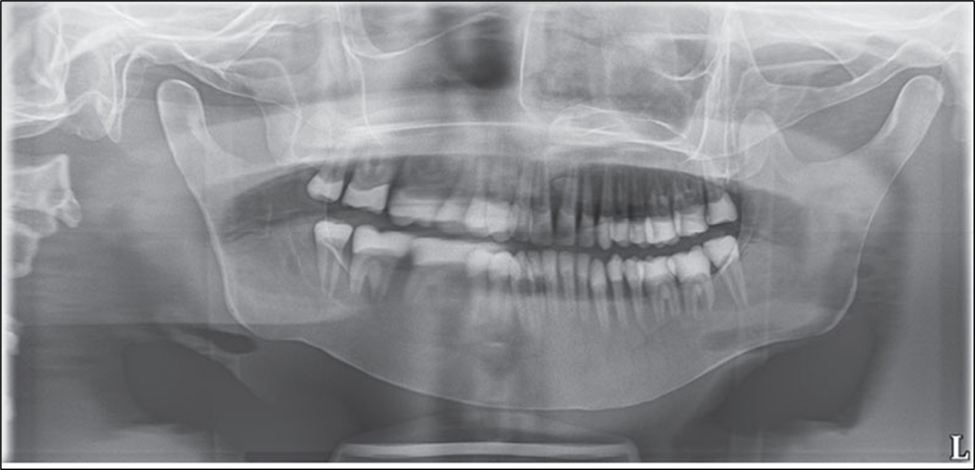
Lips & Tongue
Error: tongue not in contact with roof of palate
result: obstruction of apices of max teeth by a radiolucent shadow
correction: have pt swallow and hold tongue to palate
Error: lips not closed around block
result: obstruction of anterior teeth by a radiolucent shadow
correction: have pt close lips around block
Common Panoramic Errors: Positioning Errors
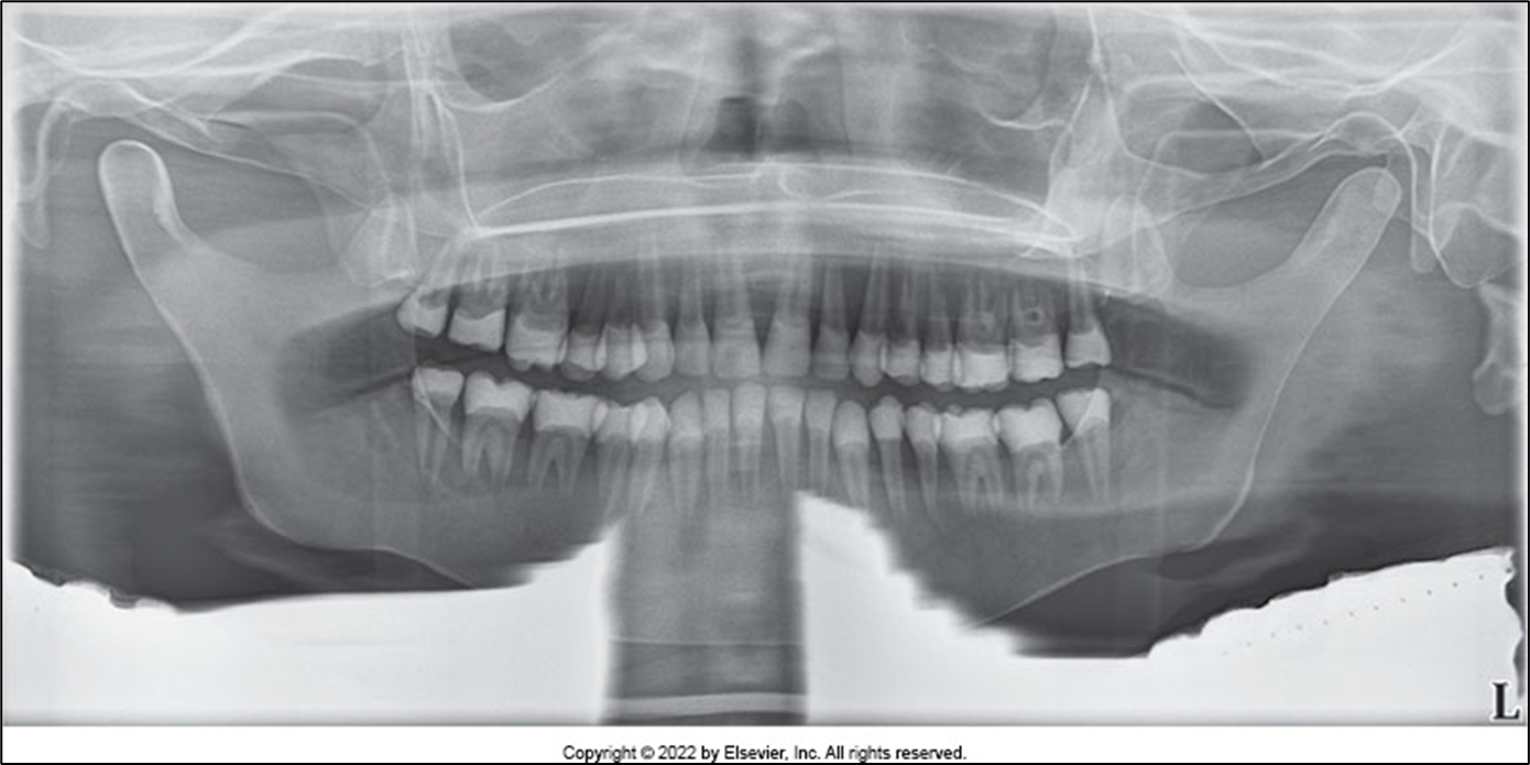
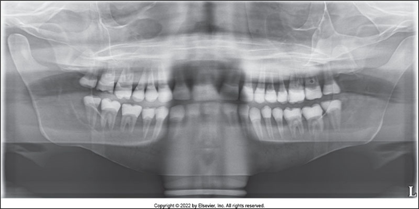
Chin Tipped UP
error: the patient’s chin is tipped UP; the Frankfort Plane is NOT PARALLEL to the floor
result: “reverse smile line,” condyles may not be visible, hard palate may obstruct the roots of max teeth, max incisors are blurred and magnified, loss of details around incisor teeth
correction: position Frankfort Plane PARALLEL TO THE FLOOR
Common Panoramic Errors: Positioning Errors
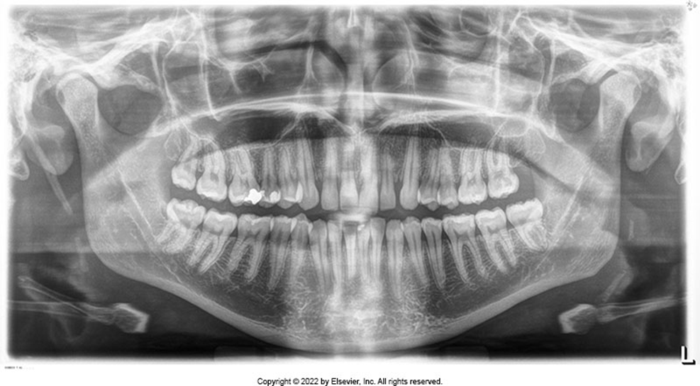
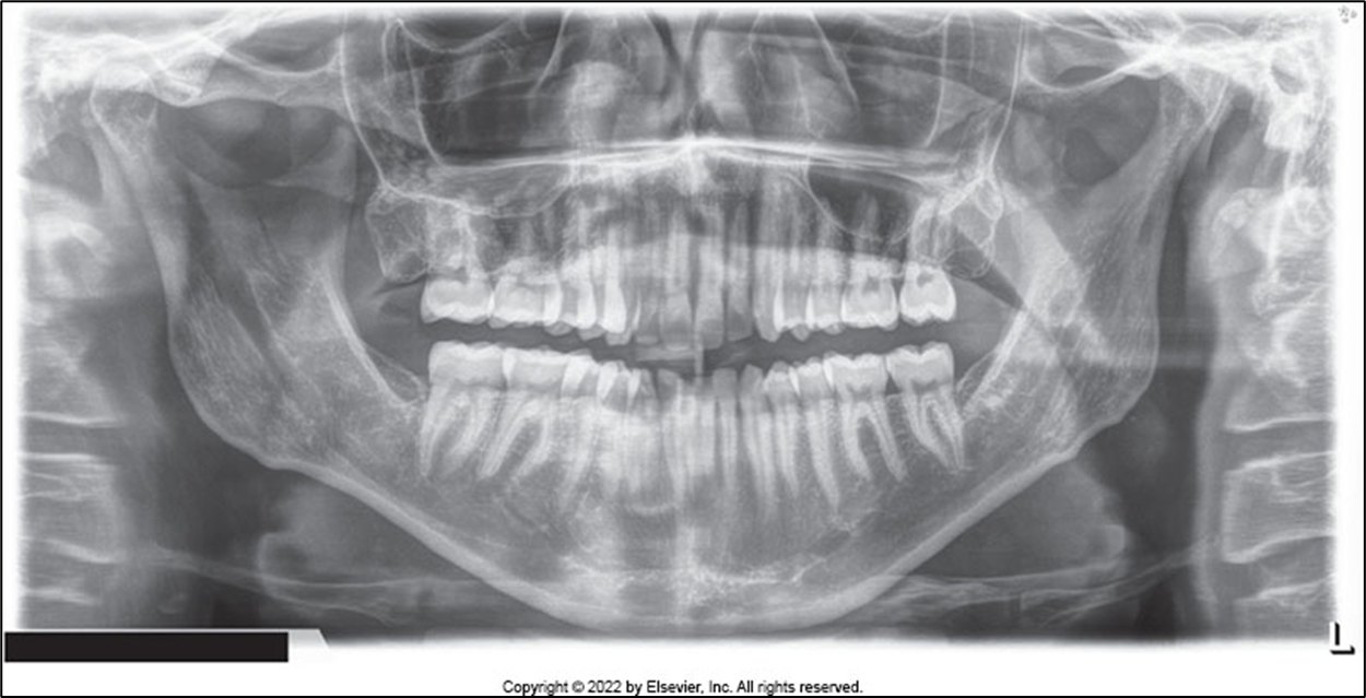
Chin Tipped DOWN
Error: pt chin is tipped DOWN; Frankfort Plane is NOT PARALLEL to the floor
result: “exaggerated smile line,” condyles will not be high or not visible, the hyoid bone forms a single wide line instead of a double image, loss of detail in the apical region of ant teeth
correction: Frankfort Plane is PARALLEL TO THE FLOOR
Common Panoramic Errors: Positioning Errors
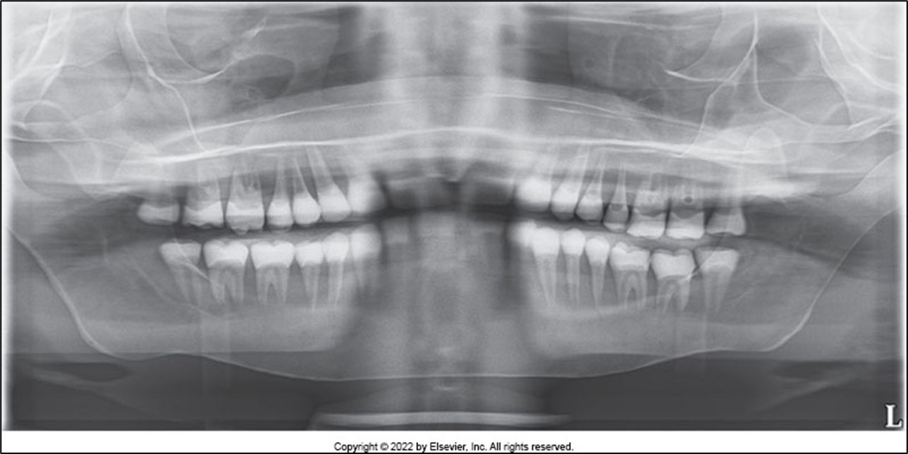
Teeth ANTERIOR to the Focal Trough
Error: teeth are ANTERIOR to the focal trough; the pt is biting in front of the groove/too far forward on the block
result: anterior teeth are “narrow” and “skinny,” premolars are overlapped
correction: make sure that pt’s ant teeth are in an end-to-end position in the groove of the block; adjust the forehead support to stabilize the pt’s head to prevent them from sliding forward on the block
Common Panoramic Errors: Positioning Errors
Teeth POSTERIOR to the Focal Trough
Error: teeth are POSTERIOR to the focal trough; patient is biting behind the groove/too far back on the block
result: anterior teeth are “wide”
correction: make sure that the pt’s anterior teeth are in an end-to-end position in the groove of the bite block; adjust forehead support to stabilize pt’s head to prevent them from sliding backward on the block
Common Panoramic Errors: Positioning Errors
Head TURNED
Error: head TURNED/NOT CENTERED; midsagittal plane is NOT PERPENDICULAR to the floor
result: teeth are unequally magnified; the side FARTHEST from the receptor appears MAGNIFIED; ex: if pt is turned to the right, their right side is closer to the receptor and the LEFT side will appear magnified
correction: make sure the patient’s midsagittal plane is PERPENDICULAR TO THE FLOOR and that the midline is CENTERED ON THE BITE-BLOCK; the light should divide the face into EQUAL PARTS
Common Panoramic Errors: Positioning Errors
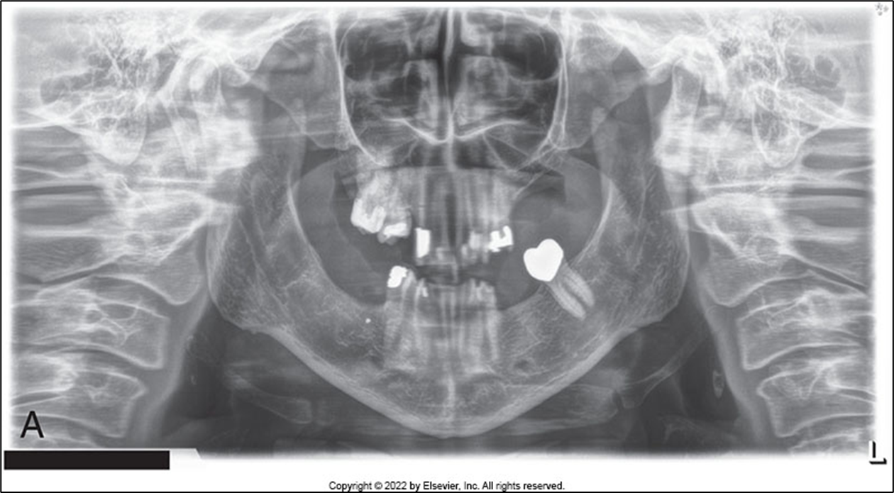
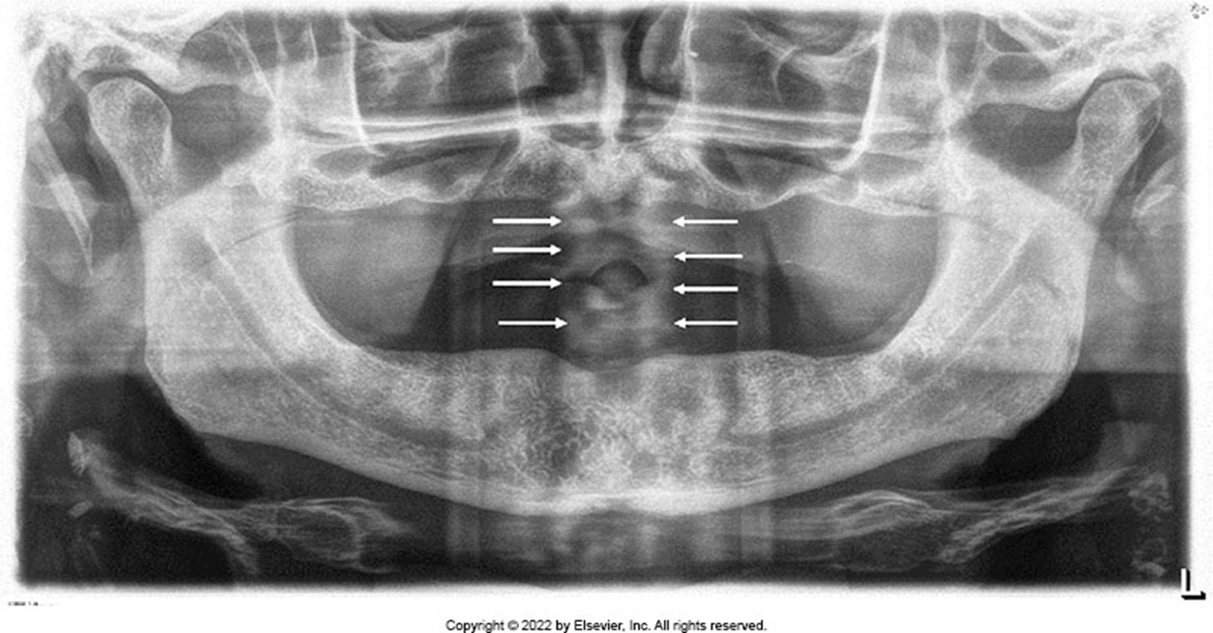
SLUMPED POSTURE
Error: the patient is not standing tall
result: cervical spine appears as a radiopacity in the center of the image and obscures teeth
correction: instruct pt to stand or sit tall with their shoulders back; elderly pts may find it easier/more comfortable to sit in a chair during exposure
Advantages / Disadvantages of Panoramic Imaging
Advantages:
large field size
simple procedure
patient cooperation
minimal exposure
Disadvantages:
low image quality
focal trough limitations
distortion
expensive equipment
3-D Digital Imaging: CBCT
CBCT: Cone-beam computed tomography
use: to evaluate the oral and maxillofacial complex; provides more detailed information than 2-D images; primarily used in medical field but introduced to dentistry in 1999
implant placement
extraction or exposure of impacted teeth
endodontic assessment
airway and sinus analysis
evaluation of TMJ disorder
orthodontic evaluation
evaluation of lesions and abnormalities
evaluation of trauma
Process:
source of radiation rotates around the head (similar to pan imaging)
data received on the receptor is reconstructed and imported to viewing software (DICOM images)
data is viewed in three planes:
Axial (Transverse) Plane
Coronal Plane
Sagittal Plane
Field of View (FOV): the area that can be captured when performing imaging procedures; region of interest of the patient anatomy
AAOMR recommends that images be interpreted by a board-certified maxillofacial radiologist / dentist who has adequate training / experience
Advantages:
lower radiation dose than medical CT scans
quick scanning time
accurate measurement of anatomic structures
images shared electronically and easily saved
Disadvantages:
streak artifacts from metallic restorations can obscure surrounding anatomy
selection size of the FOV
cost of equipment and training needed for imaging software
lack of training in interpretation of image data