Ch28 Pregnancy & Development
 Call Kai
 Learn
Learn Practice Test
 Spaced Repetition
 Match
Match Flashcards
 Knowt Play
1/89

There's no tags or description
Looks like no tags are added yet.
Name | Mastery | Learn | Test | Matching | Spaced | Call with Kai | Chat |
|---|
No analytics yet
Send a link to your students to track their progress
90 Terms
phases of development
prenatal: fertilization → birth
neonatal: birth → 28 days
perinatal: around birth
postnatal: 28 → year
embryology
study of embryonic development
first 2 months after fertilization
fetal development
start of ninth week → birth
prenatal development
embryonic + fetal development stages
postnatal development
birth → maturity
gestation
time in prenatal development
divided into 3 three month trimesters
teratogens
cause malformations by affecting embryo during periods of rapid growth, development, or specialization
drugs, viruses, radiation
first trimester
rudiments of major organ systems appear
second trimester
body shape and proportions start to change
development of organs and organ systems
third trimester
rapid fetal growth and adipose tissue deposition
organ systems are fully functional
four stages of 1st trimester
cleavage
implantation
placentation
embryogenesis
1st trimester: cleavage phase
sequence from immediately after fertilization until contact with the uterine wall
zygote → pre-embryo → blastomere → blastocyst
1st trimester: implantation
blastocyte attaches to endometrium of uterus
1st trimester: placentation
blood vessels form around blastocyst
placenta develops
1st trimester: ebryogenesis
formation of viable embryo
establishes foundation for all major systems
differentiation
selective changes in gene activity result in different types of cells
induction
chemical substances released from cells to affect differentiation of other embryonic cells
inheritance
transfer of genetically determined characteristics from one generation to the next
fertilization
fusion of haploid gametes to produce diploid gamete
in uterine tube
vehicles of chromosome delivery
spermatazoon and oocyte
capacitation
increases motility of spermatozoa
thinned head of sperm to release digestive enzymes in acrosome
FPP fertilization promoting peptide
produced by prostat gland to control capacitation
relationship of capacitation and female reproductive system
conditions in female reproductive tract facilitate capacitation
acrosome
contains enzymes hyalurodinase (follicle bond breaker) and acrosin (proteolytic)
layers sperm penetrate moving into oocyte
corona radiata
zona pellucida
acrosomal reaction
calcium dependent exocytosis of sperm when it releases enzymes and fuses with plasma membrane of egg
fast block to polysomy
plasma membrane is depolarized so that no more sperm can fuse with the egg
slow block to polyspermy
calcium released from ER stimulate cortical granules to fuse with egg plasma membrane and release enzymes
enzymes destroy sperm binding proteins and harden plasma membrane
female pronucleus
nuclear material remaining in ovum after occyte activation
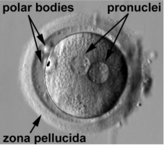
male pronucleus
swollen pronucleus of spermatozoon
migrates to center of cell

amphimixis
fusion of male and female pronucleus
fertilization // conception
meiosis II finishes
cleavage
series of cell divisions resulting in daughter cells
after fertilization
name of cells from day 0 - day 3
blastomere
identical cells produced by cleavage divisions
name of cells from day 3 - 6
morula
pre-embryo is solid ball of cells
uterus reached on day 4
blastocyst
formed by blastomeres
hollow inner cavity called blastocoel
trophoblast
outer cells that separate blastocoel and outer world
provide nutrients to developing embryo
inner cell mass
clusters at one end of the blastocyte and will later become the embryo
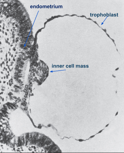
implantation
7 days after fertilization
blastocyte adheres to uterine lining
trophoblast divides rapidly to form many layers
cellular trophoblast
cells closest to interior of blastocyst
syncytial trophoblast
outer layers of cells that fuse together while they burrow into endometrium
secrete hyaluronidase to erode through uterine epithelium
how is amnion and amniotic cavity formed
epiblast (inner disc) cells and hypoblast (outer disc) separate

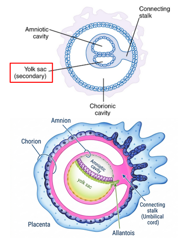
yolk sac
supplies nutrients and primitive blood circulation in first 2-3 weeks before placenta is formed
becomes part of embryonic digestive
incorporated into umbilical cord
chorion
outermost extraembryonic membrane
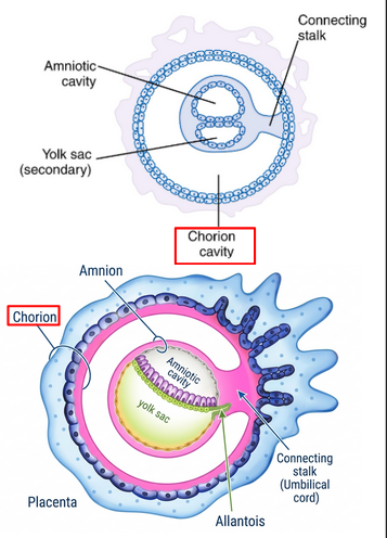
chorionic cavity
space between chroion and embryo
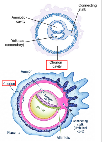
chorionic vili
projections that grow out of syntotrophoblast and invade endometrium via enzyme degradation

embryonic blood vessels
form along connecting stalk which attaches embryo to developing placenta

umbilical cord
forms from folding amnion which envelops connecting stalk
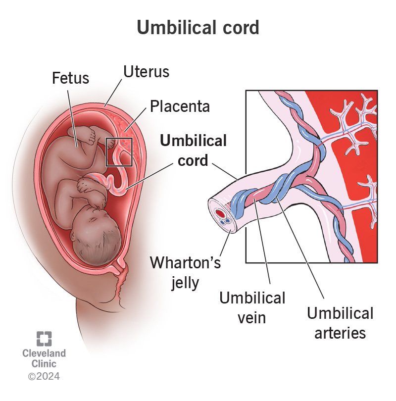
allantois
finger like projection that forms during 3rd week between yolk stalk and connecting stalk

structure that develops into urinary bladder
allantois

structures that combine to form the umbilical cord
yolk sac and stalks of allantois
structures that make up the extra-embryonic membranes
chorion
amnion
yolk sac
allantois
support and nourish developing embryo

placenta
disc shaped area where chorion contacts developing uterine wall
connects embryo to maternal blood

structures in the umbilical chord
2 umbilical arteries
1 umbilical vein
allantois and yolk sac
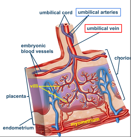
portions of the placenta
embryonic and maternal portions
placental membrane
separates embryonic blood from maternal blood
allows gas exchange between embryo and mom

what filters waste of the fetus
capillaries in chorionic villi
prevent maternal cytotoxic T cells from destroying fetus

summary of first trimester
summary of first trimester


gastrulation
primitive streak forms along 2 layered embryo to form a three layered structure
forms primary germ layers
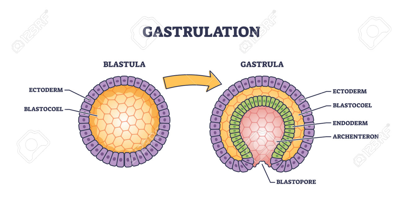
endoderm
becomes digestive, liver, pancreas, inner lungs
adjascent to yolk sac

mesoderm
becomes circulatory system, lungs, epithelium, skeletal, and muscular system
NOTOCHORD
middle layer
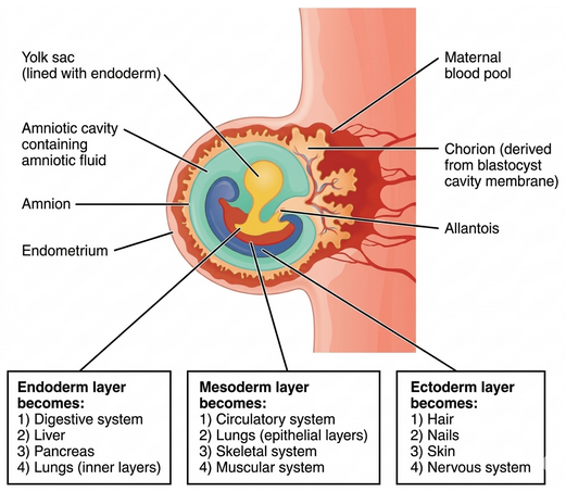
ectoderm
hair, nails, skin, nervous system
cells of epiblast that remain

embryogenesis
body of embryo separates from embryonic disc
organs form
folding and differential growth of embryonic disc projects into amniotic cavity
2 projections that form the body during embryogenesis
head fold
tail fold
neurulation
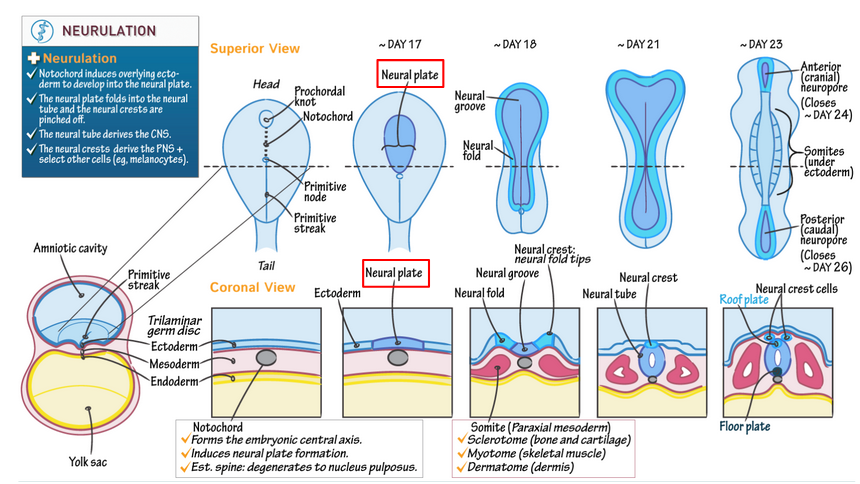
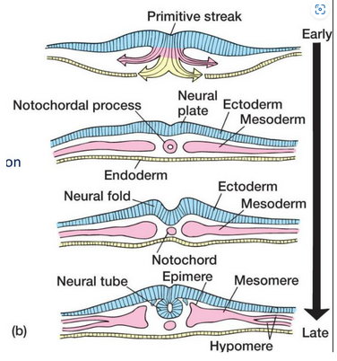
nervous system develops from ectoderm
tissues along length of embryo form neural plate
neural folds converge to form neural tube
neural tube divides into brain structures at week 4-5
organogenesis
organ formation
second trimester
fetus grows faster than surrouding placenta
third trimester
organ systems are ready
growth rate slows
weight gain
mothers organs are displaced
maternal adaptations
increased respiratory and tidal volume
increased blood volume
increased GFR
increased nutrient and vitamin intake
enlarged uterus and mammary glands
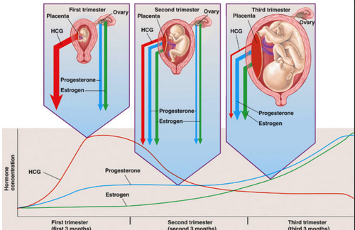
progesterone
released by the placenta
inhibitory effect on uterine smooth muscle to prevent extensive, powerful contractions
oppositions to progesterone
rising estrogen levels
rising oxytocin levels
prostaglandin production
false labor
occasional spasms in uterine musculature cause not regular or persistent contractions
true labor
results from biochemical and mechanical factors
continued due to positive feedback
where do labor contractions start
begin in myometrium (muscular layer of uterine wall)
partution
forcible expulsion of the fetus
contractions
begin at top of uterus and sweep in wave toward cervix
occur at strong, regular intervals and increase in force and frequency
change position of fetus to move it toward cervical canal

stages of labor
dilation - 8 hours
expulsion - 2 hours
-delivery-
placental - 1 hour

dilation stage of labor
begins with onset of true labor
cervix dilates to 10 cm
fetus moves toward cervical canal
amniochorionic membrane ruptures
contractions increase steadily
expulsion stage
cervix dilation is complete
contractions reach maximum intensity
continues until fetus is emerged from vagina
placental stage
muscle tension builds in walls of partially empty uterus to tear connection between endometrium and placenta
afterbirth, expulsion of placenta
loss of blood
episiotomy
incision thorugh perineal musculature in the case where vaginal canal is too small to pass fetus
cesarean section
removal of infant through incision in abdominal wall
needed when complications arise
premature labor
true labor begins before fetus has finished developing
newborns chances of survival directly correlate to body weight at delivery
immature delivery
refers to fetuses born at 25-27 weeks of gestation
most die despite intensive neonatal care
survivors have high risk of developmental abnormalities
premature delivery
refers to birth at 28-36 weeks
newborns have a good chance of surviving and developing normally
neonatal period
transition from fetus to neonate
neonate
newborn
systems begin functioning independently
colostrum
initial secretion from mammary glands during first 2-3 days after birth
mostly proteins and antibodies to aid neonate until immune system is functional
mucins to inhibit rotaviruses
hormones involved in hormone requirement
prolactin: milk production from myoepithelial cells
oxytocin: moves milk to lactiferous ducts via contraction
contents of breast milk
water, proteins, amino acids, lipids, sugars, salts
lysozymes with antibiotic properties
milk let-down reflex
mammary gland secretion triggered when infant sucks on nipple
continues until weaning (1-2 years)