growth and development
 Call Kai
 Learn
Learn Practice Test
 Spaced Repetition
 Match
Match Flashcards
 Knowt Play
1/218

There's no tags or description
Looks like no tags are added yet.
Name | Mastery | Learn | Test | Matching | Spaced | Call with Kai | Chat |
|---|
No analytics yet
Send a link to your students to track their progress
219 Terms
growth is associated with an increase in ___ or _____ but ocasionally associated with increase in ________.
size
number
complexity
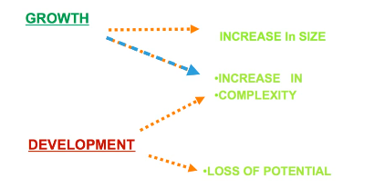
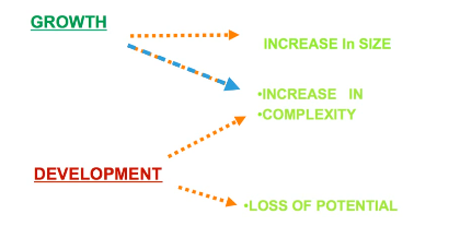
‘development’ is a term for an increasing degree of…
organisation/complexity
loss of potential
physiological and behavioural
why are the concepts of growth and development important?
helps us to understand when working with children what normal dental, facial and psychosocial growth and development looks like
helps to understand aetiology of malocclusion
find ideal tx timing
can be aimed at manipulating growth
understand factors influencing tx stability
growth studies may either be _____ or ___-_______ in design
longitudinal
cross-sectional
give details about cross-sectional studies
data is taken from different time points on different groups
eg) rather than same individual at 9/10/11 yrs old, data is taken from a group of 9 year olds, 10 year olds and 11 year olds
quick and easy
but there is individual variation
give details about longitudinal studies
follows the same individual over time
provides information about individual variation
long and expensive
list and elaborate on the 2 basic approaches to studying physical growth
MEASUREMENT APPROACHES
craniometry
anthropometry
cephalometric radiography
3D radiography (computed tomography)
EXPERIMENTAL APPROACHES
vital staining
implant radiography
give details about craniometry (measurement approach)
based on measurements of human skulls
enabled better understanding that distortions of skull can occur during growth
cross sectional study
give details about anthropometry (measurement approach)
measurements on living individuals
various landmarks on skull were transferred to overlying soft tissue points
varying soft tissue thickness introduces error source if goal is to measure facial skeleton growth
produced longitudinal data
cranial and facial growth knowledge comes from this approach
give details about cephalometric radiology (measurement approach)
slows direct measurement of skeleton bc soft tissue thickness can be ignored
technique depends on precisely orientating the head so all radiographs are reproducible (if yes then studies can be longitudinal)
lateral ceph’grams of the same person can be superimposed based on stable anatomical landmarks
allows growth to be visualised
give details about cephalometric radiology (measurement approach)
routinely used in orthodontics
monitor growth
diagnostic records
DISADVANTAGES
exposure to xrays
2d image of 3d structure
give details about 3d radiography (measurement approach)
computed tomography (CT)
reconstruct facial images in 3d
but issues) expensive, large radio dose
cone beam ct (CBCT)
much less cost and radio dose
on the same person images can be superimposed
give details about vital staining (experimental approach)
dyes that stain mineralising tissues are injected into animals
bone that was growing when the injection happened is stained and the amount of growth between 2 injections can be visualised
inadvertently seen in humans in tetracycline staining
give details about implant radiology (experimental approach)
metal pins are implanted into the skeleton and visualised with radiographs
used by bjork
responsible for understanding of jaw growth patterns
to identify abnormal growth, you must know…
timing of tx
that growth and development are different concepts
methods for studying growth (measurement and experimental)
what is a growth pattern?
changes in proportional relationships over time
cephalocaudal gradient of growth
axis of increased growth from head toward the feet (near the brain grows first and faster)
occurs within head and face (craniofacial structures)
mandible tends to grow more and later than the maxilla
scammon’s curve
the curve shows the growth of neural tissues is high first
genital grows exponentially during puberty
so not all tissue systems of the body grow at the same rate
changes in growth pattern would indicate some alteration in expected changes in body proportions
growth variability
everybody different in the way they grow
important to know if a kid is just a midget or they have a condition
variability in growth should be viewed as deviations from the usual pattern
expressed quantitively
eg standard growth charts
standard growth charts
can be used to determine whether growth is normal/abnormal
most used is height/weight chart
variability between genders
variability within each gender at the same age
other chart for other ethnicities too
above 97th or below 3rd percentile (h/w chart) may be abnormal or need test
where does variability in growth arise from?
normal variation
heredity
influences outside the norm (eg illness)
timing effects
growth timing
variability in timing arises because the same event happens for different people at different times
particularly noticeable during puberty (growth spurts)
generally, girls have their growth spurts 2 years ahead of boys (earlier maturation, shorter as plates close)
ortho tx also done earlier for girls
puberty for boys starts later and extends longer
timing of puberty
girls - menarche signals that PHV (peak height velocity) has occurred
boys - voice change coincides with PHV/PHV has occurred
studies show that PHV coincides with peak mandibular growth - perfect time for ortho growth mods
the _____ the onset of puberty, the _______ the adult size and vice versa
earlier
smaller
why should age be measured biologically and not chronologically?
chron. isnt good bc of variability in timing of growth
age should be measured biologically
achievement of certain development markers
s*xual development age
skeletal development age
dental development age
(i had to censor it bc otherwise it won’t let me share this set with others lmao)
skeletal (biological) age
hand and wrist radiographs - lots of markers
radiograph shows underlying ossification stages and morphological changes in multiple bones
sequence of ossification consistent among individuals
ossification associated w pubertal growth spurt
subjective and tricky - need atlas all the time
methods of reading hand and wrist radiographs
greulich and pyle
fishman’s skeletal maturity indicators (SMI)
smi 4-7 associated with high velocity of growth
eg) take RG and 1, 2, 3, 4 and 5 ossified → probably in high velocity of growth
skeletal age with lateral cephalograms
done instead of hands
for ortho records
shows cervical vertebrae (skeletal growth)
cervical stages (C2, 3 and 4)
assesses the presence of concavity in inferior border
different shapes in cervical vertebral body
cervical stages (skeletal growth)
CS 2 indicates that peak growth at adolescence is in 1 year
Cs 3 indicates that it is less than 1 year til peak growth
CS 3-4 coincides with peak mandibular growth - good time to begin tx, best time to do so
CS 5-6 exhibits some mandibular growth
dental age - techniques to describe dental age
eruption sequence
circumpubertal stage
calcification
eruption sequence of dental age
associates eruption of certain teeth and corresponds it to chronological age
age 6 - associated with first perm molars erupting
tooth emergence can be influences by multiple factors
early extraction
impacted teeth
crowding
tooth calcification of dental age
estimated from calcification based on atlas approach/scoring system
atlas - radiographically observing morphologically distinct changes of tooth mineralisation and comparing them to tables to estimate chronological age
scoring - assigning score based on mineralisation
tooth calcification scoring system
use quadrant 3 teeth
demirjian, goldstein, and tanner method
OPG analysis of quad 3 teeth
stages were developed from A-H based on shape criteria and proportion of root length relative to crown height
scores summed to give dental maturity score
limitations - need chart and x-ray
for quick use, usually use dental age (eruption)
basics of bone growth
cartilage within craniofacial structure acts similar to soft tissue (scaffold for bone later)
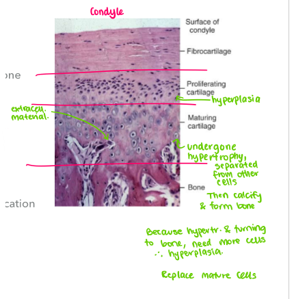
growth of soft tissues occur by a combo of hyperplasia (++ cells) + hypertrophy (++ cell size)
when this happens within tissues, called ‘interstitial growth’
secretion of extracellular material can also accompany IG (hypertrophy + plasia main characteristic)
bone formation occurs by…
endochondral ossification
intramembranous ossification
endochondral ossification
process where cartilage replaced by bone
cartilage grows interstitially while bone replaces it (hyperplasia/trophy)
when calcification happens (hard tissue formed), IG cannot occur (bc cells calcify so its hard and can’t expand)
growth of bone happens via…
direct addition on free surfaces (cells in periosteum)
replacement of soft tissue that grew before calcification
how does endochondral ossification in a long bone happen?
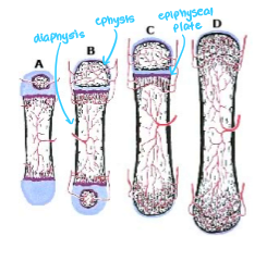
bone forms on the surface of the cartilage
invasion of blood vessels that produce hollowed out centre w bone formation in that area
ingrowth of blood vessels leads to an ossification centre in cartilaginous caps on either end
band of cartilage remains btwn diaphysis and epiphyses (epiphyseal plates) - continuous cartilage
bone grows longer as cart. of epi plate grows, matures and is replaced by bone
rate replacement exceeds rate of cart. growth
cart. is replaced and bone cannot grow longer
what bones/formations are formed by endochondral ossification?
what is the line of fusion btwn bones in cranial base called?
cranial base
mandible
synchondrosis (similar to epi. plate but its cartilage btwn 2 diff bones)
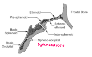
what is intramembranous ossification?
process of bone dev. from fibrous membranes
mesenchymal cells form template of future bone
mes. cells differentiate into osteoblasts at ossification centre
osteoblasts secrete extracell. matrix and deposit calcium - hardens matrix
what does intramembranous ossification form?
cranial vault
maxilla
mandible
junction btwn 2 bones in cranial vault = ‘suture’
what are the differences between synchondroses and sutures?
synchondroses
filled with cart.
makes bone by cart. replacement (endochondral)
capable of active independent bone growth
sutures
filled with periosteum and connective tissue (without cart. first)
makes bone without making cartilage first
can only react to its surroundings - only makes bone if sutures are pulled apart
as bone is under a constant dynamic state, it undergoes _______ and _______. this allows bone to adapt to… and is important for…
remodelling (helps maintain shape)
displacement
mechanical stresses
maintaining homeostasis
what is bone remodelling?
involves a combo of apposition of new bone to free surfaces
resorption of bone in other areas
occurs on inner + outer surfaces of each bone
allows change in size/shape and relocation of bone
what is displacement? how many types are there?
involves movement of the whole bone
mediated by soft tissues which apply external forces upon bone
compensatory growth at suture maintains articulation w bones
2 types: primary and secondary displacement
what is primary displacement?
refers to changes in position of bone by its enlargement
eg maxilla articulates w cranial base, so sutures in superior and posterior areas can deposit bone - pushes maxilla down and forward
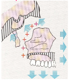
what is secondary displacement?
happens when bone position changed bc of growth of adjacent bone
eg cranial base grows, moving maxilla down and forward, second degree of maxilla displacement
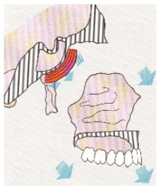
list the parts of the craniofacial complex
cranial vault
cranial base
nasomaxillary complex
mandible
alveolar processes
give details about the cranial vault (craniofacial complex)
frontal, parietal, squamous part of temporal and occipital bones
grows by intramemb oss
sutures (primary)
inner + outer surfaces remodelling
reacts to brain growth (deposits and resorbs)
at bird, sutures separated by connective tissue (fontanelles) allows defamation of vault during birth
after birth, apposition of bone occurs along edges and spaces
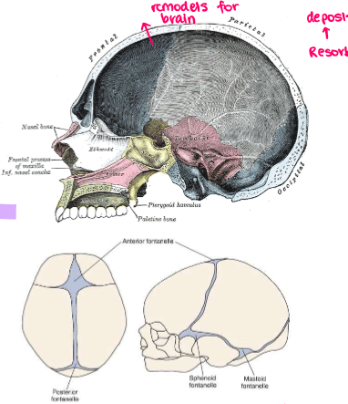
give details about the cranial base (craniofacial complex)
middle cranial fossa
sphenoid
temporal
occipital bone
growth occurs by endochondral ossification
synchondroses
surface remodelling
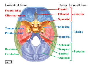
where are the main growth sites of the cranial base?
spehno-occipital synchondrosis
inter-sphenoid synchrondosis
sphene-ethmoidal synchondrosis
cranila base articulates with the maxilla and mandible
growth of the cranial base will affect the maxilla and mandible (secondary displacement)
give details about the nasomaxillary complex
maxilla
nasal
palatal (anatomy)
growth happens by intramemb oss
nasomax complex grows downward and forward from cranium
pos + sup growth of max sutures (primary displ - allows down+frwrd movement of maxilla)
secondary displace. from growth of cranial base
surface remodelling
why would you use the palatal suture in ortho?
when pt has periosteum and separated left and right by connective tissue, bone gets deposited and maximum size is increased
if narrow arch, use expander
pt has key to unwind expander to split suture, eventually will fuse and then cannot be undone (surgery to break suture)
nasomaxillary complex and surface remodelling
allows bone to keep shape while growing
front surface of max resorbed
bone added to tuberosity and sutures
bone deposited at tuberosity lengthens and widens the maxilla posteriorly
remodelling changes in opposite direction to translation
maxilla being moved forward more than its being reduced on anterior surface
nasomax complex and translation of palate
palate remodelled in same direction as it is translated
bone removed from floor of nose and added to roof of the mouth
palate being translated down and forward with the rest of the maxilla
secondary displacement of nasomax complex
helps with growth of maxilla
AP growth of cranial base contributes to max being translated anteriorly
cranial base stops growing around age 7 (2nd displacement no more after age 7)
give details about the mandible (craniofacial complex)
anatomy
condyle
body
growth occurs by
endochondral ossification (condyle)
intramemb oss (body)
surface remodelling
secondary displacement
mandible grows down and forward relative to cranial base
condyle and cartilage
cart. here not epi plate or synchondrosis
hyperplasia + trophy, and endochondral replacement happen here
condylar growth occurs in up and back direction
multidirectional adaptive growth via endochondral ossification
how does the mandible grow?
body of mandible grows longer by periosteal apposition on pos surface
ramus grows higher by endochondral replacement at condyle, accompanied by surface remodelling
anterior part of ramus is resorptive
secondary displacement from cranial base growth evident but effects are less than maxilla
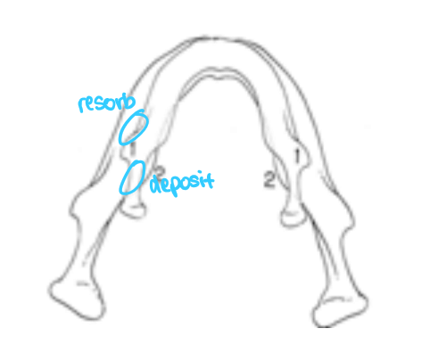
give details about the alveolar process (craniofacial complex)
as mandible grows downward away from maxilla, space is created btwn jaws
teeth must continue to erupt to maintain contact
eruption brings alv bone with it
give details about craniofacial growth
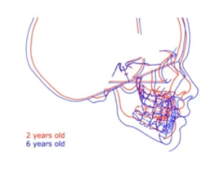
cephalometric superimposition of the same individual at 2 and 6 yrs of age
superimp - over stable landmark (sella turcica)
cranial base lengthened (brain mass inc. during this time - enlarged cranial vault)
maxilla and midface - pushed forward
mandible - moved down and forward
face grew more than cranium
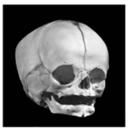
newborn skull
reflects cephalocaudal gradient of growth
underdevelopment of the mandible
ramus very short
nasal floor very close to eyes
maxilla not well developed compared to cranium BUT ahead of the mandible
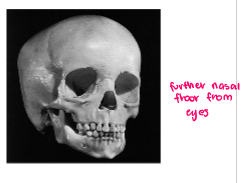
age 4 skull
nasal floor a bit more apart from eyes
cranium still larger tahn face
face has developed vertically and AP plane of space
max and mand have…
expanded laterally
downward and forward
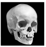
age 6 skull
displays cephalocaudial gradient of growth
cranial base nearly complete (completes at age 7)
face grown more than cranium
max and mand develop more
alveolar process has developed
cranial vault growth occurs largely at the…
sutures with some surface remodelling
cranial base growth occurs by…
endochondral replacement and some apposition at the sutures laterally
maxilla growth occurs by…
combo of suture growth and surface remodelling
mandible grows by…
apposition and remodelling resorption of the ramus and by endochondral replacement at the condyles
alveolar bone growth occurs as the…
teeth erupt to maintain contact during growth
facial growth and development is dependent on the interplay between…
genetic potential
environmental influences
define ‘site’, ‘centre’, ‘mode’, ‘mechanism’ and ‘determinant’
site: location at which growth occurs
centre: location of internally controlled growth that has the ability to grow on its own (contains info to control growth)
all centres are sites but not all sites are centres
mode: type of bone formation (endochon/intramem)
mechanism: method where skeletal changes are produced
determinant: controlling factor
list the 3 theories on how the craniofacial skeleton grows
suture theory - harry sicher
cartilage theory - James scott
functional matrix theory - melvin moss
give details about the suture theory (harry sicher)
sicher observed that bone formed at sutures
pressures made by new bone formation here pushed bones apart
intramemb bones are able to determine their own growth
sutures considered growth centres
suture growth should occur largely independently of environment and is impossible to change expression of growth at sutures
evidence against sicher’s suture theory
transplantation experiments (transplant suture somewhere else and it will grow)
reaction to manipulation (if sutures growth centres, manipulation won’t change growth
transplantation experiment (evidence against sicher’s suture theory)
when sutures transplanted to different locations, tissue doesn’t grow
indicates lack of innate growth potential in suture
reaction to manipulation experiment (evidence against sicher’s suture theory)
growth at sutures will respond to outside influences under many circumstances
if facial bones pulled apart at sutures, new bone would fill it more than it would otherwise
indicates sutures are reactive rather than independently acting
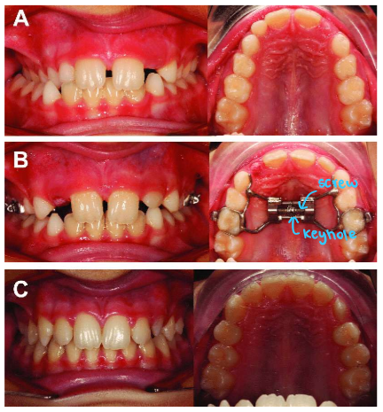
note) midpalatal suture often manipulated during ortho
midpalatal suture manipulation
image shows posterior crossbite
so they used an expander, or rapid max/palatal expansion device (RPED/RMED)
you either get skeletal or dental expansion but that depends on the timing its done and what stage the suture is at
once suture fused - permanent, so need to find good timing - find palatal suture ossification in biological age, before pubertal growth spurt, stage 3. after this - fusion of suture more tough so you just get dental movement not palatal, so need surgery to do that
cartilage theory (james scott)
cartilage does the growing and is then replaced by bone
cartilage determinant of craniofacial growth (growth centre)
cart. growth pushes bones to new positions, in response, bone will fill suture and surface remodelling can happen
cartilage theory and the mandible
mandible can be considered to be a long bone with cartilage at its ends
condylar cart. represents half an epi plate
in this scenario, cartilage should be considered a growth centre
cartilage theory and maxilla
growth of maxilla harder to explain using cartilage theory
scott made hypothesis that nasal septum was control centre for max. growth
cart. growth could easily lead to down + forward translation of maxilla (gap btwn maxilla and cranial base fills in with bone)
evidence against cartilage theory
transplantation experiment
surgical removal experiment (if you remove it, it won’t grow)
transplantation experiment and cartilage theory
epi plate cartilage → bone growth
synchondrosis cart → bone growth
nasal septal cart. → little/no growth (sometimes did, other times didn’t)
mand condyle cart. → no growth
not all skeletal cart. acts the same when transplanted
surgical removal experiment and cartilage theory
removing part of cartilaginous nasal septum causes considerable deficit in midfacial growth
but what if the surgery caused that? so its not rlllly counted as a proper experiment
achondroplasia (dwarfism) and cartilage theory
achondroplasia is inherited - epi plates cart. of long bones don’t grow as they should
short limbs, severe midface deficiency
pulls from the growth of nasal septal cartilage to open space at sutures
mandible unaffected
surgical removal experiment (cartilage theory) and the Scandinavian study
following an accident, fracture of mandibular condyle in kids results in resorption of the cartilage and condylar bone over time
75% chance condyle would regenerate and mandible would grow normally
growth at condyle is entirely reactive (so condylar cart. is not a growth centre)
not all cart. created equal in terms of importance to further grow and in terms of ability to independently grow
functional matrix theory (melvin moss)
growth of craniofacial structures occurs as a response to functional needs (mediated by soft tissues near skeletal units)
soft tissue and both bone and cart. react
growth of cranial vault illustrates this
cranial vault grows in direct response to growth of the brain
the functional matrix theory is supported by…
experiments of nature
microcephaly - small brain - size of head accurately represented size of brain
hydrocephaly - CSF accumulation - fluid accum. leads to intra-cranial pressure, small brain, enormous growth of cranial vault
functional matrix theory and maxilla/mandible
major determinant of growth is enlargement of nasal and oral cavities which grow in response to functional needs
theory doesn’t make it clear how functional needs are transmitted to tissues of mouth/nose
FMT and cartilages of cranial base, nasal septum and mand. condyles
moss believed these cartilages werent growth centres
synchondroses behave like sutures
nasal cart. removed, growth defect due to the surgery
condylar cart. reacts rather than controls growth
what does the FMT predict?
if proper function could be maintained, loss of condylar cart in a child would have no effect on mandibular growth
scandinavian study - 25% kids who suffer condylar fracture exhibit growth problem bc of function interference
ankylosis of mandible
bone of mandible reacts to soft tissue growth
determinant appears to be genetically controlled soft tissue
epigenetic - genetic control at a distance
growth of the cranium almost entirely occurs in response to…
growth of the brain
cranial base growth primarily result of endochondral growth (synchondroses) which have independent growth potential but maybe influence by brain growth
craniofacial growth and growth of maxilla/mandible
growth of max occurs from combo of growth at sutures and direct remodelling of the bone surfaces
soft tissues and nasal cartilage probably contribute to forward reposition of maxilla
mandible growth occurs by both endo prolif. at condyle and apposition and resorption of hone at surfaces
site, centre, mode, mechanism and determinant of the cranial vault
site: primarily sutures with some surface remodelling
centre: none
mode: mesenchymal
mechanism: bone fills in as sutures pushed apart
determinant: growth of brain - pressure to separate sutures
site, centre, mode, mechanism and determinant of the cranial base
site: synchondroses, sutures, surfaces (laterally)
centres: synchondroses
mode: endochondral and some mesenchymal on lateral surfaces
mechanical: cartilage push (central) and suture response (lateral)
determinant: genetic cartilage
growth of brain may cause lateral growth
site, centre, mode, mechanism and determinant of the nasomaxillary complex
site: sutures, nasal cartilage
centre: nasal cartilage
mode: endochondral (nose), mesenchymal (surfaces)
mechanisms: sutural growth, surface remodelling, push from cranial base, pull from soft tissue
translation: cranial base lengthening until age 7
apposition at sutures: pull from front (soft tissues), mid palatal suture (tension source)
surface remodelling: apposition at tuberosity, downward movement of palate/floor of nose, surface resorption (anterior surfaces)
determinant: genetic in cranial base, epigenetic factor
site, centre, mode, mechanism and determinant of the mandible
site: condyle, surface remodelling
centre: none
mode: endochondral (condyles) and mesenchymal (surface)
mechanism: pull in from front
determinant: epigenetic, soft tissue matrix
psychologists consider that there are 3 distinct mechanisms by which behavioural responses are learned…
classical conditioning
operant conditioning
observational learning
what is classical conditioning?
associating one stimulus with another
aka learning by association
occurs readily in young children
a kids vaccine at paeds can be painful/not fun
if unconditioned stimulus of pain is associated with people in white coats, sight of a white coat (conditional stimulus) will make reflex behaviour associated with pain
association btwn doc and dent can occur
but loss of conditioning can happen if the dent looks less like a hospital clinic
nothing painful should be done on the first visit
opposite of generalisation of a conditioned stimulus = discrimination
what is operant conditioning?
extension of classical
important part of communication for kids in late preschool and primary school ages
4 types
positive reinforcement
-ve reinforce.
omission
punishment
reinforcement that inc. likelihood of response is more useful in dent than omission and punishment
voice control is form of punishment (firm voice etc), reward for good behaviour
eg) +ve rein. brush teeth, get sticker
eg) -ve rein. have tanty, dont see dentist, do every time to get what they want
eg) omission. remove good stim after bad behaviour (eg go to ur room)
what is observational behaviour?
imitating observed behaviour (monkey see monkey do)
2 stages
acquisition of behaviour by observing it
performance of behaviour
executing acquired behaviour depends on many factors
characteristics of the role model
consequences of the behaviour
give details about cognitive development
cd occurs in series of distinct stages
biological adaptation to physical and sociocultural environ.
sensorimotor (birth - 2yrs)
preoperational (2-7 yrs)
period of concrete operations (7-11 yrs)
period of formal operations (11 yrs - adult)