cellular biochemistry
 Call Kai
 Learn
Learn Practice Test
 Spaced Repetition
 Match
Match Flashcards
 Knowt Play
1/11

There's no tags or description
Looks like no tags are added yet.
Name | Mastery | Learn | Test | Matching | Spaced | Call with Kai | Chat |
|---|
No analytics yet
Send a link to your students to track their progress
12 Terms

How is the transition from G1 to S phase regulated
p53 → activates p21 → inhibits CDKs → Rb remains hypophosphorylated (active) → Rb inhibits E2F → prevents G1 → S progression.
Growth factors promote the opposite effect: they activate cyclin-CDK complexes → phosphorylate and inactivate Rb → release E2F → allow G1 → S progression.

What cancers are associated with RB mutations
Mutations in the RB gene are associated with retinoblastoma and osteosarcoma

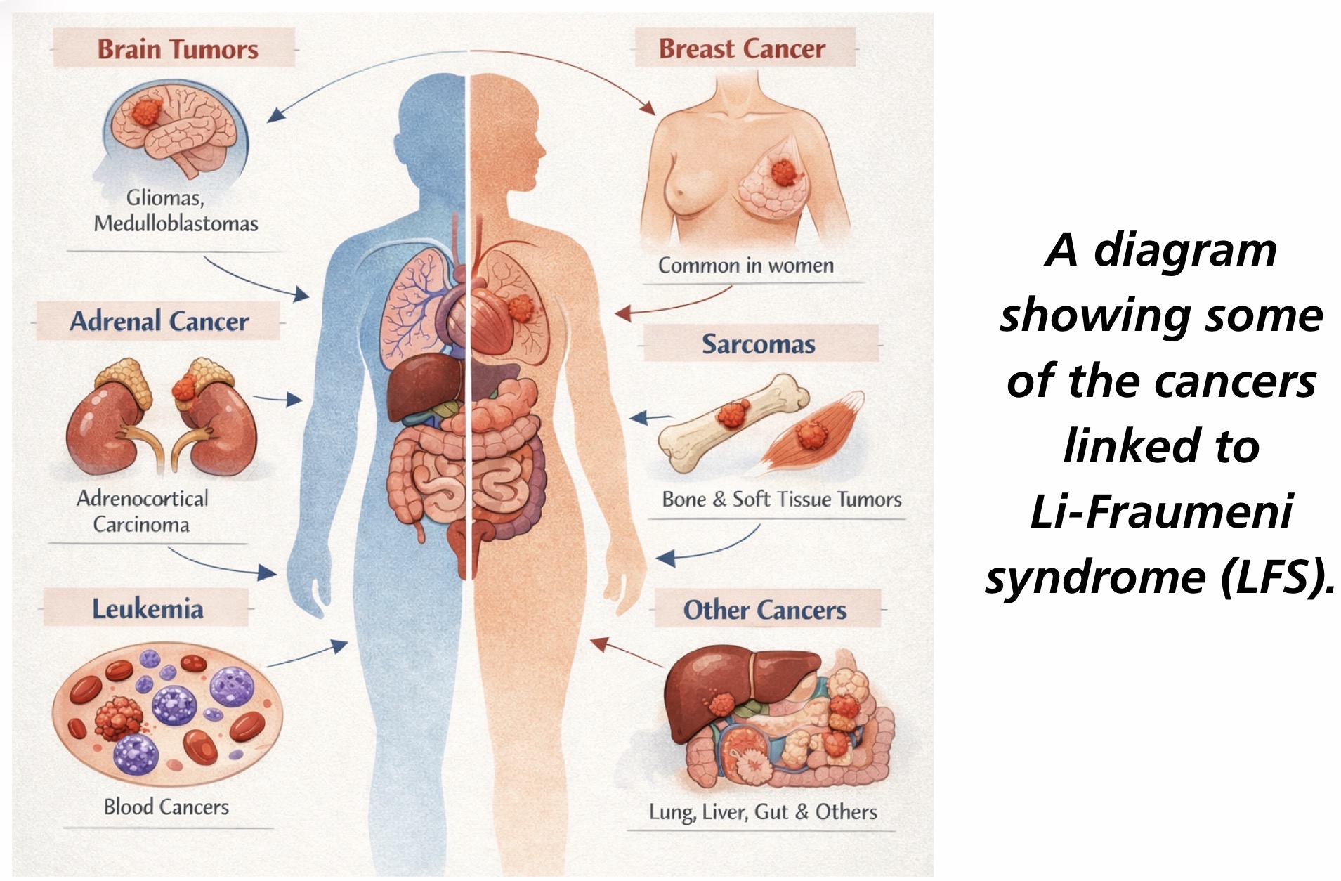
What is Li-Fraumeni syndrome and what causes it
Li-Fraumeni syndrome is caused by an inherited mutation in the TP53 tumor suppressor gene → loss of normal cell-cycle control → increased risk of multiple cancers at a young age, especially breast cancer, brain tumors, sarcomas, leukemia, and adrenocortical carcinoma.

What are the three cell types based on their ability to divide and what are examples of each
Permanent cells → stay in G0 and do not divide. Examples: neurons, skeletal muscle, cardiac muscle.
Stable (quiescent) cells → stay in G0 but can re-enter the cell cycle when stimulated. Examples: hepatocytes, lymphocytes, PCT cells, periosteal cells.
Labile cells → continuously divide and do not enter G0. Examples: bone marrow, gut epithelium, skin, hair follicles, germ cells.

Which cell type is most affected by chemotherapy
Labile cells

Where do N-linked and O-linked glycosylation occur
N-linked glycosylation → Rough endoplasmic reticulum (RER)
O-linked glycosylation → Golgi apparatus

What are the three main functions of the smooth endoplasmic reticulum (SER)
Steroid synthesis
Detoxification of drugs and poisons
Contains glucose-6-phosphatase → involved in the final step of glycogenolysis and gluconeogenesis
Which cells are rich in rough ER (RER) and smooth ER (SER)
RER → Mucus-secreting goblet cells and antibody-secreting plasma cells.
SER → Hepatocytes and steroid hormone-producing cells of the adrenal cortex and gonads.

Why is mannose-6-phosphate added to proteins in the Golgi
Mannose-6-phosphate is added to tag proteins for delivery to the lysosome
What is the pathophysiology of I-cell disease
I-cell disease is caused by a deficiency of N-acetylglucosaminyl-1-phosphotransferase → failure to add mannose-6-phosphate to lysosomal enzymes → enzymes are secreted outside the cell instead of being sent to lysosomes → lysosomes lack digestive enzymes and accumulate cellular debris

What are the main clinical features of I-cell disease
The main features are coarse facial features, restricted joint movement, and ↑ plasma levels of lysosomal enzymes. Other findings include gingival hyperplasia, corneal clouding, claw-hand deformities, and kyphoscoliosis
