LIFESCI 3K03: Brain Control: Unit 6
 Call Kai
 Learn
Learn Practice Test
 Spaced Repetition
 Match
Match Flashcards
 Knowt Play
1/41

There's no tags or description
Looks like no tags are added yet.
Name | Mastery | Learn | Test | Matching | Spaced | Call with Kai | Chat |
|---|
No analytics yet
Send a link to your students to track their progress
42 Terms
What are the three stages + areas of control of movement and what do they do?
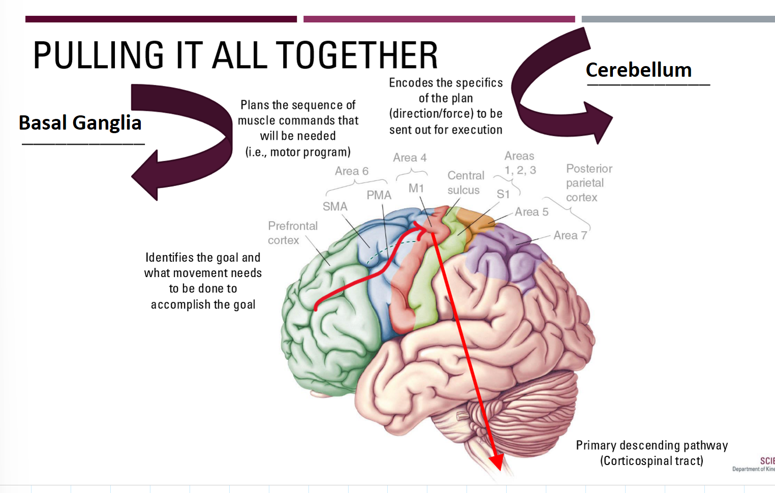
Determines what needs to be done (Ready) - Prefrontal Cortex
Identifies the goal of the movement and best strategy to accomplish this goal
Plans the specific movement (Set) - Motor Cortex
Specific sequences of muscle activations and patterns required to do the movement
Execute the plan (Go) - Spinal Cord
Activation of the motor neurons to do the movement and make minor adjustments
What is the range of complexity in the 3 stages of control of movement?

What does the Prefrontal Cortex do?
Highest in the chain of command and greatest complexity
Identifies a goal and determines what needs to be done to accomplish this
“What you need to do”
Highly connected with sensory cortex
Executive Function (EF)
Higher cognitive processes for planning, organizing, and controlling thoughts, speech, and behaviors
Involves a wide-range of skills

What are the two components of Executive Function and Movement?
Goal-directed actions
Organizing
What is the goal and how does this relate to the current sensory state?
Planning
What will need to be done to accomplish the goal?
Directing
Sending this information to the next processing station (i.e., motor cortex)
Attention
Multitasking
Allocating attention among tasks performed simultaneously
Response inhibition
Respond effectively with distractions/irrelevant information
Similar to a second level of “sensory gating” (i.e., thalamus filtering)
How is EF and aging related?
Executive function declines with age
Lesions in white matter
Loss of gray matter
Loss of dendritic branching
Changes are highly variable
Decline can be minimal in healthy aging
Influenced by things like lifestyle, education, genetics, etc.
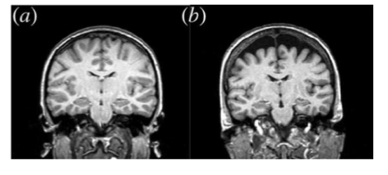
What are the age related declines related to EF?
Aging related declines in:
Overall processing speed
Problem solving ability
Organizing, planning, directing (Goal-directed actions)
Controlling attentional resources
Multitasking and response inhibition (Attention)
Declines are not to a level of dysfunction in healthy aging
What are the changes in gait parameters with age?
In general, with aging we see:
Decrease in gait speed
Decrease in step length
Increase in step time
Increase in variability in these parameters
However, healthy older adults may have little to no change
Reduced EF may be an important driver of these changes
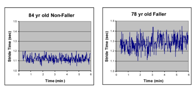
What is Gait in relation to executive function?
Gait is a complex motor task that uses executive function
Not fully managed by CPGs (especially in humans)
EF needed to plan, organize, and direct of movements
Often must also divide attention to other tasks
EF allows effective division of attention between gait and other tasks
Dual-task or Multi-tasking (e.g., Walk and talk/text)
What are the important parts of the motor cortex in relation to control of movement?
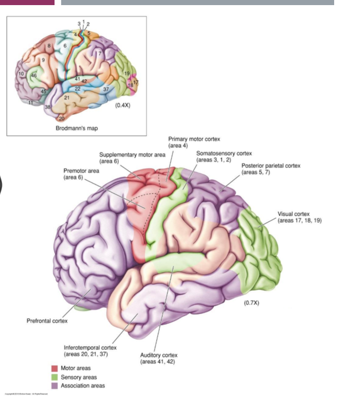
Motor cortex is involved after you have decided what you need to do, in order to decide how you will do it
Supplementary Motor Area (SMA)
Premotor Area (PMA)
Primary Motor Cortex (M1)

How are the PMA & SMA involved in the planning of movement?
Created movement plans and holds them until ready to execute
Active just BEFORE movement occurs
Up to 1s before voluntary movement
Plans must be highly integrated with sensory information
Details of coding taking place remains unclear
Premotor Area (PMA)
Selection of best motor plans based on current sensory information (e.g. picking up an object)
Supplementary Motor Area (SMA)
More complex motor sequences often with bilateral connections
May be more internally driven (remembered sequences) (e.g. performing a dance)
How was the planning of movement tested?
Through experiment with monkeys
Measuring the planning of movement
Instruction stimulus
Red light where movement will need to be
PMA neuron begins firing
Trigger stimulus
Blue light tell it to act
PMA neuron stops firing soon after the action is made
Specific to the side required for movement

What is active when planning (or even watching) movement?
“Mirror neurons” exist in the PMA
Respond when movement is imagined or watched
May be part of an extensive brain system for understanding actions and intentions of others
Monkey’s motor neurons didn’t activate when seeing the peanut being picked up by a tool instead of the monkey hand or human hand
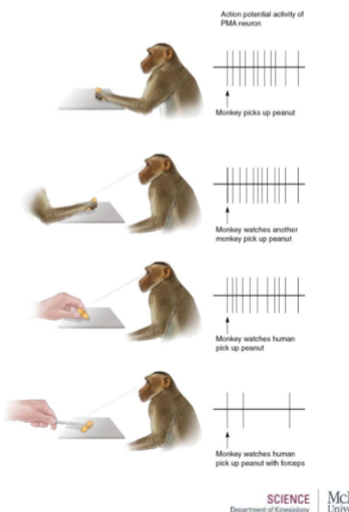
What does the primary motor cortex (M1) do in relation to movement?
Transforms the motor plan into specific movement patterns
Motor map masks the deeper complexity
Complex and overlapping neurons work together to control specific movements
Coding related to direction and force of movement

How does the coding of movement in the primary motor cortex work?
Movement direction is encoded by the collective activity of neurons
Many neurons are active for every movement
Activity of each cell represents a single “vote”
Direction of movement is based on a tally (and averaging) of votes
A single cell’s vote = Direction Vector
Tally of all cell votes = Population Vector
So…A population vector is based on MANY individual direction vectors
What is a direction vector?
Recordings from a single cell in M1 create a direction vector
Each cell has a “preferred” direction where firing rate is highest
Firing rate reduces as movement direction changes
Direction of the arrow stays the same for a given cell, but the length changes based on the direction of movement

How do you make a population vector from 2 direction vectors?
Based on testing we know cell 1 and cell 2’s preferred directions
Measure the firing rate in cell 1 & cell 2 in response to a particular motion
Create a population vector that represents the actual movement
Population vectors are created from many cells

What forces in relation to population vectors?
Population vectors generated in the direction in which force is needed
Population vectors based on normal movement

Population vectors based on moving against a resistive force (pushing in a specific direction)

Neural decoding in M1
We have a greater understanding of signals in M1
compared to prefrontal, SMA, and PMA
Still, decoding these overlapping signals remains extremely complex:
Requires interpreting inputs from potentially thousands of neurons
Relating to multiple muscle movements
What is the basal ganglia?
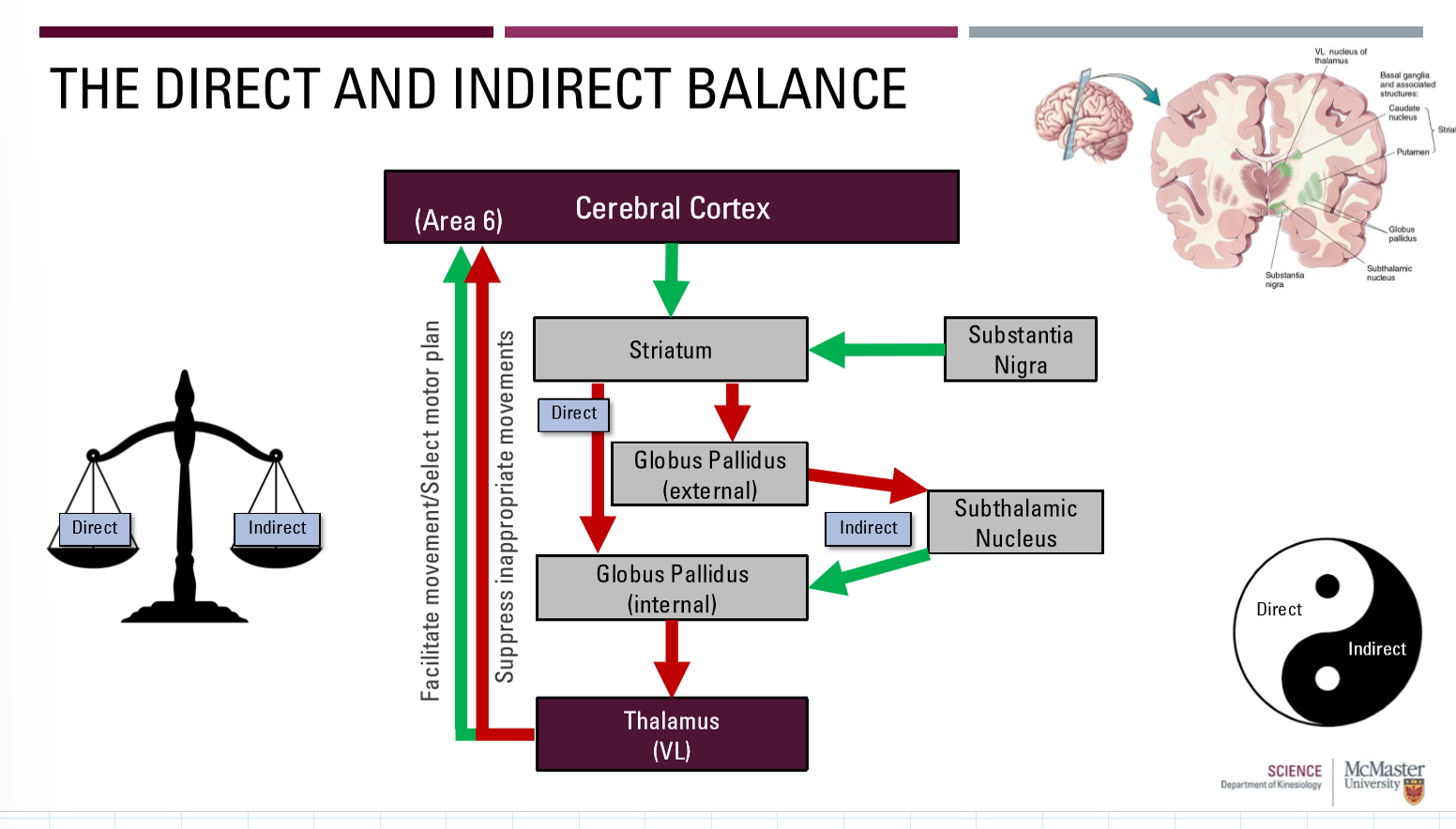
Group of subcortical nuclei which supports the selection and initiation of willed movements, while preventing unwanted movements
What are the 2 motor pathways of the basal ganglia?
Direct Pathway
Facilitates movement
Helps to select motor plans
Indirect Pathway
Inhibits movement
Help to suppress competing or inappropriate motor plans
Need a healthy balance between the two

What is the direct pathway of the basal ganglia?

What is the indirect pathway of the basal ganglia?
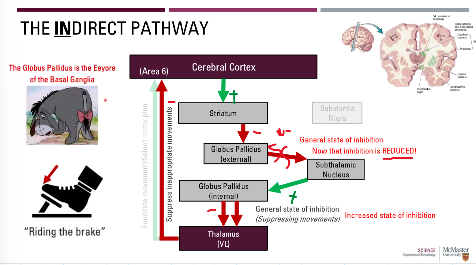
What is the direct and indirect balance of the basal ganglia?
Direct = facilitates appropriate movements (Releasing the brake)
Indirect: Inhibits inappropriate movements (Riding the brake)
What is Parkinson’s Disease?
A disorder of the direct pathway
Difficulty stimulating wanted movement
Reduced direct pathway release of thalamus inhibition
Arises from a loss of dopaminergic neurons acting on the striatum in the direct pathway
What are hypokinetic disorders?
Reduced voluntary motor activity due to a disorder of the direct pathway
Bradykinesia - slowness of movement
Akinesia - lack of movement (e.g., freezing of gait)
How does a direct pathway disorder work?
Loss of dopaminergic neurons from Cerebral Cortex to Striatum (and from the substantia nigra to the striatum)
Levodopa supplements help maintain the pathway

What is Huntington’s disease?
A Disorder of the indirect pathway
Difficulty suppressing unwanted movements
Reduced indirect pathway = reduction in thalamus inhibition
Arises from a loss of striatum neurons acting on the globus pallidus external in the indirect pathway
Rare genetic disorder (<0.01% of people)
Symptoms arise in 30s or 40s
Neuron loss will also occur in other areas of the cerebral cortex, leading to:
Dementia
Personality changes
Death (~20 years after diagnosis)

What are hyperkinetic disorders?
Disorders of the indirect pathway that involve excessive involuntary motor activity
Chorea - spontaneous and uncontrollable movements
How does an indirect pathway disorder work?
Loss of neurons from striatum to GPe
Final overall inhibition cannot occur

What is the cortico-cerebellar loop and what are the 3 parts?
Proper execution of planned, voluntary, multi-joint movements
Necessary to fine tune the sequences (i.e., timing) of muscle contractions
3 parts:
Cortex to cerebellum
Within cerebellum
Cerebellum to cortex
What is the cortico-ponto-cerebellar pathway of the cortico-cerebellar loop?
Sensory and motor cortex axons form massive projection on pons
Pontine nuclei relay information to cerebellar cortex

What is ‘within the cerebellar cortex’ of the cortico-cerebellar loop?
Granule cells
Most numerous cells in the cerebellum
Excitatory/inhibitory output on Purkinje cells
Purkinje cells
Largest cells in cerebellum
Received thousands of synaptic inputs
Excitatory/Inhibitory output to deep cerebellar nuclei
Deep cerebellar nuclei
Excitatory or inhibitory output to thalamus

What is the ‘cerebello-thalamo-cortical pathway’ in the cortico-cerebellar loop?
Deep cerebellar nuclei relay to thalamus (VL)
VL relays information back to M1
Basal Ganglia was area 6 (premotor)
Here, M1 (area 4) signifies last chance tuning (timing, direction, force)

What results from cerebellar lesions?
Ataxia: uncoordinated and inaccurate movements
Summary of Brain Control
What are the two major groups of pathways in descending spinal tracts?
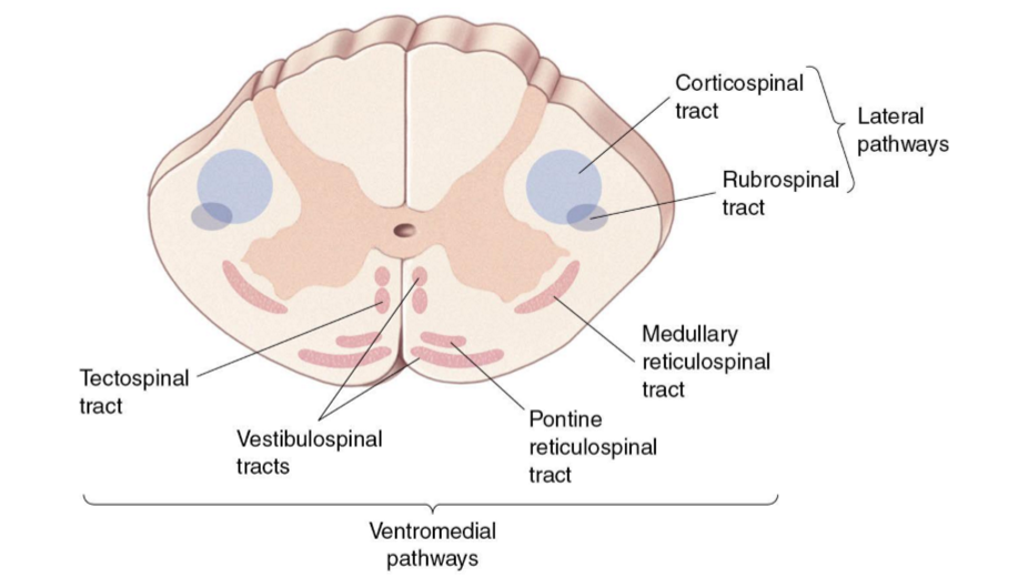
Lateral motor pathways
Commands for voluntary movements
Ventromedial motor pathways
Posture and reflex movements
What is the corticospinal tract (CST)?
Primary pathway for voluntary motor control
Neck to feet
One of the largest and longest pathways
Pyramidal cells arising from the motor cortex
M1, but also premotor and other areas
Can be further divided into:
Lateral (limbs) vs. anterior (trunk, neck, shoulders)
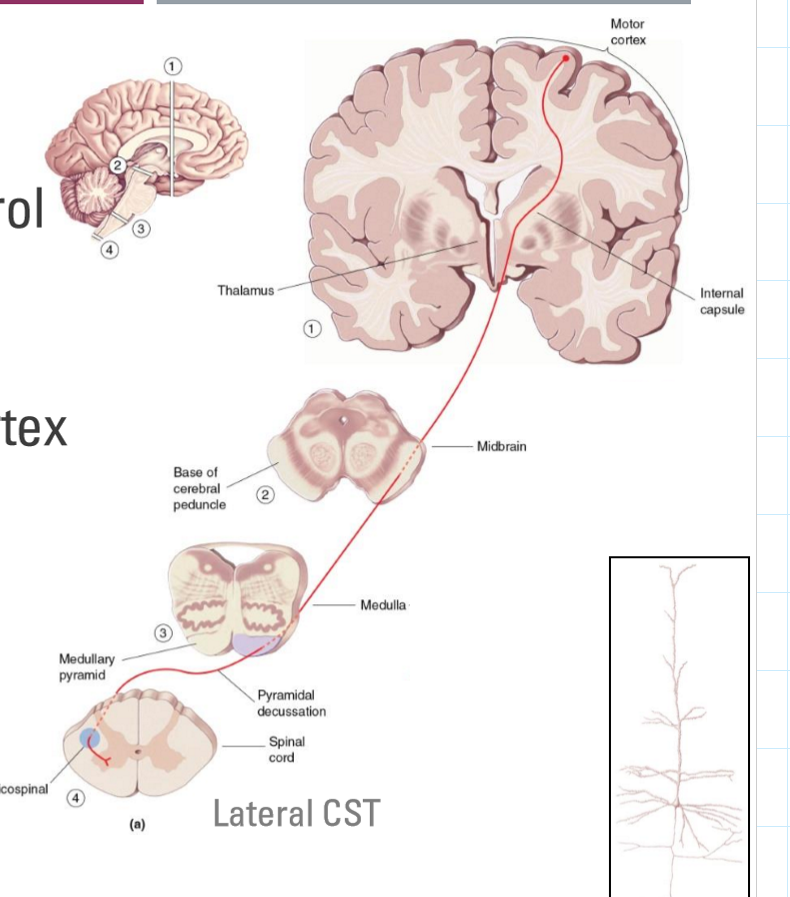
Lateral vs. Anterior CST
Lateral CST
90% of CST axons
Decussate in medullary pyramids
Control proximal/distal muscles (i.e., limbs)
Anterior CST
10% of CST axons
Decussate within spinal cord
Control axial muscles
Check slides for all diagrams

What is the lateral vs. anterior corticospinal tract pathway? (Draw it out)

How are the three different pathways impacted in Brown-Sequard Syndrome (Draw)?
