sheep brain
1/39
Earn XP
Description and Tags
AP Lab #3
Name | Mastery | Learn | Test | Matching | Spaced | Call with Kai |
|---|
No analytics yet
Send a link to your students to track their progress
40 Terms
Meninges (brain covering)
Dura Mater, arachnoid mater, pia mater
Dura Mater
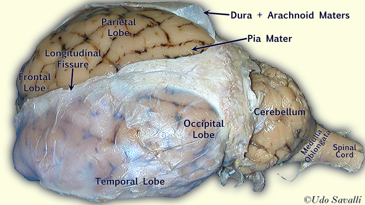
The outermost, thickest, and toughest layer.
In a sheep brain, it forms a tough, leathery covering that must be carefully removed to access the brain underneath.
Arachnoid mater
The middle layer, which is thin and delicate.
This layer is separated from the dura mater by the subdural space and from the pia mater by the subarachnoid space, which is filled with cerebrospinal fluid
Pia Mater
The innermost, most delicate, and highly vascular layer.
It adheres tightly to the surface of the brain, following all of its grooves and fissures.
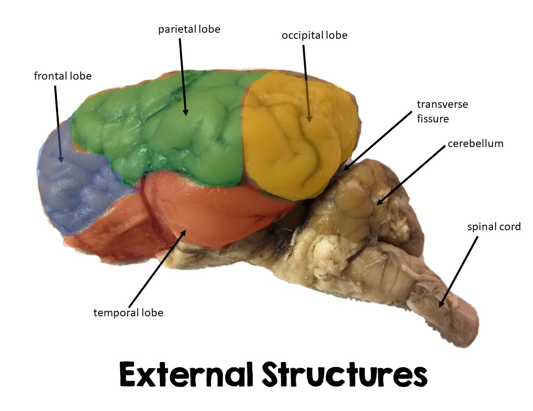
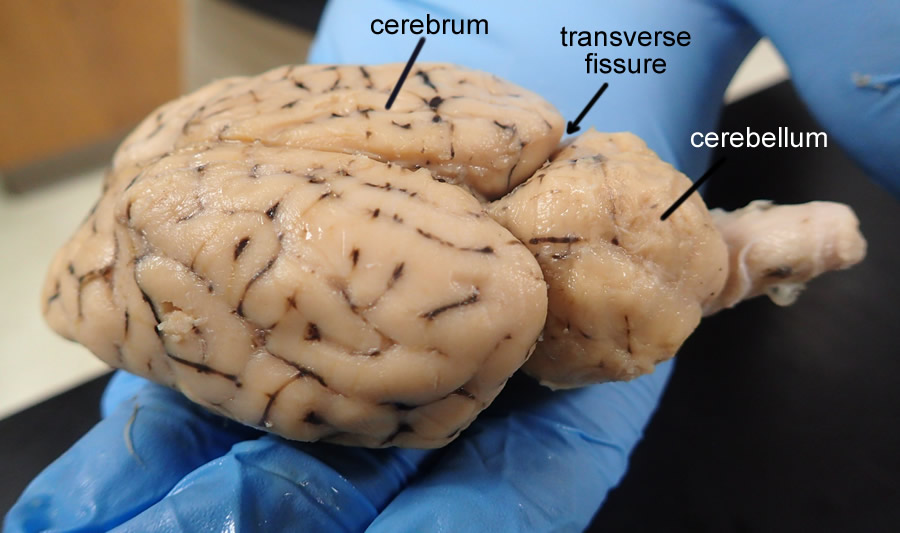
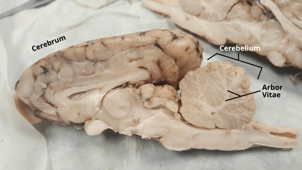
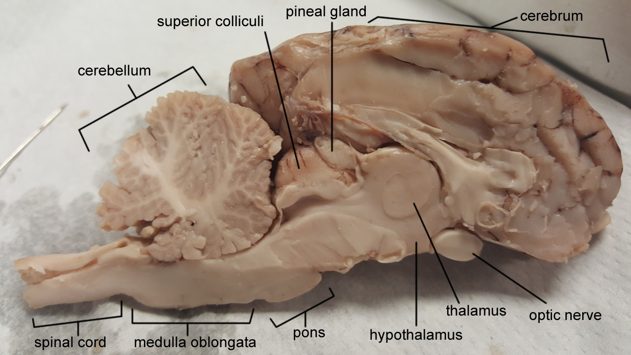
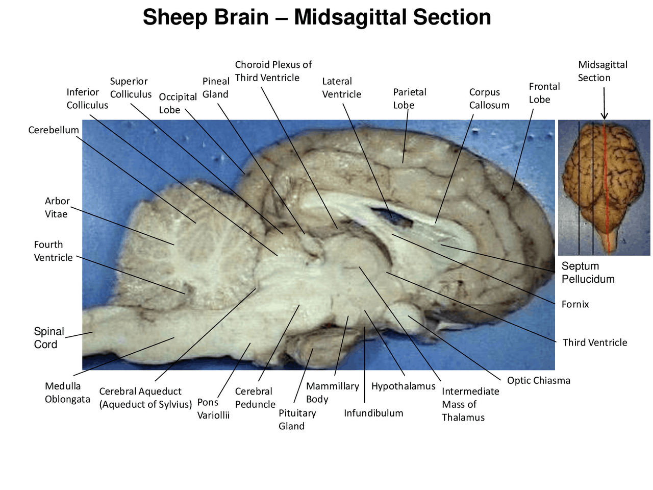
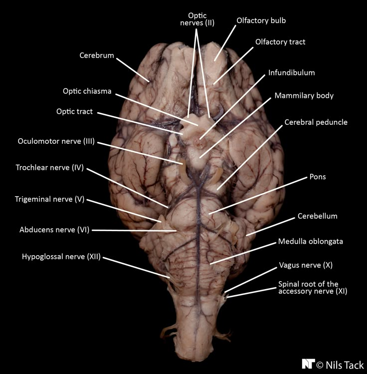
Cerebral hemispheres
cerebrum
conscious brain - the larger of the two, in the front
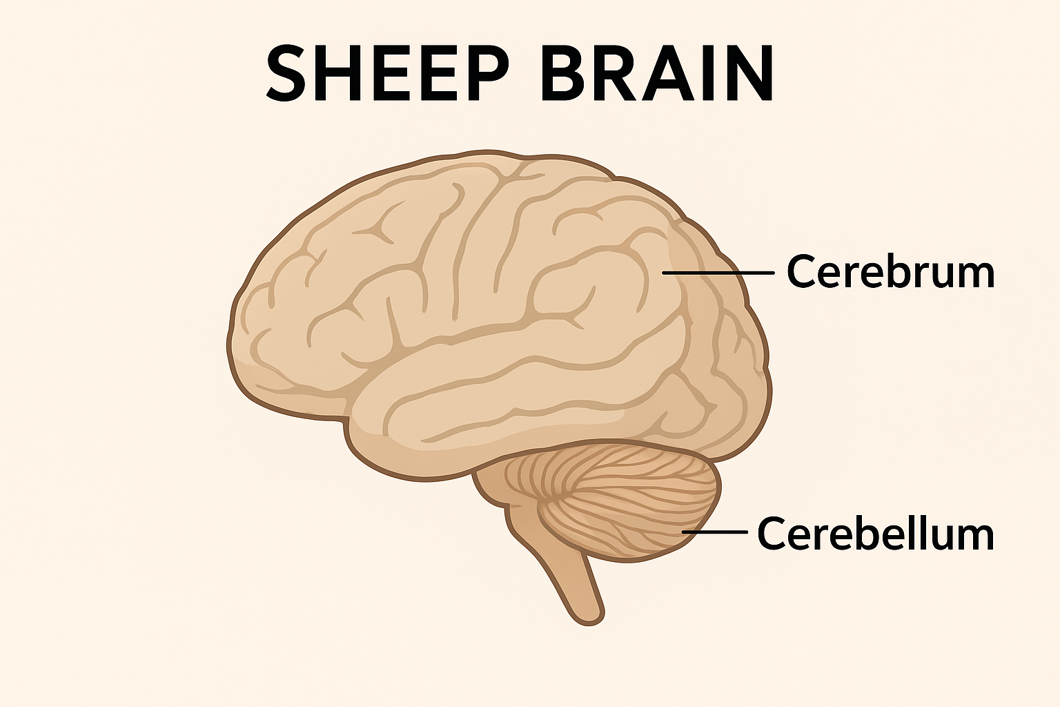
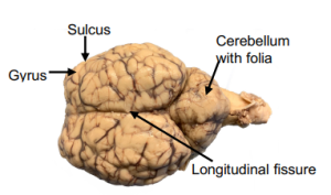
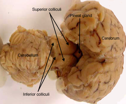
cerebellum
smaller, back part, lots of folds, coordinates muscle movements and muscel memory

gyri
the ridges - bumps
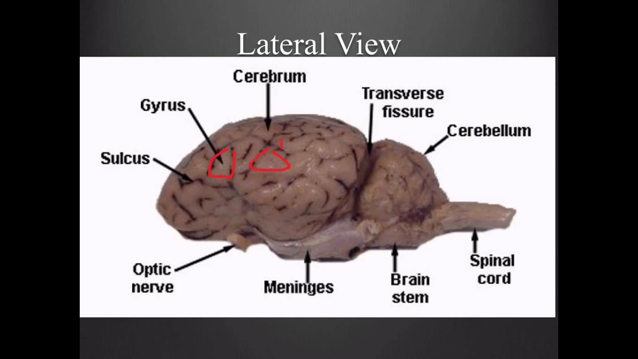
sulci
the grooves - lines
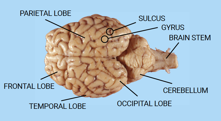
frontal, parietal, temporal, occipital lobes
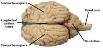
longitudinal fissure
line in the middle, separates 2 cerebrum
transverse cerebral fissure
separates cerebrum and cerebellum
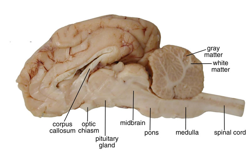
midbrain
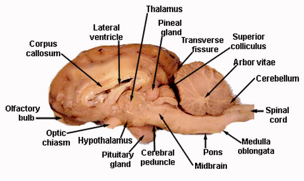
pons
“bridge that links medulla oblongata to thalamus

medulla oblongata
houses autonomic centers

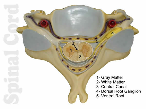
spinal cord
pituitary gland
“master” gland of endocrine system - on the middle of bottom side of brain

cerebral cortex
grey matter contains neuron cell bodies; site of the “conscious mind”
-cerebral cortex is the outer layer of the cerebrum, covering the upper part of the brain. It’s the wrinkled (convoluted) gray matter you can see on the surface of the large, elongated hemispheres.
-folded into gyri (ridges) and sulci (grooves)
White matter
bundles of myelinated axons called tracts in CNS
arbor vitae
white matter
corpus callosum
connects left and right brain hemispheres

fornix
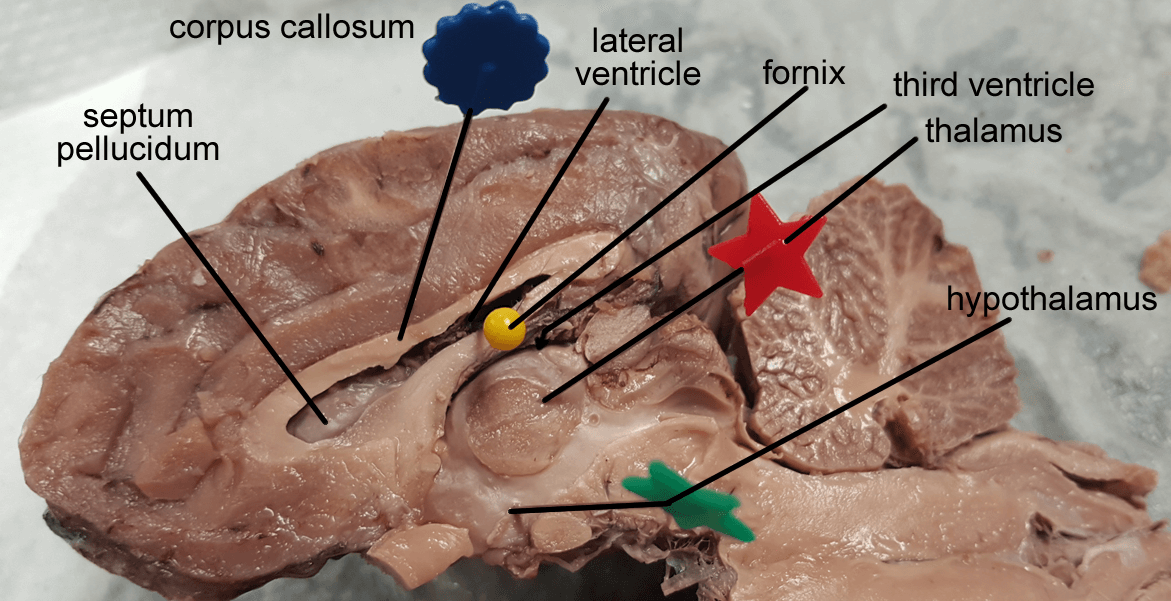
thalamus
gateway/relay station to/from cerebral cortex

hypothalamus
controls pituitary gland and vital autonomic centers of medulla oblongata
infundibulum
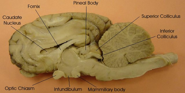
optic chiasm

choroid plexus
produces cerebrospinal fluid
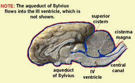
third, fourth ventricles and the cerebral aqueduct
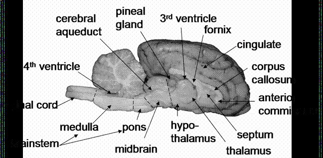
pineal gland
controls sleep/wake cycles
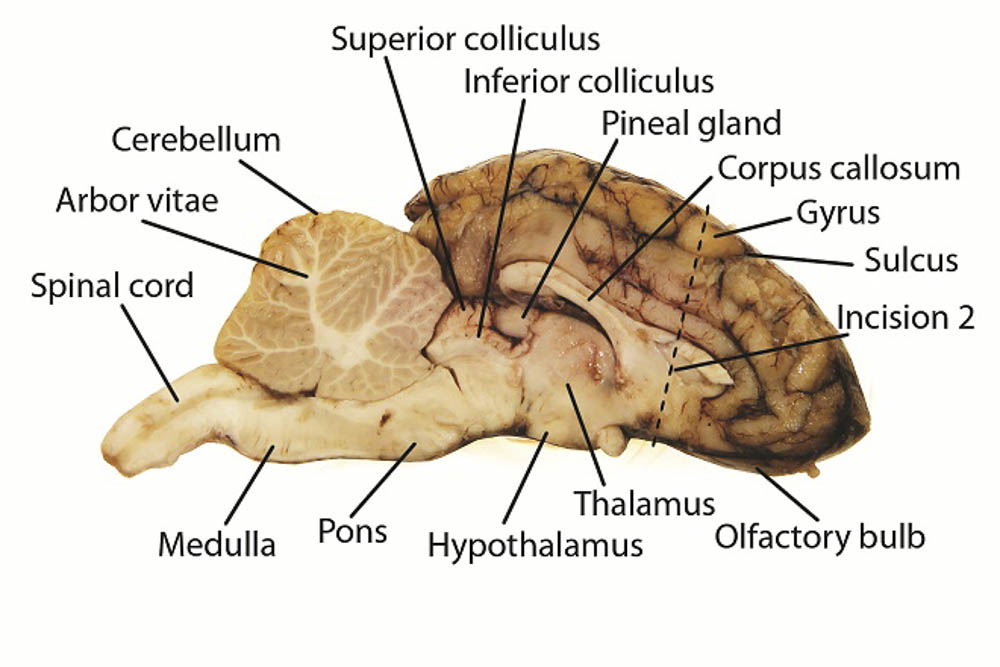
inferior colliculi
auditory reflexes

superior collicili
visual reflexes
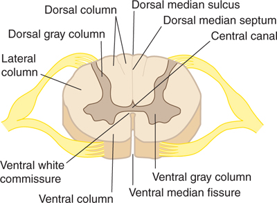
dorsal horn
the upper wings of the butterfly; they receive sensory input from the body.
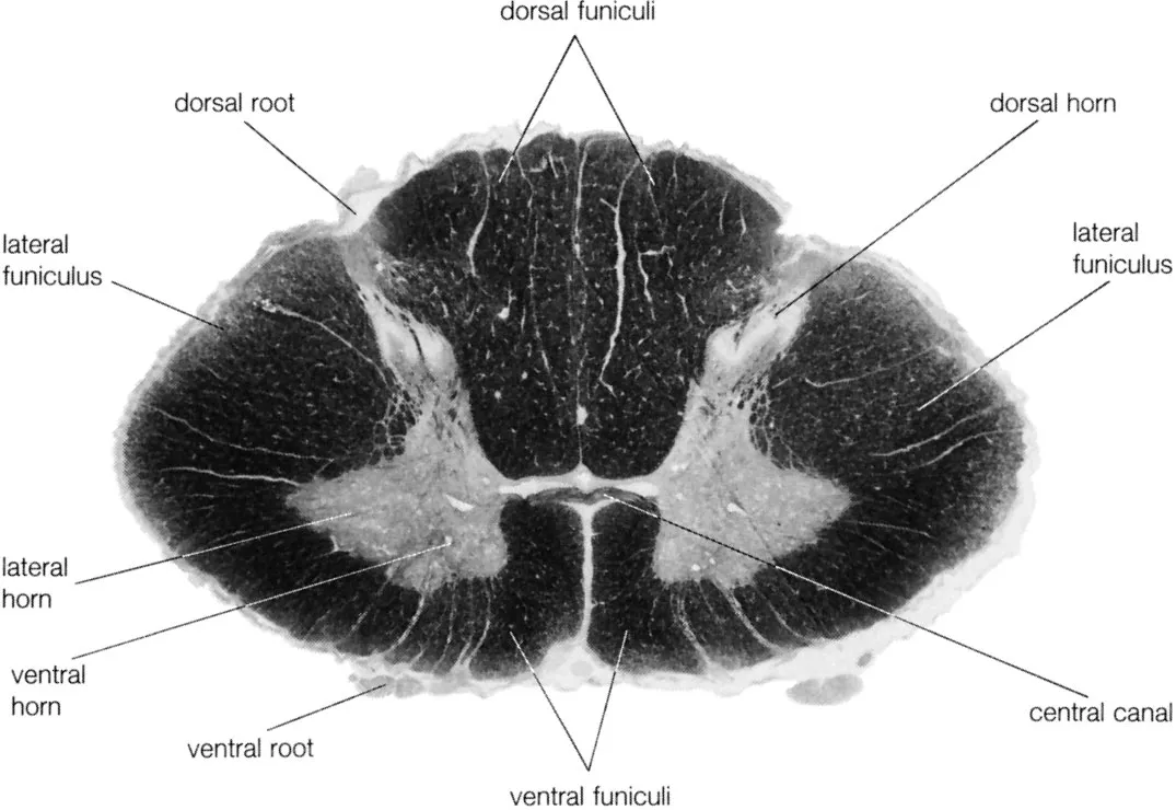
lateral horn
small bumps or extensions on the sides of the gray matter (present only in the thoracic and upper lumbar regions); they contain neurons of the autonomic nervous system.

ventral horn
the lower wing of the “butterfly” — toward the front of the cord.

ventral median sulcus
If you look at the spinal cord from the front, you’ll see a deep groove running down the midline — that’s the ventral median sulcus.
On a cross-section, it appears as a wide V-shaped or deep indentation on the bottom side of the cord (the dorsal side has a narrower groove called the dorsal median sulcus).
central canal
dorsal root
The dorsal root is located posteriorly (toward the back).
It enters the dorsal (posterior) horn of the gray matter in the spinal cord.
dorsal root ganglion
spinal nerve
dorsal median sulcus
The dorsal median sulcus is a shallow groove along the midline of the posterior (dorsal) side of the spinal cord.