Diag/Inter Acute - Module 1 - Week 1 - Pulmonary System Anatomy and Physiology
1/94
There's no tags or description
Looks like no tags are added yet.
Name | Mastery | Learn | Test | Matching | Spaced | Call with Kai |
|---|
No analytics yet
Send a link to your students to track their progress
95 Terms
What are the boney structures of the thoracic cavity?
Thoracic cavity consists of ribs laterally (12 pairs of ribs, 7 true, 3 false, and 2 floating), vertebrae dorsally, and sternum ventrally.
What seperates the lungs from each other?
The mediastinum
What is the shape of the lungs?
They are cone shaped.
What is the highest point of each lung called?
The apex
What does the base of each lung rest on?
The diaphragm
What are the three borders of the lungs?
-Anterior
-Posterior
-Inferior
What are the three surfaces of the lungs?
-Costal
-Medial
-Diaphragmatic
Which lung is larger? and the location of what structure causes the size difference?
The right lung is larger than the left due to the location of the heart.
How many lobes does each lung have?
The right lung has three lobes and the left lung has two lobes
-Right has a superior (upper), middle, and an inferior (lower) lobe
-Left has a superior (upper) and an inferior (lower) lobe
How many fissures does each lung have? What are they called? and what do they seperate?
The right lung has two fissures that seperate the lobes. The horizantal fissure seperates the superior lobe from the middle lobe. The oblique fissure seperates the middle lobe from the inferior lobe.
The left lung only has one fissure as there are only two lobes. The oblique fissure seperates the superior lobe from the inferior lobe.
How many segments does each lung have?
The right has 10 segments and the left has 8 segments
(Note sometimes, it is said that each lung has 10 segments. The discrepancy is due to the fact that in the left superior lobe, the apical segment and posterior segment are typically combined into the apicoposterior segment, and in the left inferior lobe, the anterior basal segment and the medial basal segment are typically combined into the anteromedial basal segment).
What segments are in the right upper (superior) lobe that are important for postural drainage? (3)
-Apical segment
-Anterior segment
-posterior segment
What segments are in the left upper (superior) lobe that are important for postural drainage? (3)
-Apical segment
-Anterior segment
-posterior segment
What segments are in the right lower (inferior) lobe that are important for postural drainage? (4)
-superior segment
-anterior basal segment
-lateral basal segment
-posterior basal segment
What segments are in the left lower (inferior) lobe that are important for postural drainage? (4)
-superior segment
-anterior basal segment
-lateral basal segment
-posterior basal segment
What is the functional tissue of the lungs called? and what does it consist of?
It is called the paranchyma and consists of alveoli, bronchioles, and interstitial tissue.
What is the Hilum of each lung?
It is a wedge shaped depression on the medial surface of each lung where the nerves, vessels, and primary bronchi penetrate the parenchyma of each lung.
What are the roots of each lung? (6)
The roots are the structures that enter the lung in the Hilum.· They consist of:
-The main bronchus,
-pulmonary artery,
-pulmonary veins (superior and inferior),
-bronchial arteries and veins,
-pulmonary nerve plexus,
-and lymph vessels
What are the pleurae?
The Pleurae is a thin membrane that covers each lung. There are three layers to the pleura: the visceral pleura, pleural cavity, and a parietal pleura.
What is the visceral pleura?
serous membrane that adheres directly to the lungs
What is the pleural cavity?
The fluid filled space between visceral and parietal pleura.
What is the parietal pleura?
Lines thet pulmonary cavity
What is the major function of the respiratory system?
Gas exchange. The replenishment of the bloods oxygen supply used for oxidative energy production and the removal of carbon dioxide from the returning venous blood that was manufactured as a waste product.
Aside from gas exchange, what are the other functions of the respiratory system? (6)
-fluid exchange,
-maintenance of a relatively low volume blood reservoir,
-filtration,
-metabolism,
-regulation of acid-base balance,
-and temperature homeostasis.
What are the two different types of rib motion?
The two types of rib motion are the bucket handle and the pump handle motions. Basically, the lower ribs move like a bucket handle where they move primarily laterally and superiorly as the chest expands. The upper ribs move like a pump handle where the ribs move primarily anteriorly and superiorly. The middle ribs are a transition between the two and move in all three planes fairly equally.
What innervates the lungs?
Stimulation of Cranial Nerve X - The vagus nerve and stimulation of the sympathetic nerves.
What does stimulation of cranial nerve X - the vagus nerve cause in the lugns? (3)
-bronchial constriction,
-dilation of pulmonary arterial smooth muscle,
-and increased glandular secretion.
What does stimulation of the sympathetic nerves cause in the lugns? (3)
-bronchial relaxation,
-constriction of pulmonary arterial smooth muscles,
-and decreased glandular secretion.
What structures make up the upper respiratory tract? (3)
-Nose or mouth
-Pharynx
-Larynx
What is the function of the structures within the upper respiratory tract?
-The nose or mouth is the entry point into the respiratory system. The nose filters, humidifies, and warms the air.
-The pharynx is the common area used for both the respiratory system and the digestive systems.
-The larynx connects the pharynx to the trachea including the epiglottis and vocal cords.
What are the epiglottis and vocal cords? and what are their functions?
-The epiglottis is a flap of tissue within the larynx. Its main function is to close over the trachea while you're eating and drinking to prevent food/water from entering the airway.
-The vocal cords are two muscular bands inside your voice box that produce the sound of your voice.
What is the lower respiratory tract?
The lower respiratory tract extends from the level of the true vocal cords in the larynx to the alveoli within the lungs and is broken down into two components: the conducting airways and the respiratory unit.
What structures make up the conducting airways of the lower respiratory tract? (4)
-Trachea
-mainstem and lobar bronchioles
-segmental and subsegmental bronchioles
-terminal bronchioles
What is the function of the conducting airways of the lower respiratory tract?
Only transport air, there is no gas exchange occurring in these structures.
What structures make up the respiratory unit of the lower respiratory tract? (4)
-the respiratory bronchioles,
-alveolar ducts,
-alveolar sacs,
-and alveoli
What is the function of the respiratory unit of the lower respiratory tract?
Diffusion of gas occurs through all of those structures.
What are the primary muscles of inspiration?
diaphragm and external intercostals
What nerves innervate the diaphragm?
the phrenic nerve (C3, C4, and C5),
What is the primary function of the diaphragm?
The primary function is respiration. Contraction creates flattening of the diaphragm which facilitates expansion of the thoracic cavity leading to increased volume of the cavity which in turn decreases the intrathoracic pressure allowing the lungs to expand and inspiration to occur.
Aside from respiration, what are the other functions of the diaphragm? (3)
-postural control muscle,
-a GI muscle (anti-reflux muscle and lower GI motility muscle),\
-a venous return muscle (helps blood return to the heart through the inferior vena cava through a straw like effect.)
What innervates the external intercostal muscles?
Intercostal nerves (T1-T11)
What is the function of the external intercostal muscles?
Contraction pulls the lower ribs up and out toward the upper rib which elevates the ribcage and expands the chest.
What are the two primary muscles/muscle groups of expiration?
-Abdominals (Rectus abdominus, transverse abdominis, internal obliques, and external obliques)
-Internal intercostals
What innervates the abdominal muscles?
Thoracoabdominal Nerves (T7-T11)
What is the respiratory function of the abdominal muscles?
Raise intraabdominal pressure when expulsion of air is required (i.e. coughing and huffing). Pressure generated within the abdominal cavity is transmitted to the thoracic cage to assist in emptying the lungs
What innervates the internal intercostal muscles?
intercostal nerves (T1-T11)
What is the respiratory function of the internal intercostal muscles?
Compress the thoracic cavity by pulling the ribs together
What are the secondary muscles of inspiration? (9)
-Sternocleidomastoid
-Scalenes
-Serratus Anterior
-Pectoralis major
-Pectoralis minor
-Trapezius
-Erector spinae group
-Laissimus Dorsi
-Serratus posterior superior
What innervates the SCM and Traps?
Innervated by cranial nerve XI - the spinal accessory nerve
What is the respiratory function of the SCM?
Elevates the sternum, increasing the anteroposterior diameter of the chest
What is the respiratory function of the traps?
Elevates the thoracic cage.
What innervates the scalenes?
Anterior rami of C3-C8 (cervical and brachial plexus)
What is the respiratory function of the scalenes?
Function as a unit to elevated and fix the first and second ribs
What innervates the serratus anterior?
Innervated by the long thoracic nerve (C5, C6, and C7)
What is the respiratory function of the serratus anterior?
Serratus anterior can only assist when the rhomboids stabilize the scapula in adduction allowing the serratus to expand the rib cage by pulling the origin toward the insertion.
What innervates the pectoralis major?
The medial and lateral pectoral nerves (clavicular head C5 and C6, Sternocostal head C7, C8, and T1)
What innervates the pectoralis minor?
Medial pectoral nerve (C6-T1)
What is the respiratory function of the pectoralis major and minor?
When the UE is fixed, they can pull on the anterior chest wall, lifting the ribs and the sternum, and facilitating an increase in the anteroposterior diameter of the thorax.
What innervates the erector spinae group?
Posterior rami of spinal nerves
What is the respiratory function of the erector spinae group?
Extend the thoracic spine and raise the rib cage to allow greater expansion of the thorax.
What innervates the latissimus dorsi?
Thoracodorsal nerve (C6-C8)
What is the respiratory function of the latissimus dorsi?
Posterior fibers pull the trunk into extension
What innervates the serratus posterior superior?
intercostal nerves 2-5
What is the respiratory function of the serratus posterior superior?
Raise the ribs to which it is attached (ribs 2 to 4 or 5) and expand the chest.
What is breathing/ventilation?
The mechanical movement of gases in and out of the lungs. It is a continuous and rhythmic process. Inhalation and exhalation alternate to maintain a steady exchange of oxygen and carbon dioxide in the blood stream, supporting the body's metabolic needs.
What is inspiration? and how does it occur?
Inspiration is an active process that requires contraction of muscles (diaphragm and external intercostal muscles). The diaphragm contracts and moves downward and therefore increasing the volume of the thoracic cavity. External intercostal muscles contract and lift the ribcage upward and outward, further expanding the thoracic cavity. The combined action of the diaphragm and the external intercostal muscles increases the volume in the thoracic cavity. This increasing volume causes a decrease in the pressure within the lungs making it lower than the atmospheric pressure. The pressure difference is what causes air to enter through the nose and/or mouth and rush into the lungs.
What is expiration? and how does it occur?
Expiration is a passive process that occurs when inspiration is complete. The elastic fibers in the lung tissue, as well as the elastic recoil of the chest wall (due to the relaxation of the muscles of inspiration), cause the chest cavity to decrease in size/volume. This decrease in the volume of the thoracic cavity leads to an increase in pressure within the lungs, making it greater than the atmospheric pressure. As a result, air is forced out of the lungs through the airways.
What are the physical properties of the lungs? (3)
compliance, elasticity, and surface tension
What is compliance in terms of the lungs?
Allows lung tissue to stretch during inspiration
What is elasticity in terms of the lungs?
Elastic recoil of the lungs allows passive expiration to occur
What is surface tension in terms of the lungs?
In the lung, a thin film of fluid on the alveolus has a surface tension, which is caused by water molecules at the surface being relatively more attracted to other water molecules than to air. This surface tension acts to collapse the alveolus and increase the pressure air within the alveolus. Surface tension forces within the alveoli allow the lung to get smaller during expiration.
What is surfactant? and how does it affect the surface tension?
Surfactant is a lipoprotein that lowers the alveolar surface tension at the end of expiration and thereby prevents the lung from collapsing. It is crucial for efficient and effortless breathing.
What is the tidal volume (TV)?
the total volume inspired and expired with each breath during normal quiet breathing.
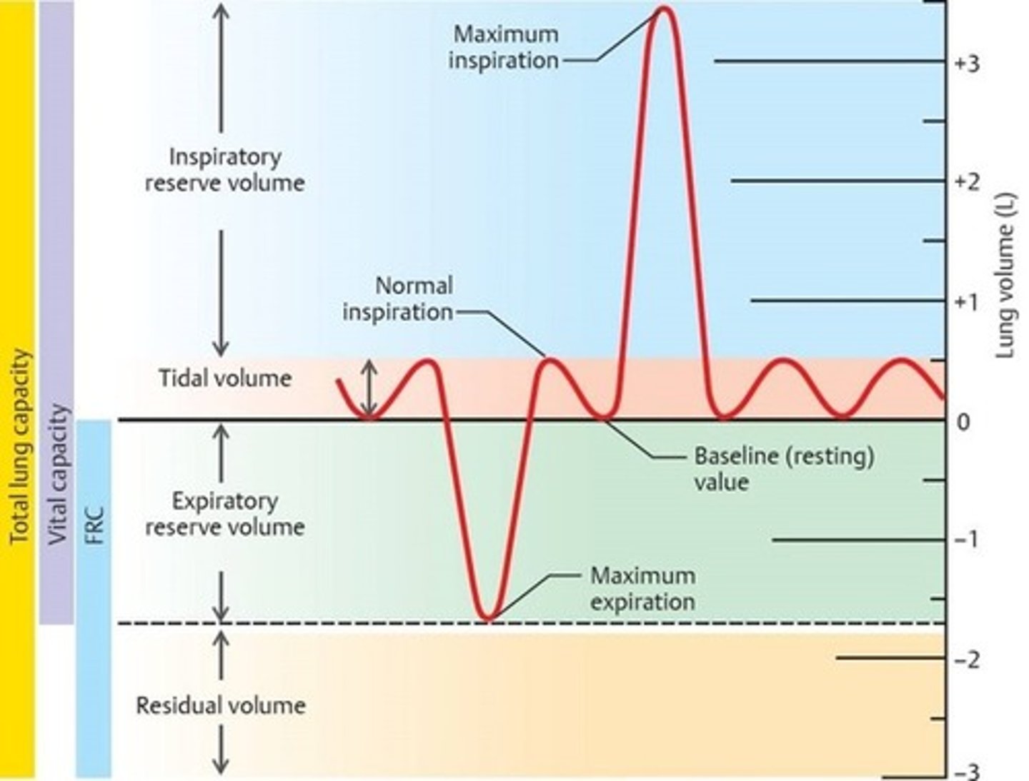
What is the Inspiratory Reserve Volume (IRV)?
The maximal volume of air that can be inspired after normal tidal volume inspiration.

What is the Expiratory Reserve Volume (ERV)?
The maximal volume of air that can be exhaled after normal tidal volume exhalation.

What is the Inspiratory capacity?
The maximal amount of air a person can inhale after a normal tidal exhalation

What is the vital capacity (VC)?
The maximal amount of air that can be inhaled following a maximal exhalation.

What is the Functional Residual Capacity (FRC)?
The volume of air in the lungs after normal tidal volume exhalation.

What is the Residual Volume (RV)?
the volume of air remaining in the lungs at the end of a maximal expiration.

What is the Total Lung Capacity (TLC)?
The maximal volume to which the lungs can be expanded. It is the sum of all the lung volumes (RV+ERV+TV+IRV)

What are the receptors that help control ventilation and what do they do?
Receptors assist in adjusting the ventilatory cycle by sending information to the controller. Receptors include: Baroreceptors, chemoreceptors, irritant receptors, and stretch receptors.
What are the central control centers of ventilation? and what do they do?
Central control centers evaluate the receptors information. They send messages out to the ventilatory muscles to alter the respiratory cycle in order to maintain adequate alveolar ventilation and arterial oxygenation. The control centers include: Cortex, Pons, Medulla, and Autonomic Nervous System.
How is the limbic system involved in respiration?
When done in a consistent way, conscious breathing stimulates the limbic part of the brain. The limbic network of the brain (amygdala, hippocampus, and hypothalamus) provides the structures for feeling and expression of emotion. Due to this, breathing can help control emotions and calm the nervous system.
How are joint and muscle receptors involved in respiration?
Receptors within peripheral joints and muscles of the extremities respond to changes in movement and cause an increase ventilation. Exercise causes a 2 fold increase in minute ventilation.
What is respiration?
Respiration is the process of gas exchange in the lungs facilitated through the process of simple diffusion.
What is diffusion?
Diffusion is the exchange of gases to and from blood at the alveolar capillary membrane.
What is perfusion?
The process of blood flow to the tissues or organs, delivering oxygen and nutrients while also removing waste products, including carbon dioxide.
What are the two requirements for diffusion to occur?
1) alveolar ventilation - Air brings oxygen to the lungs.
2) Pulmonary perfusion - there must be blood to receive the oxygen and give up the carbon dioxide.
Why is the diffusion/perfusion relationship important?
The relationship between diffusion and perfusion is critical for maintaining the body's oxygen and carbon dioxide balance. Oxygen is inhaled, diffuses across the alveolar-capillary membrane into the bloodstream, and is then carried to tissues by perfusion. Conversely, carbon dioxide produced by tissues diffuses into the bloodstream and is transported back to the lungs for removal via the process of perfusion. An imbalance in either diffusion or perfusion can lead to respiratory and circulatory problems, such as hypoxia (inadequate oxygen supply) or hypercapnia (excessive carbon dioxide levels).
Why is positioning important for optimal respiration or gas exchange?
The distribution of gas (ventilation) and blood (perfusion) at the level of the alveolar-capillary interface must be matched. Body positioning plays an important role in the distribution of ventilation and perfusion to different aspects of the lung. The greatest volume of pulmonary blood flow will normally occur in the gravity-dependent areas of the lung.
What is proning?
In general, there is greater perfusion and ventilation in the upper lung. Therefore, to improve ventilation to the posterior bases, one must position an individual in prone. For example, a patient with adult respiratory distress syndrome (ARDS).
What is Pectus Excavatum?
Pectus excavatum is a common congenital deformity of the anterior wall of the chest in which several ribs and the sternum grow abnormally. This produces a caved-in or sunken appearance of the chest. It is present at birth, but rapidly progresses during the years of bone growth in the early teenage years.
What is the opposite of pectus excavatum?
pectus carinatum (bulging sternum)
What structures would be impacted by pectus excavatum? (5)
-Bigger sternal angle
-decreased lung volumes
-altered sternum and ribs
-changese in length tension relationship of respiratory muscles
-Possible hypertrophy of secondary muscles of respiration
What are some pulmonary complications of pectus excavatum, specifically referencing the rib movement, muscles, and lung volumes?
-kyphosis scoliosis
-increased lumbar lordosis
-Decreased lung volumes (decreased total lung capacity, vital lung capacity, maximal voluntary lung capacity)