12 Lead EKG / Ischemia (DM)
 Call Kai
 Learn
Learn Practice Test
 Spaced Repetition
 Match
Match Flashcards
 Knowt Play
1/41

There's no tags or description
Looks like no tags are added yet.
Name | Mastery | Learn | Test | Matching | Spaced | Call with Kai | Chat |
|---|
No analytics yet
Send a link to your students to track their progress
42 Terms
What can we get from a 12-lead EKG
MI Infract Location
Coronary artery involved
Axis; hemi-blocks
V-Tach from Asystole
Specific BBB location
Ventricle / Atrial Hypertrophy
Drug / Electrolyte disturbances
Raise suspicion for pericarditis, and acute PE
What is needed to compare two 12-lead EKG
Consistent placment
Steps to Applying 12 Lead EKG
Place limb leads on the limbs
Place the V leads
Where do the limb leads go
Arms = Below the shoulder anywhere
Legs = Below inguinal fold (Anterior) or Gluteal Fold (Posterior)
Where do the V leads go in 12 - lead
V1 = 4th ICS, right of sternum
V2 = 4th ICS, left of sternum
V3 = Halfway between V2 and V4
V4 = 5th ICS, left MCL
V5 = Level with V4, left AAL
V6 = 5th ICS, left MAL
Why do we clean / remove hair from the 12-lead placment
Prevents artifact
What are the leads on a 12-lead EKG
I, II, III
avR, avL, avF,
V1, V2, V3
V4, V5, V6
What are the characterisitcs of a normal 12-lead EKG
Sinus Rhythm
Upright P waves
withing PRI limits
Rate = 60-100 bpm
Normal Axis
Normal QRS
Normal ST segement
Normal QTi
No pathologic Q waves
Normal T wave
No ectopy
What aspects of the EKG reading are important to identify MI and ischemia
J point
ST segement
J point
Where QRS and ST segement meet
If monitoring ST segments, what must be done
Print the strip to interrupt
No video
A depressed ST segement can indicate
Late ischemia
An elevated ST segment can indicate
Myocardial injury
An inverted T wave can indicate
Early ischemi
How do we determine ST segement elevation/depression
Compare the J point to isoelectric line
If J point is higher than isoelectirc = Elevation
If J point is lower than isoelectric = Depression
How do the type of leads differ in the view of the heart
Limb = Looks through the heart (Wide view)
Precoridal (V leads) = Look at the heart (Narrow view)
What leads look at the inferior wall
Left Leg
II
III
aVF
What leads look at the septal wall
V1
V2
What leads look at the anterior wall
V3
V4
What leads looks at lateral wall
I
avL
V5
V6
What are the quality types of 12-lead EkG
Monitor = Adds tight filters to the printout
Diagnostic = Adds loose filters to the printout
Where does 12-lead EKG have a bad time looking at
Right side of the heart
When should we RV involvement of ischemia on inferior wall MI on 12-lead
Dyspnea with clear loungs
JVD
Normal or hypotension
What type of MI affects the right side
Inferior Wall MI
How do we do we run RV EKG
Move V4 to the right side and re-run
What can cause ST elevations outside of MI
LV Hypertrophy
LBBB
Pericarditis
Benign Early Repolarization
Ventricular Pacemakers
Sgarbossa Criteria
A list of crtiera for dx of STEMI with LBBB / Pacer
ST elevation > 5 mm that is in the opposite direction of a predominantly negative QRS complex
ST depression > 1 mm in leads V1,V2,V3
ST elevation > 1mm that is in the same direction of predominantly positive QRS
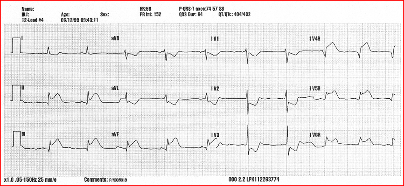
Inferior Wall MI w/ RV involvement
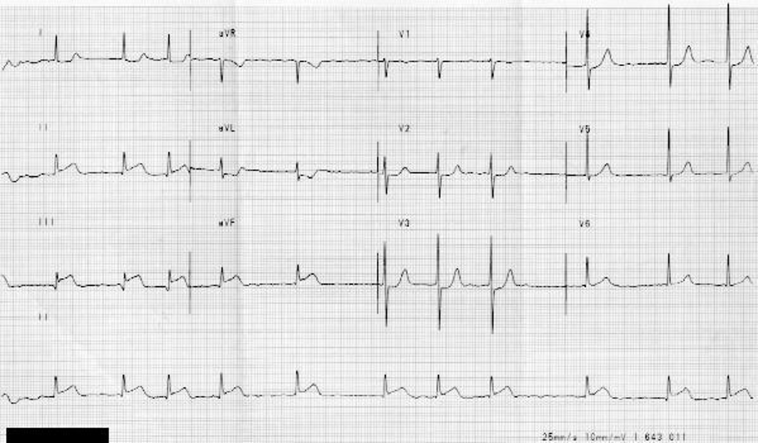
Inferior Wall MI
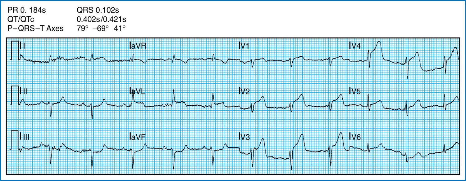
Left Main MI
Elevatiion V1,V2,V3,V4 = LAD
Elevation I, aVL, V5, V6 = LCX
LAD + LCX = Left Main
What leads look at LAD
V1
V2
V3
V4
What leads look at Left Circuflex
I
avL
V5
V6
What leads look at RCA
II
III
avF
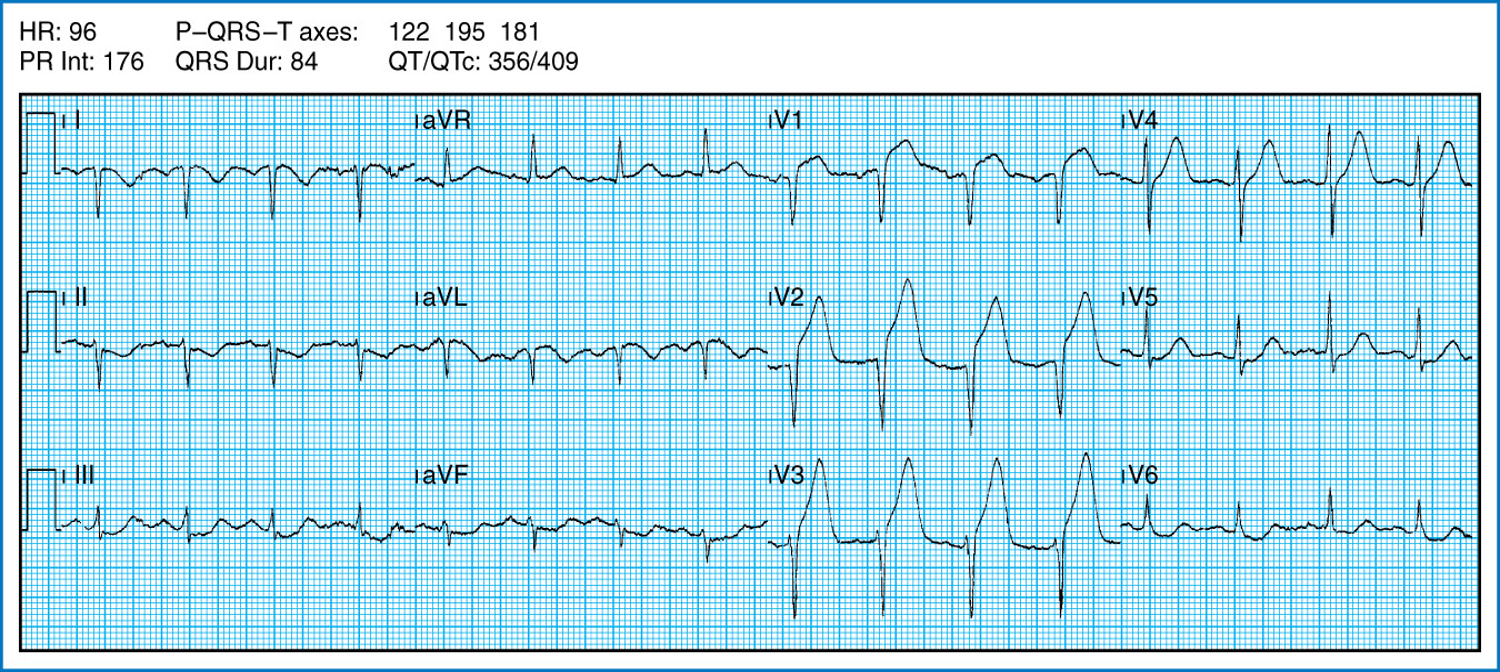
Left arm and right arm lead reversed = + aVR
LAD Lesion (Anterioseptal MI)
What type of coronary lesion is not balloon
Left ,ain
What coronary lesion is the widowmaker
Left Main
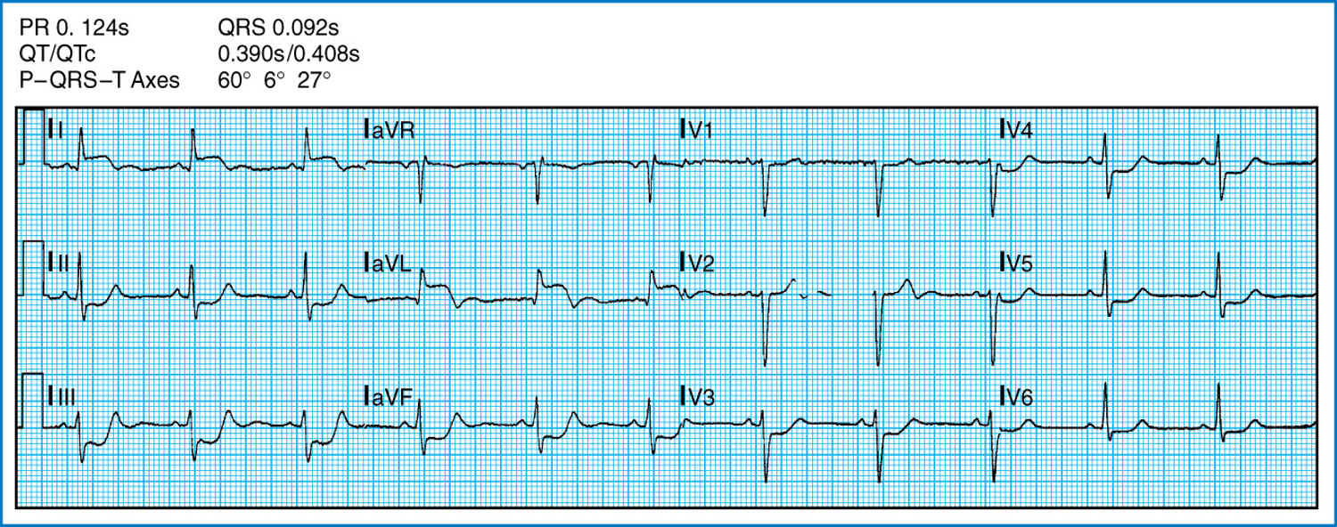
High Lateral Wall (Elveation in I and aVL)
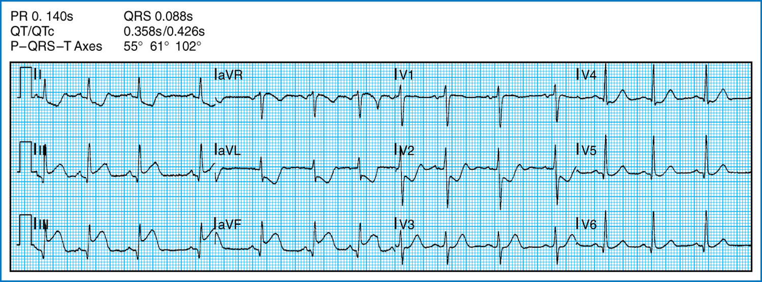
Inferior Wall MI
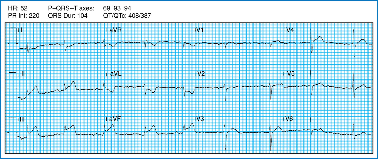
Inferior Wall MI (RCA)
II, III, aVF
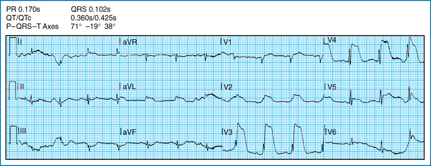
Anterior MI
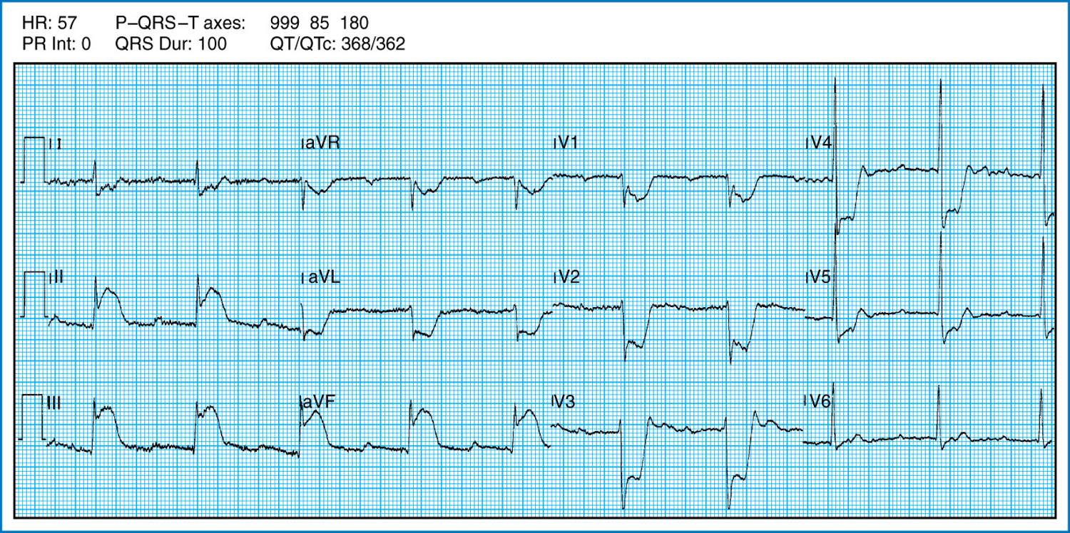
Inferior w/Posterior MI
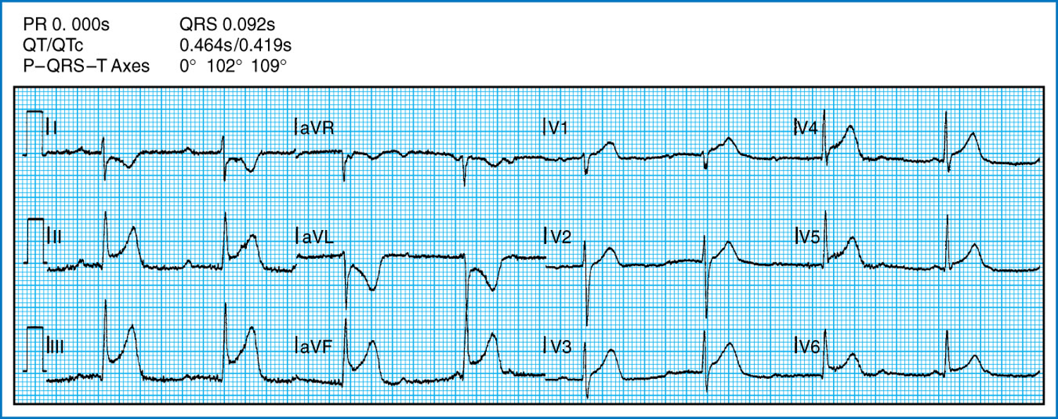
Inferior Wall w/ RV