Histology Lecture #27: Female Reproductive System II
 Call Kai
 Learn
Learn Practice Test
 Spaced Repetition
 Match
Match Flashcards
 Knowt Play
1/119

There's no tags or description
Looks like no tags are added yet.
Name | Mastery | Learn | Test | Matching | Spaced | Call with Kai | Chat |
|---|
No analytics yet
Send a link to your students to track their progress
120 Terms
- Paired muscular, tubular organs
- supported by ligaments and mesenteries
- highly mobile
- 10-12 cm long
- open into the peritoneal cavity.****
What are the uterine tubes (oviducts/Fallopian tubes)?
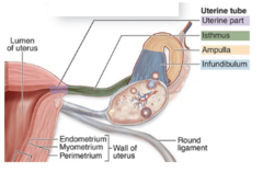
1. Infundibulum with fimbriae
2. ampulla (usual site of fertilization)
3. isthmus
4. uterine (intramural) part.
What are the regions of the uterine tubes?
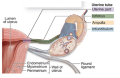
Folded mucosa.
What is the structure of the mucosa in the uterine tubes?
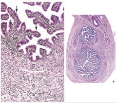
Thick, well-defined muscularis with interwoven circular (or spiral) and longitudinal smooth muscle fibers.
How is the muscularis of the uterine tubes described?
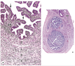
Thin serosa, which is visceral peritoneum covered by mesothelium.
What covers the uterine tubes externally?
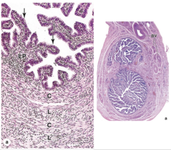
Numerous branching folds;
- more prominent in the ampulla
- smaller near the uterus
- absent in the intramural part.
What are the characteristics of the uterine tube mucosa?
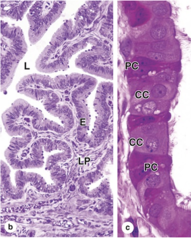
Simple columnar epithelium
What type of epithelium lines the uterine tube mucosa?
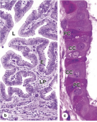
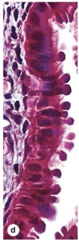
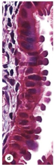
1. Ciliated cells
2. secretory peg cells
What are the two main cell types in the uterine tube mucosa?
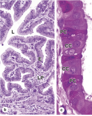
Their ciliary movements sweep fluid (and the oocyte) toward the uterus.
What is the function of ciliated cells in the uterine tube?
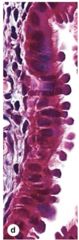
Estrogen causes cilia to elongate.
How does estrogen affect ciliated cells in the uterine tube?
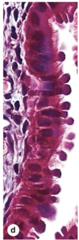
They become hypertrophic (enlarged).
How do ciliated cells change during the ovarian follicular phase?
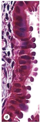
They undergo atrophy and lose their cilia.
What happens to ciliated cells during the luteal phase?
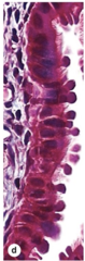
Non-ciliated cells.
What type of cells are secretory peg cells in the uterine tube?
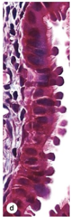
Dark staining, often with an apical bulge into the lumen.
How do secretory peg cells typically appear under a microscope?
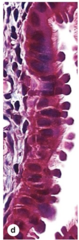
They secrete glycoproteins, forming part of the nutritive mucous for the oocyte.
What is the main function of secretory peg cells?
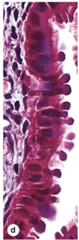
They undergo hypertrophy (enlargement).
How do secretory peg cells respond to estrogen?
They undergo atrophy.
What happens to secretory peg cells during the luteal phase?
Thick muscular walls.
What is a key characteristic of the uterus wall?
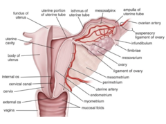
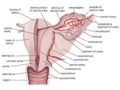
1. Body
2. fundus
3. isthmus
4. cervix
What are the main parts of the uterus?
Body
Which part of the uterus is the largest and is entered by the uterine tubes?
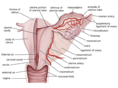
Fundus
Which part of the uterus is curved and superior to the uterine tubes
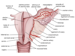
Isthmus
Which part of the uterus is narrow and connects the body to the cervix?
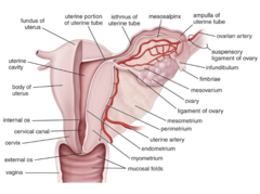
Cervix
What is the cylindrical part at the end of the isthmus?
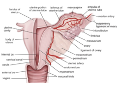
The passage within the cervix connecting the uterine lumen to the vagina.
What is the cervical canal?
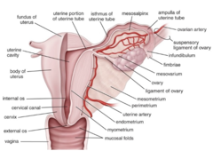
The opening of the cervical canal into the uterine lumen.
What is the internal os?
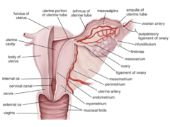
The opening of the cervical canal into the vagina.
What is the external os?
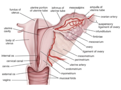
1. Perimetrium
2. myometrium
3. endometrium
What are the three layers of the uterine wall?
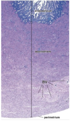
The outer connective tissue layer of the uterus, mostly serosa covered by mesothelium, continuous with ligaments; adventitia is present in some areas.
What is the perimetrium layer of the uterine wall?
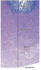
The very thick middle layer of the uterus composed of smooth muscle.
What is the myometrium of the uterine wall?
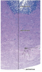
The inner mucosal layer of the uterus lined by simple columnar epithelium and supported by lamina propria of loose connective tissue.
What is the endometrium of the uterine wall?
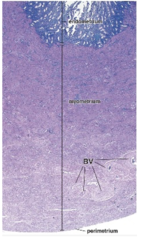
All layers (perimetrium, myometrium, endometrium) are continuous with the wall of the Fallopian tubes.
How are the layers of the uterine wall related to the Fallopian tubes?
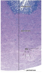
The myometrium.
Which layer of the uterine wall is the thickest?
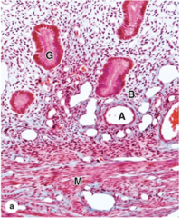
Bundles of smooth muscle interwoven with connective tissue containing venous plexuses and lymphatics.
What is the composition of the myometrium?
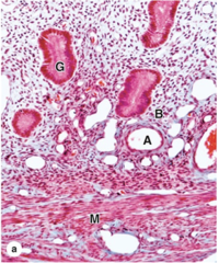
Interwoven layers, with inner and outer layers parallel to the long axis of the uterus.
How are the smooth muscle bundles arranged in the myometrium?
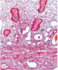
Hyperplasia and hypertrophy of smooth muscle cells, and increased collagen production.
How does the myometrium change during pregnancy?
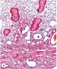
Shrinkage and apoptosis of smooth muscle cells, removal of excess collagen, and the uterus returns to normal size.
What happens to the myometrium after parturition?
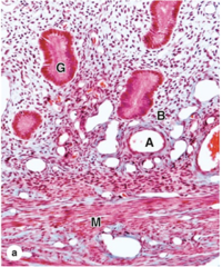
Simple columnar epithelium.
What type of epithelium lines the endometrium?
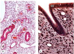
1. Ciliated cells
2. secretory cells (which also line the uterine glands).
What are the two main cell types in the endometrial epithelium?
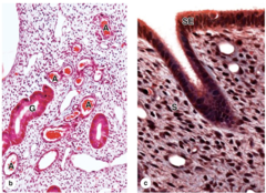
They penetrate the entire endometrium.
How far do the uterine glands penetrate the endometrium?
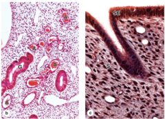
Mostly non-bundled type III collagen, abundant fibroblasts, and ground substance.
What is the composition of the lamina propria of the endometrium? (3)
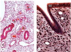
1. Basal layer
2. functional layer
What are the two zones of the endometrium?
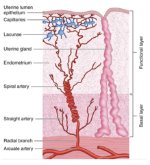
1. Adjacent to the myometrium
2. more highly cellular lamina propria
3. contains the basal ends of uterine glands
4. does not change during the menstrual cycle.****
Where is the basal layer located and what are its characteristics?
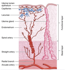
1. Superficial
2. Lamina propria is spongier
3. More ground substance
4. Contains most of the length of the uterine glands
5. Lined by the simple columnar epithelium
6. Changes during menstrual cycle
Where is the functional layer located and what are its characteristics?
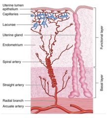
1. Straight arteries (supply the basal layer)
2. Spiral arteries (supply the functional layer)
Which arteries branch from the arcuate arteries to supply the endometrium? (2)
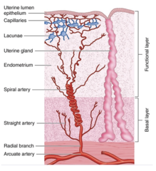
The basal layer.
Which endometrial layer is supplied by straight arteries?
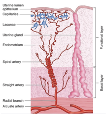
The functional layer
Which endometrial layer is supplied by spiral arteries?
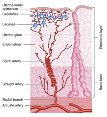
1. Longer
2. Progesterone-sensitive*****
3. Branch into numerous arterioles
4. Supply a rich superficial capillary bed
5. Includes many dilated, thin-walled vascular lacunae
What are the characteristics of spiral arteries?
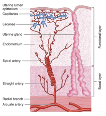
Growth and differentiation of epithelial cells and associated connective tissue.
What occurs in the endometrium during the menstrual cycle?
Female sex hormones.
What controls the menstrual cycle?
Pituitary gonadotropins.
What triggers cyclic changes in ovarian hormone levels?
It undergoes changes in response to fluctuating ovarian hormones.
How does the endometrium respond to the menstrual cycle?
About 28 days, though it can vary.
What is the average duration of the menstrual cycle?
To the years when the menstrual cycle occurs.
During which years is female fertility restricted?
Estrogen
Which hormone drives the proliferative phase of the menstrual cycle?
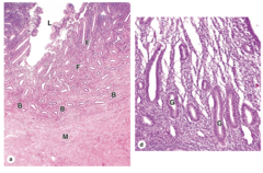
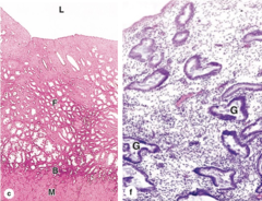
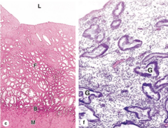
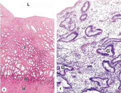
It regenerates, with proliferation and migration of cells at the base of the glands and reepithelialization of the surface exposed during menstruation.
What happens to the functional layer of the endometrium during the proliferative phase?
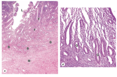
Simple columnar epithelium.
What type of epithelium lines the endometrium during the proliferative phase?
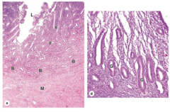
Relatively straight tubules with nearly empty lumens.
How do the glands appear in the proliferative phase?
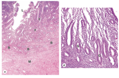
Mitosis of fibroblasts and epithelial cells.
What cellular processes occur in the proliferative phase?
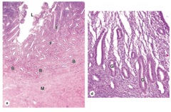
Spiral arteries lengthen, and a superficial microvasculature is formed.
How does the vasculature change during the proliferative phase?
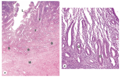
About 2-3 mm.
What is the approximate thickness of the endometrium at the end of the proliferative phase
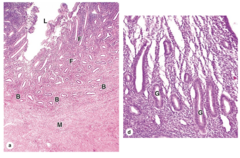
Progesterone
Which hormone drives the secretory phase?
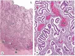
After ovulation.
When does the secretory phase occur?
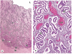
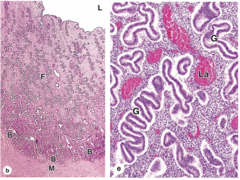
Glands secrete and accumulate glycogen, their lumens dilate, and they become tortuous.
What happens to the endometrial glands during the secretory phase?
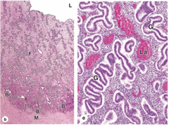
Superficial vasculature dilates, forming vascular lacunae.
How does the endometrial vasculature change during the secretory phase?
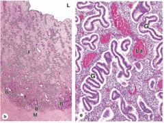
About 5 mm (maximum thickness).
What is the approximate thickness of the endometrium at the end of the secretory phase?
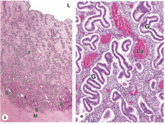
It becomes edematous.
How does the stroma change during the secretory phase?
When there is no fertilization, leading to deprivation of sex hormones.
When does the menstrual phase occur?
Dropping levels of progesterone.
What triggers the menstrual phase?
They undergo spasms, with increased synthesis of prostaglandins by arterial cells, causing vasoconstriction and hypoxia.
What happens to the spiral arteries of the functional layer during the menstrual phase?
They release cytokines, increasing vascular permeability and promoting leukocyte infiltration.
How do hypoxic cells in the functional layer contribute to menstruation?
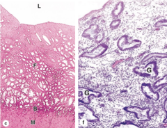
They release collagenase and metalloproteinases, degrading basement membranes and other extracellular matrix components.
What is the role of leukocytes during the menstrual phase?
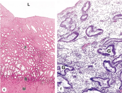
It detaches and is shed.
What happens to the functional layer of the endometrium during menstruation?
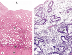
The basal layer is almost unaffected.
How is the basal layer affected during menstruation?
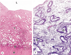
Reduced to a thin layer.
What is the state of the endometrium after menstruation?
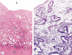
Simple columnar epithelium with a thick lamina propria.
What type of epithelium lines the endocervical mucosa?
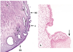
- Large, branched cervical glands that secrete mucus
- no spiral arteries
- does not change or shed during the menstrual cycle.
What features are found in the endocervical mucosa?
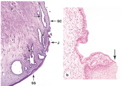
Nonkeratinized stratified squamous epithelium, continuous with vaginal epithelium.
What type of epithelium lines the exocervical mucosa?
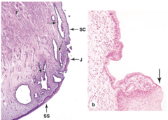
At the junction of endocervical mucosa and exocervical mucosa, outside the external os.
Where is the transformation zone of the cervix located?
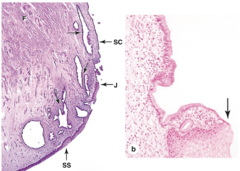
Fibromuscular tissue, less dense than the myometrium.
What is the composition of the deeper cervical wall?
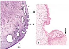
Because the cervical mucosa is exposed to the vagina, which can introduce pathogens.
Why are leukocytes present in the cervical mucosa?
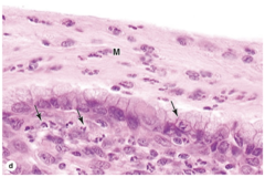
Neutrophils and other leukocytes.
Which types of leukocytes are found in the cervix?
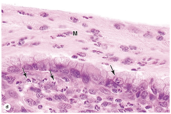
In the lamina propria, intraepithelially, and in the mucous layer.
Where in the cervical tissue are leukocytes located?
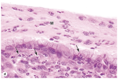
No, the vagina has no glands.
Does the vagina contain glands?
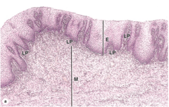
1. Mucosa
2. Muscular layer
3. Adventitia
What are the three main layers of the vaginal wall?
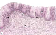
Nonkeratinized stratified squamous epithelium.
What type of epithelium lines the vaginal mucosa?
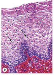
Glycogen, which is estrogen-dependent.
What do vaginal epithelial cells synthesize and what regulates this process?
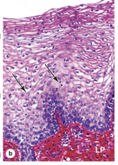
It is rich in elastic fibers and contains numerous narrow papillae.
What is characteristic of the lamina propria of the vaginal mucosa?
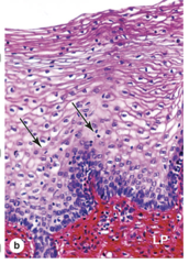
Neutrophils and other leukocytes.
What types of immune cells are relatively abundant in the vaginal mucosa?
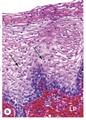
1. Circular bundles around the mucosa
2. thicker longitudinal bundles closer to the adventitia.
What are the two layers of smooth muscle in the vaginal muscular layer?
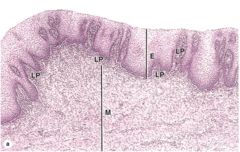
The longitudinal bundles located near the adventitia.
Which smooth muscle layer of the vagina is thicker?
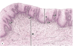
Connective tissue rich in elastic fibers, containing an extensive venous plexus, lymphatics, and nerves.
What characterizes the adventitia of the vagina?
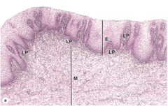
Compound tubuloalveolar exocrine glands.
What type of glands are the mammary glands?
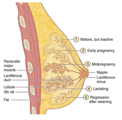
Dense connective tissue with abundant adipose tissue.
What separates the lobes of the mammary glands?
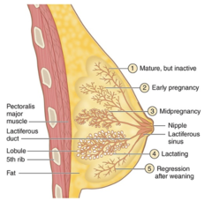
A single gland.
What does each lobe of the mammary gland represent?
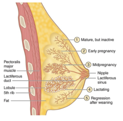
The excretory ducts of each lobe (gland).
What are lactiferous ducts?
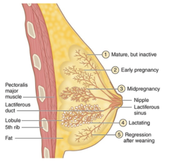
Age, sex, and physiological status.
What factors influence the major histological changes in mammary glands?
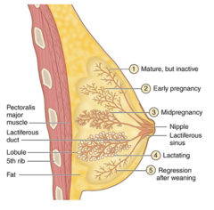
Only lactiferous sinuses with small branching tubules.
What structures are present in mammary glands before puberty?
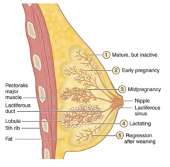
Yes, both have only lactiferous sinuses and small branching tubules.
Do both sexes have the same mammary gland structures before puberty?
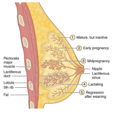
Accumulation of adipose tissue and elongation of the duct system.
What mainly causes breast growth in females at puberty?
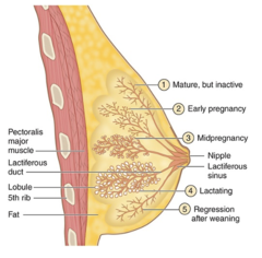
Increased adipose tissue and duct system elongation.
Which two changes in puberty contribute to breast enlargement?