Human Physiology Exam 1 (L1-L6)
 Call Kai
 Learn
Learn Practice Test
 Spaced Repetition
 Match
Match Flashcards
 Knowt Play
1/155

There's no tags or description
Looks like no tags are added yet.
Name | Mastery | Learn | Test | Matching | Spaced | Call with Kai | Chat |
|---|
No analytics yet
Send a link to your students to track their progress
156 Terms
Two components of extracellular fluid
Plasma, interstitial fluid
Intracellular fluid
Cytoplasm, fluid contained within plasma membrane of all body cells
There is direct contact between cells and blood
FALSE. Oxygen and nutrients are dissolved in plasma, not blood.
There are X types of cells. The typical human cell is Y microns in size.
~200, 10-20
Cells are held together by three different means.
1) The extracellular matrix
2) Cell adhesion molecules in the plasma membrane
3) Specialized cell junctions
Extracellular matrix (ECM)
A meshwork of proteins in a watery like substance that serves as a biological “glue”. Secreted locally by cells. Most abundant in connective tissue.
Basal lamina (basal membrane, basement membrane)
Thin mat of extracellular matrix that separates epithelial sheets (or other varieties of cells) from connective tissue
The four main functions of the basal membrane
BArry MEets FIona IN the basal membrane.
1) barrier
2) mechanical support for epithelia
3) filtering in kidney cells
4) influence cell polarity
Cadherins are cell adhesion molecules
N-terminal (side-to-side) cadherin repeats of adhered cells associate with each other with relatively low affinity. However, large numbers of interactions —> strong, stable adhesion (VELCRO PRINCIPLE)
Three types of specialized cell junctions
A) Desmosomes
B) Tight junctions
C) Gap junctions
Desmosomes
Cell junction in tissues that stretch (skin, heart, uterus); spot rivets, connect cells w/out direct contact
Tight junctions
Cell junction to form distinct, leak-proof regions, found primarily in epithelial tissue
Apical plasma membrane v.s. lateral plasma membrane v.s. basal plasma membrane
Tight junctions serve to separate different regions of the membrane. Prevent transmembrane proteins from moving outside designated region.
Apical - free surface, external
Lateral - sides, neighboring cells
Basal - bottom, basal lamina
Gap junctions (communicating junctions)
Cell junctions formed by connexons found most often in cardiac and smooth muscle (can be found in nonmuscle cells); make channels that allow for direct cell communication/exchange of nutrient molecules

Three types of carrier-mediated active transport
1) uniport
Coupled transport:
2) symport
3) antiport
Three major types of protein filaments in cytoskeleton:
1) actin filaments
2) microtubules
3) intermediate filaments
Cristae (mitochondria)
Folds of the inner membrane, containes proteins used in cellular respiration
Matrix (mitochondria)
Region enclosed by the inner membrane
Why do mitochondria need O2?
Final e- acceptor
Three cellular activities that require ATP
1) Synthesis of new chemical compounds
2) Membrane transport
3) Mechanical work
ATP Production
Glycolysis, CAC, ETC
Glycolysis
Glucose —> pyruvate, 2 NADH and 2 ATP
Citric acid cycle
Pyruvate is modified into acetyl-CoA, which enters cycle in mitochondrial matrix. Source of NADH and FADH2 for ETC. CO2 produced. ATP produced.
Electron transport chain
E- carriers in inner membrane (cristae). ATP is synthesized using E released by e- as they transfer to O2. (generate proton gradient, ATP synthase phosphorlates ADP)
Three types of intracellular communication
1) gap junctions
2) cell-to-cell binding
3) extracellular chemical messengers
Cell-to-cell binding
Surface molecules on two different cells bind to one another

Extracellular messengers: General flow of information
Receptor binding —> signal transduction —> cellular response

Chemical extracellular messengers can be water or lipid soluble
Water soluble: peptide/protein hormones, neurotransmitters, local mediators
Lipid soluble: steroids, thyroid hormones, NO2
Hormones
Extracellular chemical messengers that are carried by the blood to DISTANT target cells
Neurotransmitters
Extracellular chemical messengers released from neuron into synapse to reach NEARBY target cell
Local mediators
Autocrine and paracrine, self/local. Cytokines, nitric oxide, growth factors
Down-regulation of receptors
Excess of extracellular messengers can result in decrease number of target cell receptors
Up-regulation of receptors
Deficient messengers can result in increase of number of target cell receptors
Relay proteins
Secondary, intracellular messengers
Four conserved components of intracellular signal transduction pathways
1) second messengers
2) protein kinases and phosphatase
3) GTP binding proteins
4) protein ubiquitylation and degradation
Second messengers
Small, intracellular molecule lvls inc/dec upon binding of ligand. Short-lived. Activates downstream proteins.
Second messengers: cAMP
Activates PKA
Second messengers: cGMP
Activates PKG, open cation channels in rod cells
Second messengers: diacylglycerol (DAG)
Activates PKC
Second messengers: IP3
Opens Ca2+ channels in the ER
Second messengers: Ca2+
Produce/increase cell damage
Protein kinase
Activates target protein via transfer of phosphate to substrate
Reversible, so that future signals can be received
Protein phosphatases
Mediate removal (inactivation) of protein via removal of phosphate from substrate
Undoes what a kinase does
Phosphorylation is the most important post-translational modification of proteins. The two major types of kinases = two major types of phosphatases
STY
1) Serine/threonine
2) Tyrosine
GTPase superfamily of signal transduction proteins
Guanine nucleotide binding proteins. Two classes:
1) Trimeric (G-α proteins are associated with G protein-coupled receptors)
2) Monomeric (small, Ras superfamily)
GTPase switch proteins (G-proteins)
switch between active (phosphorylated, GTP) and inactive (bound by GDP) states
G-proteins are regulated by guanine nucleotide exchange factor (GEF) and GTPase-activating protein (GAP)
GEF —> activator (think: excite)
GAP —> inactivator (think: active to inactive)
Three main classes of cell surface receptors
1) GPCRs
2) Enzyme-coupled
3) Ion-channel-coupled
GPCRs flow
The inactive form of GPCR:
G-protein is bound by ? and ? is phosphorylated.
The ? subunit, dissociates and binds the target protein.
GTP is ?.
The complex reassociates with the G-protein, and it returns to its ? state.
The inactive form of GPCR: G-protein, α-GDP, γ subunit, β subunit
G-protein is bound by ligand and GDP is phosphorylated (activated).
The α-GTP subunit dissociates and binds the target protein.
GTP is dephosphorylated (GDP).
The complex reassociates with the G-protein, and it returns to its inactive state.

The GPCR that is coupled to ? activates the secondary messenger cAMP.
Reminder: cAMP activates…
adenylyl cyclase
protein kinase A
Enzyme-coupled receptor responses are typically ?, but highly ?.
Enzyme-coupled receptor responses are typically slow, but highly sensitive.
Main types of enzyme-coupled receptors
1) Kinase itself: receptor tyrosine kinase family
2) Associate and activate a kinase
3) Guanylyl cyclase receptors: cGMP activates protein kinase G
Ion-channel-coupled receptors are responsible for the rapid transmission of signals across
neuron synapses
(When ligand binds receptor, ion channel opens)
Signal termination
All pathways must eventually be terminated:
1) Dissociation of messenger from receptor
2) Inactivation of signal transduction pathway components
Nervous v.s. endocrine system
1) Messenger molecules
2) Site of mediator action
3) Target cells
4) Onset time
5) Duration
1) neurotransmitter, local; hormone, far
2) Close; far
3) muscle, gland, neuron; cells throughout
4) FAST ms; varies
5) generally briefer, ms; generally longer s to d
All cells establish a membrane potential.
TRUE. Uneven distribution of ions —> polarized separation of charges across membrane.
Key ions involved in membrane potentials
Na+ is predominantly OUTSIDE
K+ predominantly INSIDE
Cl-
Generating a membrane potential
1) Membrane has no potential.
2) Pump artificially generates potential.
3) Charges align along membrane.
Rank the membrane permeability (hi to lo) of the folllowing ions
Na+, K+, A-
K+, Na+, A-
Slightly permeable, almost impermeable, not permeable
Rank the extracellular membrane concentration (hi to lo) of the folllowing ions
Na+, K+, A-
Na+, K+, A-

Resting membrane potential
-70 mV
Membrane electrical states: polarization
Membrane potential ≠ 0 mv
Membrane electrical states: depolarization
Membrane potential becomes less negative (polarized)
Membrane electrical states: repolarization
Membrane potential returns to resting potential after having been depolarization
Membrane electrical states: hyperpolarization
Membrane becomes more negative (polarized) than at resting potential
Graph the types of changes in membrane potential

Because the ions regulating potential cannot permeate the plasma membrane, they must cross via channels or carrier-mediated support. List the two types of membrane channels and their subtypes.
Leak channels
Gated channels
voltage-gated
Chemically-gated
Mechanically-gated
Thermally-gated
The two basic forms of electrical signals in neural communication
Graded potentials: short distance
Action potentials: long distance
What makes a potential “graded”?
Duration of signal is directly proportional to the strength and duration of the triggering event. Dies out over short distances. No refractory period.
Current flow during a graded potential
Triggering event opens Na+ channels, Na+ flows intracellularly and partially neutralizes negative charge. Depolarization spreads across membrane, resulting in movement of the current.
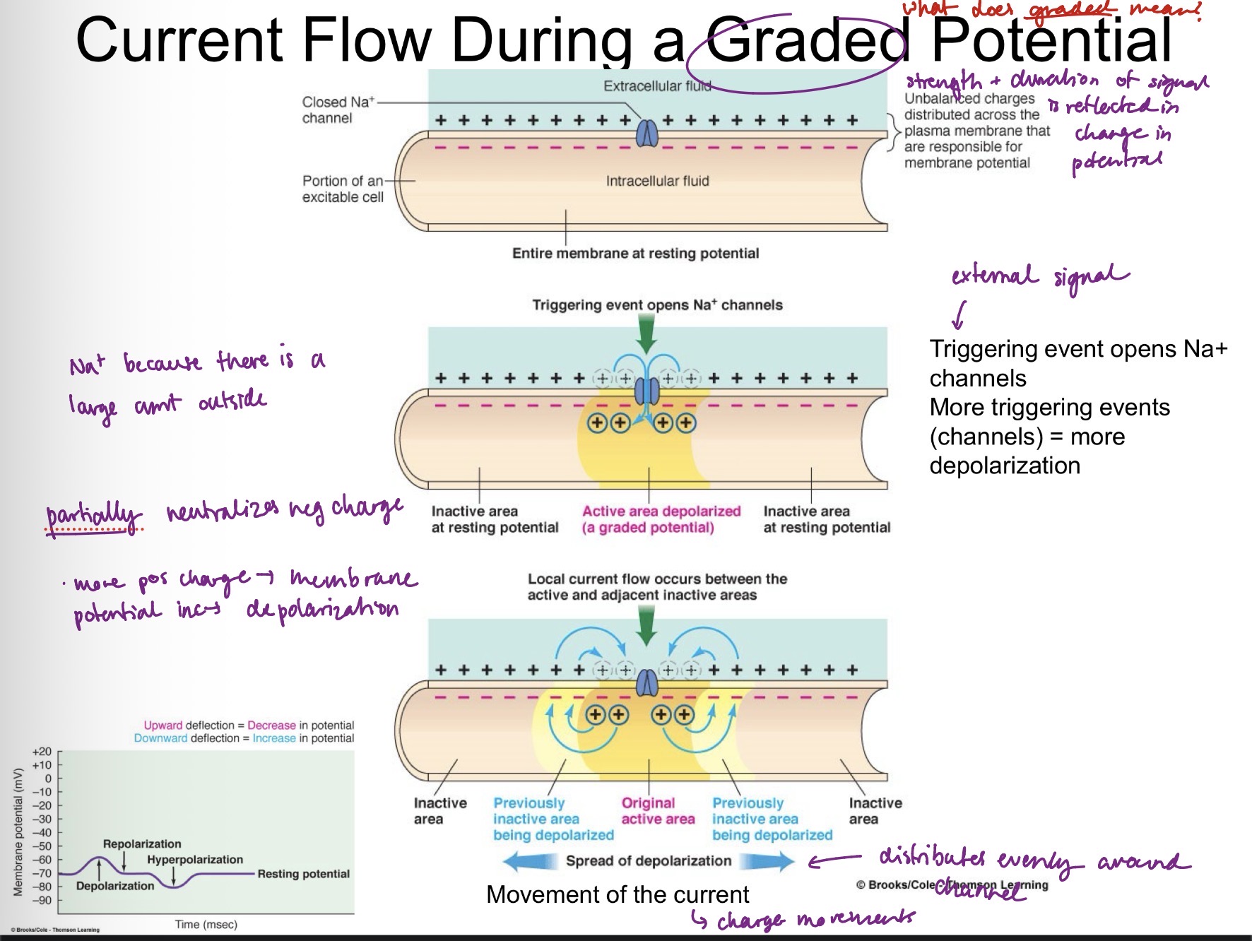
5 examples of graded potentials
PREPS
Postsynaptic
Receptor
End-plate
Pacemaker
Slow-wave
When a graded potential reaches a certain threshold, it can initiate an Action Potential
Brief, rapid, large (100 mv) changes in membrane potential during which potential actually reverses. Trigger involves a small portion of excitable membrane. Do not decrease in strength as current travels.

Once initiated, action potentials are conducted throughout a nerve fiber. Action potentials are propagated from
The axon hillock to the axon terminals
Draw the signal traveling through the neuron.
Dendrites —> cell body —> axon hillock —> axon + myelin sheath —> axon terminals

List the input zone, trigger zone, and output zone of the neuron
Dendrites, axon hillock, axon terminals
Voltage-gated sodium channel
Resting potential: all channels are closed
Reach threshold: channel opens
Action Potential: Influx of Na+ begins to neutralize negative charge —> depolarization
Peak: Na+ inactivaion gate begins to close, K+ gate opens
Repolarization: Efflux of K+
Hyperpolarization: Na+ inactivation gate begins opens, Na+ activation gate closes, K+ gate closes (gates are capable of reopening) —> restoration of resting potential

Propagation of action potentials: contiguous conductino
Local current flow that depolarizes adjacent inactive area from resting to threshold
Propagation of action potentials: contiguous conductino
Local current flow that depolarizes adjacent inactive area from resting to threshold
Refractory period
Ensures that the action potential is unidirectional. “Backward” current flow does not re-excite previously active area. (Recall voltage-gated channels. Gates are closed, and it takes time for them to reopen.)
All of None Law of action potentials
Stimulus must depolarize to threshold (-55/-50 mV) to induce action potential. Unlike graded potentials, the amplitude does not vary in response to signal strength.
How does stimulus strength relate to action potential?
Increase in stimulus strength —> more frequent action potentials
Myellin
Composed of lipid, insulates axon. Formed by oligodendrocytes of CNS or Schwann cells of PNS.
Nodes of Ranver
non-myelinated areas of the axon, exposed to the extracellular fluid
Propagation of action potentials: Saltatory Conduction
Action potential impulses jump from node to node, drastically increasing the speed of conduction (50X). Bypasses need to regenerate action potential.
The three termination sites of neurons
1) Muscle-cell contraction
2) Gland-cell secretion
3) another neuron-synapse
Synapses and chemically-gated ion channels
Action potentials reach axon terminals.
Voltage-gated Ca2+ channels open.
Ca2+ binds to sensor protein n cytoplasm.
Ca2+-protein complex stimulates fusion and exocytosis of neurotransmitter (to post synaptic cell).
Two types of synapses
Excitatory and inhibitory
Central nervous system (CNS) consists of
Brain and spinal cord
The peripheral system consists of the
Afferent division and efferent division
Afferent division (PNS)
Carries sensory and visceral stimuli to the CNS
Efferent division (PNS) and its two divisions
Carries information away from CNS to effector organs, consists of somatic nervous system and autonomic nervous system
Autonomic nervous system consists of two further subdivisions
Sympathetic and parasympathetic nervous systems
Interneuron (association neurons)
Found in the CNS, lies between afferent and efferent neurons (peripheral responses to peripheral information). Associated with abstract phenomena (mind, emotion). 99% of all neurons are interneurons.

Describe the connection of sensory, somatic motor, and autonomic motor neurons to the spinal cord.
Sensory neurons are directly connected to the spinal cord. Termini are closely associated with the dendrites of interneurons.
Somatic motor neurons are directly connected to the spinal cord via their cell bodies.
Autonomic motor neurons are associated with efferent neurons whose cell bodies are embedded in the spinal cord.

The CNS is mostly (90%) composed of glial or neuroglial cells. Definition and four types?
Connective tissue of the CNS, do not initiate or conduct nerve impulses.
1) astrocytes
2) oligodendrocytes
3) microglial cells
4) ependymal cells
Astrocytes
4 main functions?
Neurotransmitter?
Other roles?
Main “glue” of CNS. Direct neural fetal development, establishment of blood brain barrier, brain injuries and neural scar formation.
Glutamate receptors, communicate by gap junction.
Learning and memory, thrombospondin enhances synapse formation/transmission. Take up excess K+ from brain ECF.
Reminder: Oligodendrocytes form X around axons in CNS?
Myelin sheaths
Microglia
Immune defense cells of CNS (can relate to autoimmune/neurodegenerative disease). At rest, release growth factors.
Ependymal cells
NEURONAL STEM CELLS. Line internal, fluid-filled cavities of CNS. In ventricles of brain, help form and circulate cerebrospinal fluid.
Protection of CNS
Bony structures: cranium (skull) and vertebral column
Meninges (membranes): Dura, arachnoid, pia maters
Cushioned by cerebrospinal fluid
Blood-brain barrier