ILS 5: Nervous System & Histology
 Call Kai
 Learn
Learn Practice Test
 Spaced Repetition
 Match
Match Flashcards
 Knowt Play
1/81

There's no tags or description
Looks like no tags are added yet.
Name | Mastery | Learn | Test | Matching | Spaced | Call with Kai | Chat |
|---|
No analytics yet
Send a link to your students to track their progress
82 Terms
Telencephalon
cerebral hemispheres
“end brain”
Encephalon
“brain”
Diencephalon
thalamus, subthalamus, hypothalamus, & epithalamus
“in between the brain”
Brain stem composed of
mesencephalon (midbrain), pons, & medulla oblangata
Insular lobe function
pain perception, taste, emotion
limbic lobe functtion
learning & memory
Meninges
Dura Mater
tough, outer fibrous layer that provides protection
Arachnoid Mater
middle, web-like layer
beneath it, subarachnoid space contains CSF
Pia Mater
thin, delicate layer that closely adheres to brain surface
Gray Matter
contain neuron cell bodies, dendrites, & axon terminals
appears pinkish-grey in fresh brain tissue
forms cortex in brain
site of processing & info integration
White Matter
composed mainly of myelinated axons
connects regions of gray matter
specialized for rapid signal transmission
lies beneath cortex in brain & surrounds gray matter in spinal cord
conus medullaris
cone-shaped terminal end of spinal cord
located at L1-L2 vertebral lvl
from which cauda equina & filum terminale arise
Cauda equina
bundle of spinal nerve roots below conus medullaris
filum terminale
thin fibrous strand of pia mater that extends from conus medullaris to coccyx, anchoring spinal cord in place
Lumbar puncture
performed at L3-L4 or L4-L5 to collect CSF
Spinal cord Levels
Cervical [C1-C8]
Thoracic [T1-T12]
Lumbar [L1-L5]
Sacral [S1-S5]
Coccygeal [CO1]
Cervical enlargement
C4-T1
supplies upper limbs
Lumbosacral enlargement
L1-S2
supplies lower limbs
central canal
small ependymal-lined CSF-filled cavity
Posterior [dorsal] horn
sensory processing
anterior [ventral horn]
somatic motor neurons
intermediate zone
autonomic neurons & interneurons
somatic motor neurons are for
skeletal muscle
visceral motor neurons are for
smooth muscle, cardiac muscle, & glands
brainstem
connects cerebrum & cerebellum to spinal cord
contains ascending & descending tracts
houses nuclei & root entry/exit zones of 9 of 12 cranial nerves [NOT I, II, & XI - spinal origin]
regulates vital autonomic functions
breathing, HR, BP, consciousness
somatic sensory system
detects sensations from skin, skeletal muscle, joints, & connective tissue
visceral sensory system
detects sensations from internal organs [viscera]
visceral motor system is also known as
autonomic nervous system, ANS
frontal lobe function
motor planning & execution
parietal lobe function
processing somatosensory info
occipital lobe function
processing visual info
temporal lobe function
processing auditory info
PNS contains how many pairs of nerves
43
12 pairs of cranial nerves
31 pairs of spinal nerves
Cervical [8]
Thoracic [12]
Lumbar [5]
Sacral [5]
Coccygeal [1]
Metencephalon
pons
basilar pons
ventral [anterior] portion of pons
pontine tegmentum
dorsal [posterior] portion of pons
cervical nerves innervate
neck, shoulders, diaphragm [phrenic C3-C5], & upper limb [esp. brachial plexus]
thoracic nerves innervate
chest & upper abdomen wall, intercostal muscles, skin
lumbar nerves innervate
lower abdomen, pelvis, hip region, anterior & medial thigh, parts of lower limb
sacral nerves innervate
pelvis, genitals, lower digestive & urinary tracts, buttocks, posterior lower limb, & foot
brachial plexus
C5-T1
supplies upper limb [shoulder, arm, forearm, hand]
lumbar plexus
supplies lower abdominal wall, pelvic region, & anterior/medial thigh
spinal nerves
each one is a MIXED nerve [both sensory & motor fibers]
formed by union of 2 roots attached to spinal cord
posterior [dorsal] root
carries sensory sginals
contains dorsal root ganglion [DRG]
dorsal root ganglion [DRG]
contains cell bodies [somas] of sensory neurons [typically pseudounipolar neurons]
anterior [ventral] root
formed by nerve rootlets emerging from anterior spinal cord that converge
carries motor signals
posterior ramus
innervates muscles, joints of spine, & skin of back
anterior ramus
innervates anterior & lateral trunk & limbs
communicating rami
[T1-L2 only]
connect spinal nerves to sympathetic chain [trunk] ganglia
carry sympathetic autonomic fibers
meningeal branch
re-enters vertebral canal to innervate the vertebrae, spinal ligaments, BVs, & meninges
nerves
bundles of axons in PNS
ganglion
cluster of neuron cell bodies in PNS
nucleus
cluster of neuron cell bodies in CNS
epineurium
thick layer of dense, irregular connective tissue that encloses entire nerve
perineurium
layer of dense, irregular connective tissue that wraps the fascicle
endoneurium
delicate layer of areolar connective tissue that wraps an individual axon
multipolar neurons
many dendrites
1 axon
most common type
spinal motor neurons
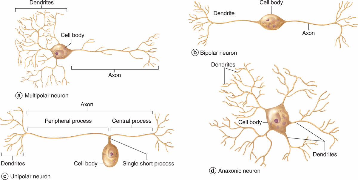
bipolar neurons
1 axon
1 dendrite
sensory neurons of retina, olfactory epithelium, inner ear
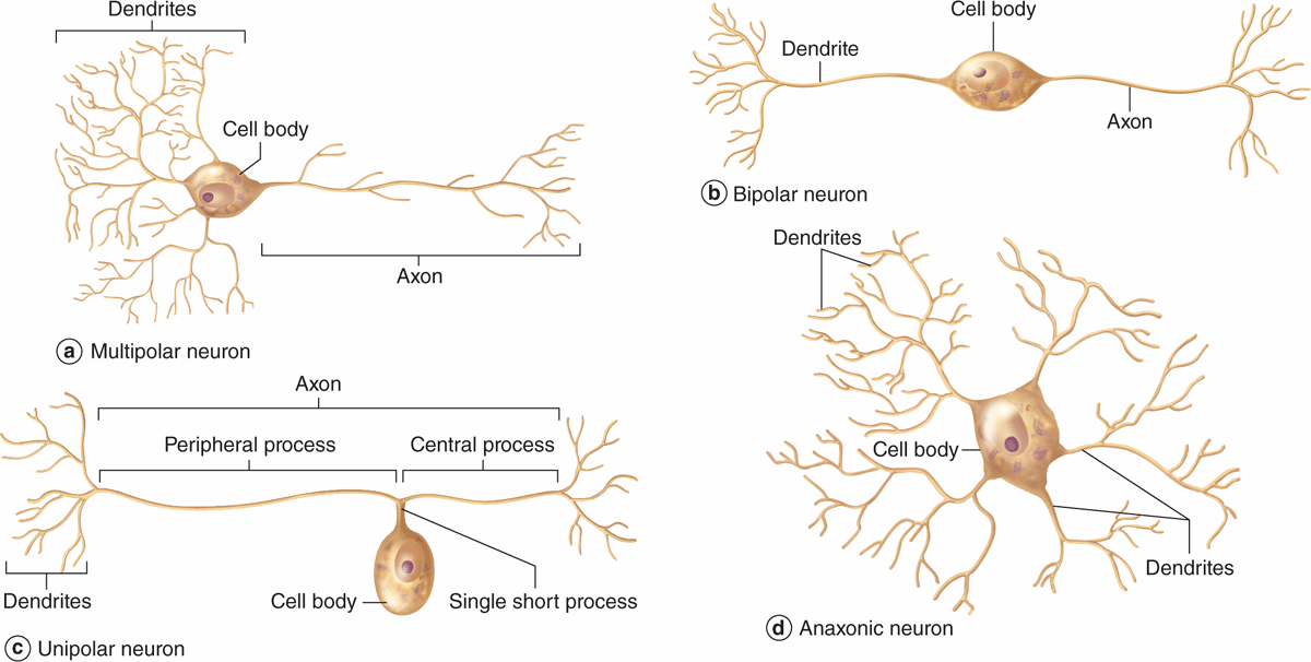
unipolar [pseudounipolar] neurons
1 process extends from cell body & splits into 2
central process → synaptic knobs of CNS
peripheral process → dendrites
all other sensory neurons
DRG of spinal cord
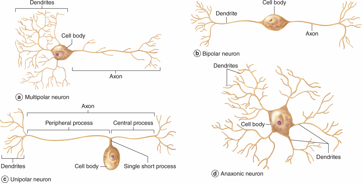
anaxonic neurons
many dendrites
no true axon
don’t produce APs [send signals via electronic conduction]
regulate electrical changes of adjacent CNS neurons
![<ul><li><p>many dendrites</p></li><li><p>no true axon</p></li><li><p>don’t produce APs [send signals via electronic conduction]</p></li><li><p>regulate electrical changes of adjacent CNS neurons</p></li></ul><p></p>](https://assets.knowt.com/user-attachments/3f9abc9e-c019-4c75-859e-0d0536410432.png)
interneurons
“association neurons”
multipolar
located within CNS
coordinate sensory & motor neurons
make up 99% of our neurons
cell body [perikaryon or soma]
acts as trophic center, producing cytoplasm for neuronal processes
large, euchromatic nucleus w/ prominent nucleolus [high synthetic activity]
nissl bodies
chromatophilic substance
basophilic regions rich in RER & polysomes
esp abundant in motor neurons
dendritic spines
small dynamic projections supported by actin filaments & change morphology continuously
dendritic spine plasticity crucial for
embryonic brain development
neural adaptation
learning & memory
axolemma
plasma membrane of axon
wallerian degeneration
when axon is severed from cell body, this occurs to the disconnected portion & is removed by phagocytic cells
anterograde transoprt
cell body → axon terminal
FAST via kinesin motor proteins
retrograde transport
axon terminal → cell body
FAST, uses dynein motor proteins
*can transport pathogens to CNS [tetanus toxin, HSV, rabies, polio]
resting membrane potential
-70mV
Na+-K+ ATP pump maintains
low Na+ inside
high K+ inside
[resting potential]
threshold
~ -55 mV
depolarization caused by
rapid influx of Na+ via voltage gated channels
Repolarization caused by
opening of voltage-gated K+ channels
Mechanism of local anesthetics
Ex. Lidocaine or benzocaine
bind to & block voltage-gated Na+ channels
prevents initiation & propagation of APs
loss of sensation
Glial cells [neuroglia]
nonexcitable support cells in CNS & PNS
same # of glial cells as neurons
mitosis, protect & nourish neruons, provide physical scaffolding for nervous tissue
Astrocytes
glial cells of CNS
perivascular feet = protective seal of BBB
>90% of tissue in some areas of brain
regulate tissue fluid composition
secrete nerve growth factors
*most brain tumors are astrocytomas: distinguished pathologically by their expression of glial fibrillary acidic protein [GFAP]
Ependymal cells
glial cell in CNS
ciliated cuboidal epithelium
line ventricles of brain & central canal of spinal cord
lack BM
produces CSF
Microglia
small macrophages in CNS [glial cell]
aid in synaptic remodeling, changing connections between neurons
*from monocytes from bone-marrow
multiple sclerosis [MS]
myelin sheaths damaged by autoimmune mech
T lymphocytes & microglia, which phagocytose & degrade myelin debris, play major roles in progression of MS
destruction actions of these cells > oligodendrocytes capacity to produce & repair myelin
symptoms
numbness, tingling, blurred/double vision, balance/walking difficulties, memory problems, extreme fatigue
oligodendrocytes
only in CNS
main glial cells in white matter
forms myelin
covers entire axon using multiple oligodendrocytes
*1 oligodendrocyte can myelinate multiple axons from diff neurons
satellite cells
glial cells of PNS
surround neuronal cell bodies in dorsal root ganglia
provide electrical insulation & structural support
regulate exchange of nutrients, ions, & wastes
Schwann cells
glial cells of PNS
produce myelin sheath
wrap around a single axon
enable saltatory conduction btwn nodes of Ranvier
Guillain-Barré syndrome [GBS]
loss of myelin disorder → loss of reflexes