Hematology: Peripheral blood smear
 Call Kai
 Learn
Learn Practice Test
 Spaced Repetition
 Match
Match Flashcards
 Knowt Play
1/39

There's no tags or description
Looks like no tags are added yet.
Name | Mastery | Learn | Test | Matching | Spaced | Call with Kai | Chat |
|---|
No analytics yet
Send a link to your students to track their progress
40 Terms
Anisocytosis
Variation in RBC size (normal 7-8 um)
Aniso: not same
Causes:
Iron-deficiency anemia→ smaller cells
Low Vit B12 (folate)→ larger cells
Kidney/ liver disease

Hypochromia
RBCs paler than usual due to less hgb
Causes:
Iron deficiency→ abnormal hgb
Sideroblastic anemia→ BM cannot incorporate iron into hemoglobin properly
Thalassemia→ Makes abnormal or less Hgb
Chronic disease that interfere w/ iron use

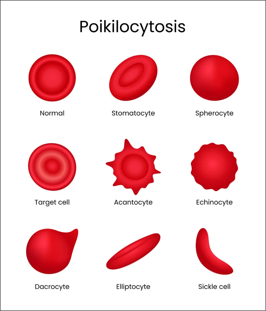
Poikilocytosis
Distorted RBC shape in 10% or more of RBCs
Poikilo: Variable
Causes
Sickle cell anemia→ crescent shaped
Thalassemia→ bullseye shape
Myelofibrosis (scar tissue buildup in BM)→ tear drop
Iron deficiency→ oval, elonagated cells
Mechanical damage→ fragmented cells
Pleocytosis
Increased Leukocytes in CSF (cerebrospinal fluid)
Pleo: plus, more
Causes:
Central nervous system (CNS) infections such as meningitis or encephalitis
Inflammatory autoimmune conditions like MS, systemic lupus
Malignancy: cancers affecting CNS, leukemia/ lymphoma spreading to fluid
Tissue damage
Types:
Lymphocytic (mononuclear): Increased lymphocytes→ from viral/ fungal infections, or chonic inflammation
Neutrophilic (suppurative): increased neutrophils→ from acute bacterial infection, or early non-infectious inflammation
Stomatocyte
Slit-like or “fish-mouth” central area of pallor in RBC
Stomato: mouth, opening
Causes:
Lab artifact: if blood smear slide dries too slow
Hereditary stomatocytosis: genetic membrane defects where RBC leak ions and water
Acquired conditions: Alcoholic liver disease, medications, low pH

Hgb C crystals
Precipitated abnormal hgb C proteins inside RBC→ causes clumping
Can be dense, rectangular
or hexagonal rod-shaped
Caused by:
Hemoglobin C Disease (HbCC): inherited homozygous autosomal recessive blood disorder (both genes)
Hemoglobin SC Disease: one HbC and one sickle cell (HbS) gene
Splenic function: spleen doesn’t properly filter them out (asplenia). Or doesn’t exist (splenectomy)

Drepanocyte
Sharp crescent, elongated RBC
Breaks down in 10-20 days→ leads to anemia
Drepano: sickle or crescent-shaped
Cause:
Abnormal HbS.
When O2 drops, HbS forms stiff rods inside the cell→ curving it

Codocyte
Appeareance:
Bullseye: dark center is hgb
Bell shaped: under electron microscope
Too much cell membrane
Causes:
Liver disease: changes lipids in blood→ extra cholesterol to RBC walls
Thalassemia: Abnormal form or not enough hgb
Hemoglobinopathies: like HgC or HgS
Iron deficiency anemia→ reduces Hgb in RBC
Asplenia→ cannot clean out abnormal cells

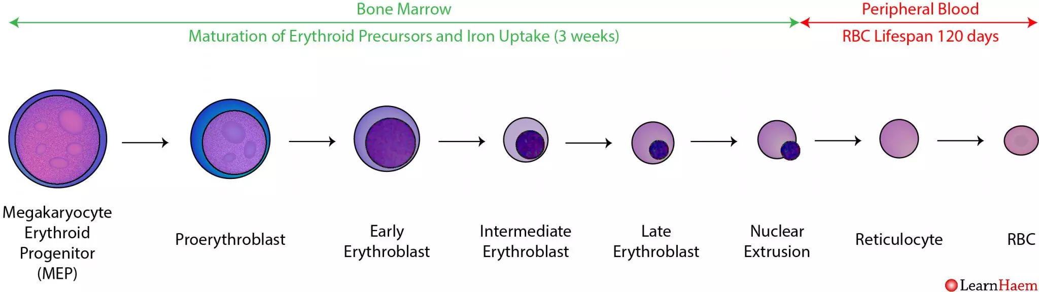
Erythropoiesis nmemonic
PB in my PORES
Proerythroblast/ Normoblast
Basophilic erythroblast
Polychromatic erythroblast
Orthochromatic erythroblast
Reticulocyte
Normal RBC

What is this?
Pronormoblast/ Proerythroblast
From CFU-GEMM→ BFU-E (Burst-Forming Unit-Erythroid) → IL-3 trigger→ CFU-E→ EPO trigger→ Pronormoblast
Nucleus takes up most of the cell
12-24um
High N:C ratio
1-2 nucleoli
Blue cytoplasm
Golgi complex visible
spread out chromatin


What’s this?
Basophilic erythro/normoblast
~20 hrs to develop
12-17um
Chromatin clumps
Accumulates more hgb and RNA
Blue cytoplasm with pink RNA
nucleus more compact
Deep purple nucleus


What’s this?
Polychromatic erythro/normoblast
~30 hours to mature
10-15um
Clumped/ condensed chromatin
Blue hgb apparent in cytoplasm
Pink RNA apparent in cytoplasm
Nucleoli not visible


What’s this?
Orthochromatic erythro/ormoblast
48 hours to mature
8-10um
Solid nucleus with clumped chromatin
Nucleus moves to the side in preparation to be launched out
Cytoplasm starts to be pink
Cytoplasm has blue tint from Hgb
No more DNA synthesis→ cannot divide

What’s this?
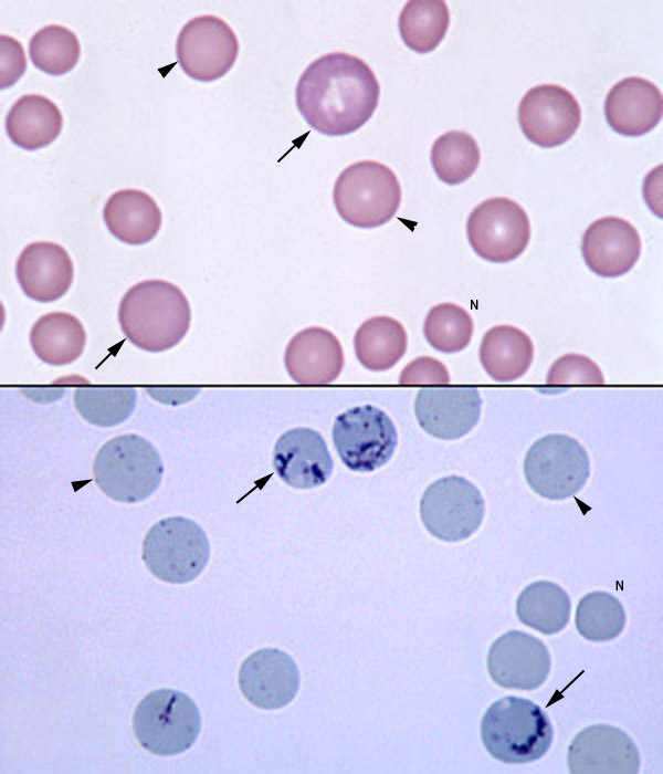
Polychromatophilic erythrocyte/
reticulocyte
115fL
Nucleus gone but has RNA supravital stains
Bluish cytoplasm
1-2 days circulation before maturing
Released in 2-3 days
Ribosomes disappear to make room for more Hgb
Granulofilamentous ribosomes
Has 2/3 of total Hgb

What’s this?
Mature erythrocyte
80-100fL
No nucleus, mitochondria, ribosomes→ no more hgb synthesis
Flexible to allow full passage of body


What’s this?
Myeloblast
From PSC→ IL-1,6,3 triggered→ Common myeloid progenitor (CMP)→ GM-CSF (granulocyte macrophage colony forming factor), IL-3 triggered→ CFU-GEMM→ GM-CSF, IL-3 triggered again→ CFU-G→ GM-CSF, IL-3 triggered again→ myeloblast
15-20um
N:C ratio 6:1
1-3 nucleoli
Oval, central nucleus
Blue cytoplasm, no granules
Chromatin red-blue mesh, no clumps
Divides 3-5 times before next stage
First recognizable cell


What’s this? (1,2,3)
Promyelocyte
12-24um
N:C ratio 3:1
Round-oval nucleus
1-2 nucleoli, fades w/ maturation
Blue cytoplasm w/ azurophilic scattered granules (non specific)
Mesh chromatin starting to aggregate at nuclear membrane

What’s this?
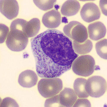
Myelocyte (neutrophilic)
GM-CSF (granulo monocytic colony stimulating factor), G-CSF (granulo colony stimulating factor) triggered
10-18um
N:C ratio 2:1
Nucleus oval or round, slight indent
nucleus off center
Nucleolus may have faded
Cytoplasm blue-pink w/ azurophilic specific granules (secondary pink granules)
Fine chromatin that’s red-blue. Starts aggregating, is granular


What’s this?
Metamyelocyte, neutrophilic
10-18 um
N:C ratio 1:1
Nucleus kidney shaped/ indented
Nucleus central or off-center
no nucleoli
Cytoplasm clear pink w/ many neutrophilic granules
Chromatin light blue-purple w/ basophilic chromatin


What’s this?
Neutrophilic band
10-16 um
N:C ratio 1:1
nucleus is horseshoe/ kidney shaped, central or off-center
no nucleoli
Cytoplasm pink with pink granules
Chromatin purple red, clumped, pyknotic (shrinking and dark staining of chromatin)


What’s this?
Neutrophilic segmented
10-16um
N:C ratio 1:3
Nucleus is 2-5 individual lobes. Central or off-center
Cytoplasm pink with pink fine granules
Chromatin purple-red and clumped

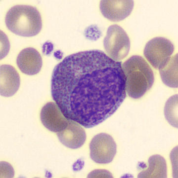
What’s this?
Myelocyte, eosinophilic
GM-CSF, IL-5 triggered
10-18um
N:C ratio 2:1
Nucleus oval or round w/ indent. off-center
may have nucleolus
Cytoplasm blue-pink
Uniform eosinophilic granules red-orange
Chromatin red-blue, fine, starts to aggregate


?
Metamyelocyte, eosinophilic
10-18 um
N:C ratio 1:1
Nucleus kidney shaped (indented), central or off-center
no nucleoli
Cytoplasm clear pink cytoplasm, eosinophilic granules red-orange
Chromatin light-blue purple and distinguishable basophilic chromatin

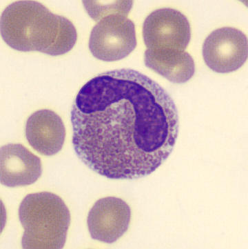
?
Eosinophilic band
10-16 um
N:C ratio 1:1
Nucleus elongated, central or off-center
no nucleoli
Cytoplasm pink w/ red eosinophilic granules
Chromatin coarse blue-purple

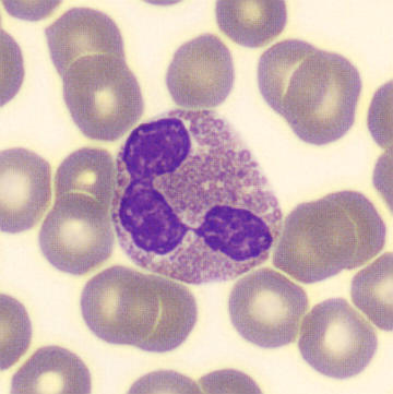
?
Eosinophilic segmented
10-16 um
N:C Ratio 1:3
Nucleus has two lobes (bilobed) thinly connected by chromatin
Cytoplasm pink, granules red and uniform
Chromatin is blue and granular


?
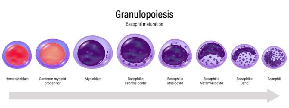
Myelocyte, basophilic
IL-3/ IL-4 triggered
10-18um
N:C ratio 2:1
Nucleus oval/ round, slight indent, off-center
May have nucleous
Cytoplasm blue-pink, granules non-uniform and coarse basophilic
Chromatin red-blue, fine, begins aggregating
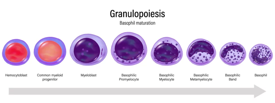

?
Basophilic metamyelocyte
10-18um
N:C ratio 1:1
Nucleus kidney shaped, central or off-center
no nucleoli
Cytoplasm pale blue, granules coarse blue-violet
Chromatin light blue-purple

?
Basophilic band
10-16um
N:C ratio 1:1
Nucleus sausage-shaped, central or off-central
no nucleoli


?
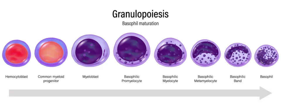
Basophilic segmented
10-16 um
N:C ratio 1:3
Nucleus has 2 lobes, central or off-center
Cytoplasm light blue, granules coarse blue-violet
Chromatin coarse purple

?
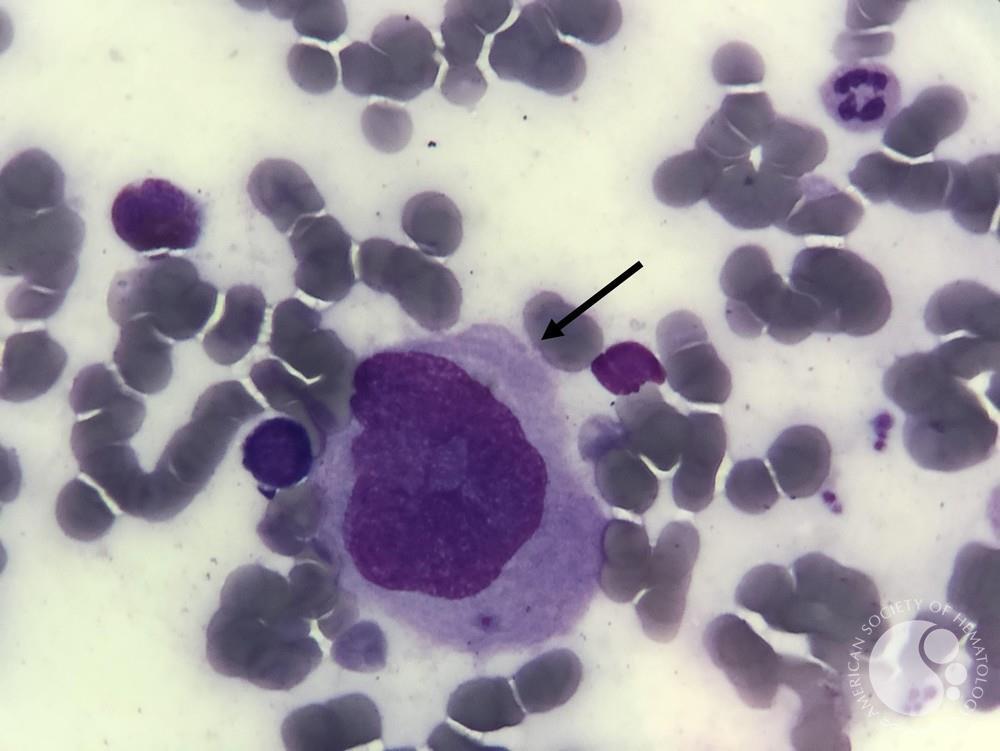
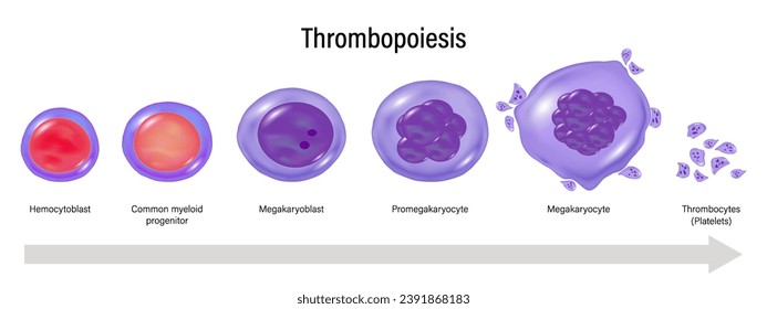
Megakaryoblast
CMP→ GM-CSF, IL-3 triggered→ CFU-Meg→ GM-CSF, IL-3 triggered→ megakaryoblast
20-45 um
N:C ratio 5:1-3:1
Nucleus round/ oval/ kidney-shaped, central or off-center
1-2 nucleoli
Cytoplasm basophilic, nongranular
Pseudopodium (cytoplasm has protrusions)
Chromatin red-purple

?
Promegakaryocyte
20-89 um
N:C ratio is 3:1-5:1
Nucleus is round/ oval/ kidney-shaped, central or off-central
0-2 nucleoli
Cytoplasm basophilic, with many pseudopodia
Granules are fine, azurophilic
Chromatin red-purple w/ increased granularity


?
Megakaryocyte
30-100um
N:C ratio 1:1-1:2
Nucleus has 2+ lobes
Cytoplasm pale blue-pink
Granules fine, azurophilic
Chromatin purple-blue, granular
No nucleoli

?
Thrombocyte/ platelet
1-4um
Cytoplasm light blue
Granules red-blue, evenly dispersed
