Lab exam notes (Bio 1100)
1/21
Earn XP
Description and Tags
L
Name | Mastery | Learn | Test | Matching | Spaced | Call with Kai | Chat |
|---|
No analytics yet
Send a link to your students to track their progress
22 Terms
Cellular Respiration
During cellular respiration, a glucose molecule is
gradually broken down into CO2 & H2O
• ATP is produced during the reactions that transform glucose
4 stages of Cellular Respiration:
• Glycolysis
• Pyruvate oxidation
• Citric acid cycle
• Oxidative phosphorylation
In the absence of O2, fermentation is required to
convert NADH back to NAD+ so that glycolysis can
continue to occur and produce ATP
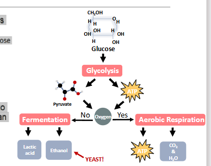
Alcohol Fermentation
Alcohol Fermentation
• EtOH + CO2 produced
• Ex. Yeast
Photosythesis-effect on strach of leaves left in the dark
One plant has been left in the dark and another in the
light for at least one week
❑ Leaves from each plant were harvested and boiled in
water then placed in hot ethanol to remove pigments
❑ Iodine was used to stain the leaves.
❑ Iodine turns black in presence of starch
Parts of mircoscope
❑ Power switch
❑ Light source
❑ Intensity control
❑ Eyepiece/ocular lens
❑ Objective lenses
❑ Stage
❑ Stage control knob
❑ Coarse focus knob
❑ Fine focus knob
Total magnification
To calculate the total magnification for any specimen or structure you are viewing, you
take the power of the ocular lens (10X) and multiply it by the power of the objective lens
you are using
field of view
Field of View (FOV) – the diameter of the lit, circular area you see when you look
through the lens of a microscope
• Represented in mm (millimetres) and μm (micrometres)
❑ Knowing the FOV allows you to determine the approximate size of a specimen that you are examining
Calculate actual size
❑Line up structure of interest with the
edge of the ocular micrometer by moving
the stage using the mechanical stage
control knobs
❑You can rotate the ocular micrometer by
rotating the ocular lens
❑Count how many small divisions your
structure takes up (this is the first part of
the formula)
Actual size = Number of small division observed x Size of smallest division (at objective used)
scale bar
Add a scale bar next to your drawing
• It should span only the size of your drawing
• It should be in the same direction as you measured your
structure using the ocular micrometer
• Label the scale bar with the actual size (not drawing size!)
calculating drawing magnification
Determine your drawing size by measuring the scale bar you just drew
next to your drawing
• Use the metric side of the ruler and convert from cm or mm to μm
Drawing Magnification = 𝐷𝑟𝑎𝑤𝑖𝑛𝑔 𝑆𝑖𝑧𝑒
𝐴𝑐𝑡𝑢𝑎𝑙 𝑆𝑖𝑧𝑒
Animals Vs.Plant cells
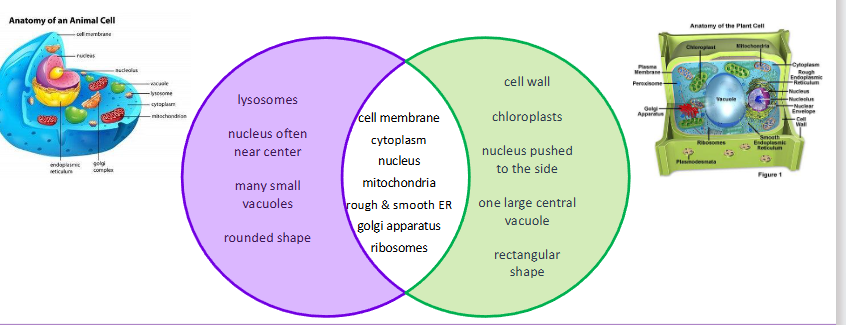
Plasmodesmata
-cytoplamic channels through cell walls
-connects the cytoplasm of adjacent cells
cyclosis
The movement of the fluid substance
(cytoplasm) within a cell
• occurs along actin filaments of cytoskeleton
• movement requires ATP
• allows movement of organelles and other
molecules to move around cell
Osmosis
Movement of water (or other solvent)
Through a semi-permeable membrane
From region of low solute concentration to
high solute concentration
To balance the concentrations of solute
Plasmoloysis
Shrinking of the cell membrane of a plant cell
In a high solute concentration (hypertonic) solution
The cell membrane pulls away from the cell wall
Water is diffusing out of the cell
Interphase
Not a phase of mitosis
• The most common state to find a cell
• Round, homogenous nucleus, nucleoli may
be present

Prophase
Chromatids starting to bundle together
• Nucleus looks like noodles or polka dots
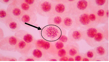
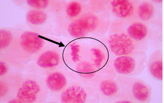
Metaphase
All the chromatids are aligned in a single plane in center of the cell
• Chromosome “tails” point outwards
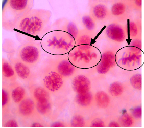
anaphase
Chromatids are being drawn to opposing
poles of the cell
• Two separate sets of chromosomes
• “Tails” point toward middle
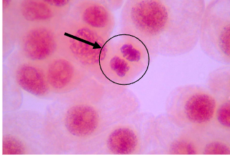
Telophase
Chromatids re-forming into nuclei at
opposing ends of the cell and membrane
begins to form between them
• 2 oval bundles of chromosomes
• Like two tight hair buns
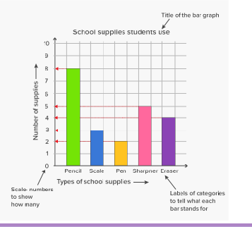
Graphing data
-even intervals
-descriptive titles
-axes labelled (units)
-range of data fills graph space
cell division in eurkoryotic cells
-meiosis
mitosis
DNA
Deoxyribonucleic acid – a nucleic acid
- Molecule that contains a genetic code – the instructions for the development and function of living things