chapter 10: CNS
 Call Kai
 Learn
Learn Practice Test
 Spaced Repetition
 Match
Match Flashcards
 Knowt Play
1/20

There's no tags or description
Looks like no tags are added yet.
Name | Mastery | Learn | Test | Matching | Spaced | Call with Kai | Chat |
|---|
No analytics yet
Send a link to your students to track their progress
21 Terms
ventricles
Ventricles = hollow spaces inside the brain that contain CSF.
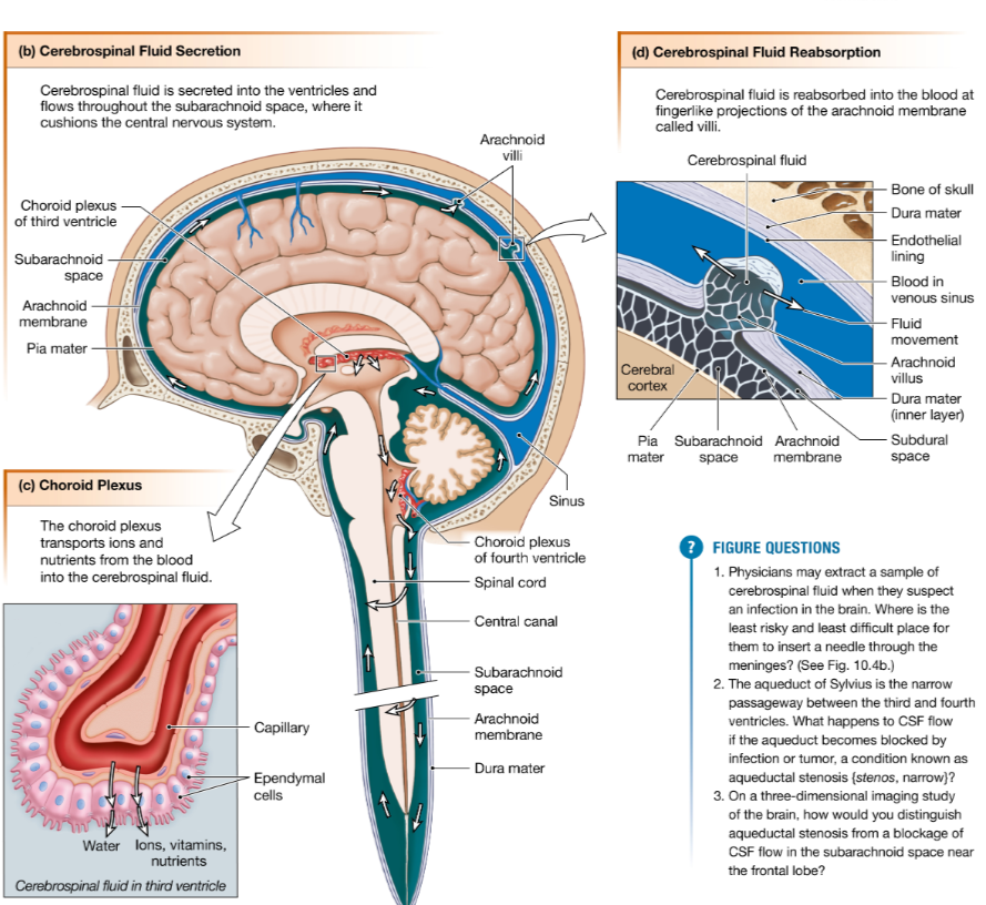
From the ventricles, cerebrospinal fluid flows into the subarachnoid space between the pia mater and the arachnoid membrane, surrounding the entire brain and spinal cord in fluid

white matter vs gray matter
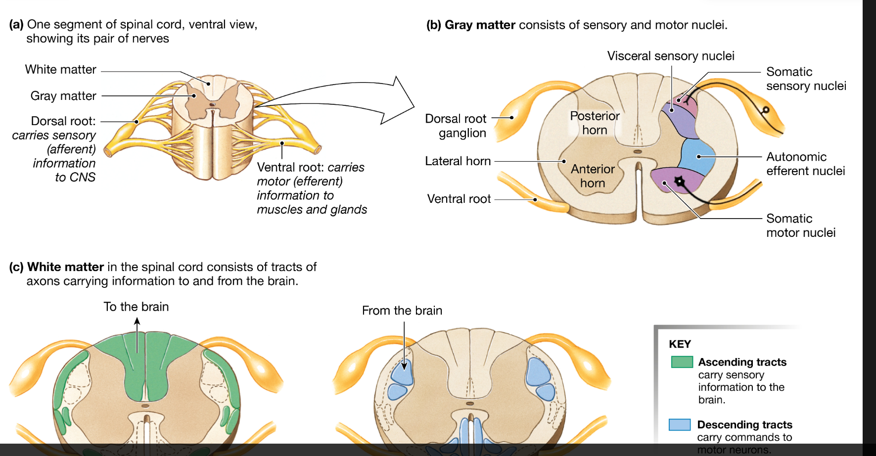
When viewed on a macroscopic level, the tissues of the CNS are divided into gray matter and white matter
White matter is mostly myelinated axons and contains very few neuronal cell bodies. Its pale color comes from the myelin sheaths that surround the axons.
Gray matter consists of unmyelinated nerve cell bodies, dendrites, and axons.
tract
Bundles of axons that connect different regions of the CNS are known as tracts.
Tracts in the central nervous system are equivalent to nerves in the peripheral nervous system.
meninges
Three layers of membrane, collectively called the meninges {singular meninx, membrane}, lie between the bones and tissues of the central nervous system.
These membranes help stabilize the neural tissue and protect it from bruising against the bones of the skeleton.
Starting from the bones and moving toward the neural tissue, the membranes are (1) the dura mater, (2) the arachnoid membrane, and (3) the pia mater
cerebrospinal fluid (CSF)
Cerebrospinal fluid (CSF) is a salty solution that is continuously secreted by the choroid plexus, a specialized region on the walls of the ventricles
choroid plexus
The choroid plexus cells selectively pump sodium and other solutes from plasma into the ventricles, creating an osmotic gradient that draws water along with the solutes
produces CSF
spinal nerve
spinal nerve is a mixed nerve containing both: Sensory (afferent) axons & Motor (efferent) axons
Each spinal region is subdivided into segments, and each segment gives rise to a bilateral pair of spinal nerves. Just before a spinal nerve joins the spinal cord, it divides into two branches called roots
dorsal root vs ventral root
The dorsal root of each spinal nerve is specialized to carry incoming sensory information.
The ventral root carries outgoing information from the CNS to muscles and glands.
dorsal horn vs ventral horn
Sensory fibers from the dorsal roots synapse with interneurons in the posterior horns (dorsal) of the gray matter. The posterior horn cell bodies are organized into two distinct nuclei, one for somatic information and one for visceral information
The anterior horns (ventral) of the gray matter contain cell bodies of motor neurons that carry efferent signals to muscles and glands. The anterior horns are organized into somatic motor and autonomic nuclei. Efferent fibers leave the spinal cord via the ventral root.
ascending tract vs descending tract
Ascending tracts take sensory information to the brain. They occupy the dorsal and external lateral portions of the spinal cord (Fig. 10.6c).
Descending tracts carry mostly efferent (motor) signals from the brain to the cord. They occupy the ventral and interior lateral portions of the white matter.
cranial nerve
Cranial nerves carry sensory and motor information for the head and neck
cerebral cortex
The cerebral cortex {cortex, bark or rind; adjective cortical, plural cortices} is the outer layer of the cerebrum, only a few millimeters thick (FIG. 10.10a).
Neurons of the cerebral cortex are arranged in anatomically distinct vertical columns and horizontal layers (Fig. 10.10b). It is within these layers that our higher brain functions arise.

sensory/motor/association areas of the cortex
sensory areas (also called sensory fields), which receive sensory input and translate it into perception (awareness);
motor areas, which direct skeletal muscle movement; and
association areas (association cortices), which integrate information from sensory and motor areas and can direct voluntary behaviors
Identify the cerebral hemispheres/cerebral cortex, pons, cerebellum, medulla, thalamus, hypothalamus, and spinal cord on a figure such as Figure 10.8c/d
ok

List three meninges of the brain, put them in order from superficial to deep, and recognize that the space between the arachnoid and pia is where CSF circulates around the brain
Starting from the bones and moving toward the neural tissue, the membranes are (1) the dura mater, (2) the arachnoid membrane, and (3) the pia mater
ok
Describe how CSF protects the brain physically
Explain or identify what the blood-brain barrier is “made of” (structurally, what allows it to be a barrier?) and which types of molecules are and are not able to cross it
Draw or label the relationships among the dorsal and ventral roots, dorsal root ganglia and dorsal and ventral horns and explain what parts of neurons each consists of, and what information each part carries
Provide or identify the major function(s) of: medulla, pons, thalamus,
hypothalamus, corpus callosum, basal ganglia/nuclei, primary motor cortex, and limbic system
Recognize that each of the senses (e.g. hearing, taste, etc.) has a dedicated region of the cerebral cortex (e.g. auditory cortex) to process its signals
Recognize that most physiological variables have a circadian rhythm