T3 YR10 W10 HBY test
1/104
There's no tags or description
Looks like no tags are added yet.
Name | Mastery | Learn | Test | Matching | Spaced | Call with Kai |
|---|
No analytics yet
Send a link to your students to track their progress
105 Terms
function of a microscope- coarse adjustment knob
knob which moves the stage up and down to help focus the specimen.
1. retrieve the microscope using one hand on the base and one hand on the arm. Place it down, unwrap the cord and plug it in then turn the microscope on.
2. check the at the condenser lens is positions just below the stage and make sure that you use the revolving nosepiece to put the scanner objective lens on.
3. move the stage up using the coarse adjustment knob then adjust the light.
4. place the wet mount slide onto the stage and make sure it’s secure using the stage clips. Then move the slide to centre it within the viewing area.
5. look through the eyepiece lens and use the coarse and fine adjustment knobs to focus the specimen.
6. once focused, use the revolving nose piece to put the low-power objective lens and repeat. However, just focus using the fine adjustment knob as it should be almost focused.
7. move to the high-power objective lens and repeat.
8. remove the wetmount slide and move the stage down using the coarse adjustment and use the revolving nose piece to put the scanner objective lens back.
9. switch off the light, unplug the microscope and wrap it around.
10. holding the base and arm, put it back
1. collect a slide, making sure that it is clean as well as grabbing a coverslip for the later steps.
2. using your pipet, extract some of the liquid and place one drop at the centre of the slide.
3. grab your coverslip and angle it 45 degrees at the end of the droplet before gently letting go.
1. collect a slide, making sure that it is clean as well as grabbing a coverslip for the later steps.
2. place the specimen at the centre of the slide
3. using your pipet, extract some of the liquid and place one drop at the centre of the slide.
4. grab your coverslip and angle it 45 degrees at the end of the droplet before gently letting go.
cells are measured in?
UM (micrometres)
endosymiotic theory (elaborated)
the theory that eukaryotic cells came from prokaryotic cells. A long time ago, only prokaryotes existed and some had the ability to photosynthesis, some had the ability to use oxygen to produce ATP energy and some were larger than others. The idea here was that the larger prokaryote engulfed the smaller ones and instead of it digesting, they lived in symbosis.
endosymbiotic theory profs
Both chloroplasts and mitochondria (C & M) had DNA different to that in the nucleus and also had DNA structure similar to Bacteria (a prokaryote)
they divide similarly to bacteria
their size is pretty much the same to bacteria
* cytoskeleton
* inclusions
* organelles
* cytoplasm
* cytosol
Golgi body
A series of flattened membranes stacked upon each other. Their role is to modify proteins and pack them in vesicles (liquid bubbles containing proteins) for secretion.
lymphatic system
one of the 11 systems consisting of lymph nodes, lymph capillaries and lymph vessels which collect and return excess fluid leaked out by the capillaries.
Lymph
watery fluid found in lymph vessels that is a pale yellow colour and moves very slowly due to having no pump.
lymph capillaris
small vessels which collect the interstitual fluid found in tissues. This interstitual fluid in capillaries is called lymph
how is lymph transported?
thorough peristaltic movements and the contractions of surrounding skeletal muscles
function of lymph vessels
to return excess fluid from the capillary to the lymph ducts which empty them into the subclavian veins and back into the circulatory system.
lymph vessels
thin walled, one-way, blind ended vessels that lie in the space of most tissues.
structure of lymph vessels
one-cell thick
like veins in the way that they are slow moving and have valves to prevent backflow
features of lymph vessels
slightly larger than capillaries
more permeable meaning that pathogens are able to seep through
lymph nodes
small bean-shaped structures found at certain intervals within the lymph vessels. They are mostly concentrated around the armpits and groin and the lymph passes through many of these before returning to the circulatory system. its role is to filter the lymph
structure of lymph nodes
they are surrounded in connective tissue which extends into the node and forms a framework.
this framework contains alot of lymphatic tissue rich in macrophages, lymphocytes and plasma cells.
lymph nodes role in immunity
within the mass of lymphatic tissue, there are macrophages which destroy pathogens found in the lymph via engulfing them through phagocytosis and breaking them down using enzymes.
during infection the production of lymphocytes increases and so lymph nodes become sore and swollen.
lymphatic organs include
spleen, thymus, tonsils, adenoids
lymph ducts
two large vessels which the other vessels connect to that bring the lymph back to the circulatory system via the left and right subclavian veins.
Prokaryote
single-celled organism that lacks a nucleus and membrane bound organelles. Due to this, they are unable to perform multiple functions at once. These include bacteria and archaea.
Eukaryote
cells found within single and multicellular organisms that contain a membrane bound nucleus and organelles. Due to this, they are able to carry out many functions. These include protists, fungi, plants and animals.
condenser lens
lense that aligns the light rays up into a straight path
protein synthesis (transcription)
DNA is found within the nucleus
DNA goes through the nucleolus where rna polymerase is stored. its role is to break apart the dna strand into RNA strands and make a replica of one of the strands by pairing its complementary nucleotides to it. This newly formed strand is calles mRNA
protein synthesis (translation)
the mRNA leaves then nucleus and finds a ribosome.
the ribosome takes this information and collects the amino acids, making a polypeptide chain. 20 of these make a protein.
protein synthesis (secretion)
the newly formed protein is transported to the golgi body.
the golgi body modifies it and packages it in vesicles ready for secretion.
red blood cells structure which help to carry out its function.
they have a bi-concave disc to increase surface area and maximise the amount of oxygen that passes through
they are small and flexible allowing them to pass through small vessels such as capillaries
they have a thin membrane allowing oxygen to easily pass through
they lack a nucleus so as much haemoglobin can fit in.
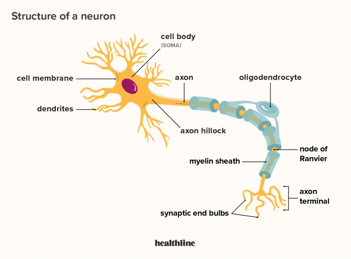
neurons structure which help to carry out its function.
the dendrites extended out allow the neuron to receive the message
the axons allow the neuron to transmit chemical and electrical signals to other cells
bone cell structure which help to carry out its function.
they contain osteocytes which help with strength and rigidity of the bone
muscle cell structure which help to carry out its function.
theyre long and cylindrical making it easier for contraction and relaxation
they contain acting and myosin which allow contraction, relaxion and produce force needed for movement of limbs.
functions of the cell membrane
to separate its components from the external environment
communicate with other cells
decide what goes in and out (semi-permeable)
phospholipids
molecules making up the cell membrane which consists of a hydrophilic head and a hydrophobic tail making it amphiphilic. phospholipids also contain a glycerol backbone which holds the tails and head together.
hydrophilic
attracted to water
hydrophobic
wants to get away from water
cholestorol
lipid made of 4 carbon rings that has the role of controlling flexibility and temperature by separating the hydrophillic heads when it is too cold to increase movement and pulling them together when it is hot to reduce movement.
channel proteins
create hydrophilic holes (channels) in the membrane to allow smaller molecules (i.e ions) through.
adhesion proteins
form junctions between adjacent cells
peripheral proteins
act as enzymes and connect to the cytoskeleton to help with cell shape
carrier /integral proteins
go through the bilayer and transport larger molecules such as glucose into and out of the cell. They also bind to larger molecules, helping them pass through the membrane.
semi-permeable membrane
a membrane that allows some molecules to pass through but not others.
what can pass the membrane (5)
small, non-polar molecules (co0,02)
small, polar molecules (water) however, hard to do so because of the hydrophobic tails
large, non-polar molecules (carbon rings) however is really hard due to size
large, polar molecules (glucose). However, needs help from protein due to size, charge and inability to pass the non-polar region.
ions (NA+). However, needs protein due to size, charge and inability to pass the non-polar region.
recognition proteins
proteins that identify your cells to prevent an immunity attack against them
receptor proteins
molecules which bind to receptor proteins to produce change to the cell.
circulatory system
body’s system which delivers oxygen and nutrients to cells, co2 and other wastes away from cells as well as transports hormones. These include the heart, blood and blood vessels.
blood
a fluid tissue consisting of 55% plasma, 41% red blood cells and 4% white blood cells.
plasma
the liquid part of blood (55%) consisting of 91% water and the remainder being dissolved substances such as ions, hormones, nutrients, gasses, plasma proteins and wastes
red blood cells (erythrocytes)
bi concave discs which carry out the main function of transporting oxygen around the body. They contain haemoglobin which carries the oxygen and when they combine it forms oxyhemoglobin which makes the cell red. It also lacks a nucleus to maximise the amount of haemoglobin which can fit in as well as making the cell more flexible.
white blood cells (leucocytes)
cells which play a major role in fighting off infection and protecting the body against infection. They are larger than erythrocytes.
Platelets (thrombocytes)
fragments of a cell with no nucleus. Their role is to stop bleeding within an area by forming a clot there.
Arterioles
small arteries which give blood to the capillaries. When the oxygenated blood leaves the heart through the aorta, the artery will distribute into many medium-sized (distributing) arteries. These distributing arteries will then branch out into the artierioles which then give blood to the capillaries.
arteries
blood vessels which carry blood away from the heart to the capillaries within tissues.
artery structure
endothelial lining which lines the inside of the artery and is what is in contact with the blood
the tunica media which is smooth muscle that can contract and relax
the tunica externa which is the thick, muscular layer. The walls of the arteries need to be so thick and elastic to withstand the blood pressure.
why don’t arteries have valves?
due to the immense blood pressure
vasodilation
when the muscle walls relax, increasing diameter and allowing more blood to flow.
vasoconstriction
when the muscle walls contract, decreasing diameter and allowing less blood to flow.
veins
vessels which carry blood to the heart from the tissues.
venules
the smallest veins which carry blood from the capillaries to the larger veins
structure of venules
endothelium (inner lining of the venule)
tunica externa (thin layer of inelastic tissues)
structure of veins
endothelium (inner lining of the venule)
tunica media (think layer of smooth muscle)
tunica externa (thin layer of inelastic tissues)
why do veins have valves?
to prevent back flow as the blood pressure isnt as great.
capillaries
small vessels which connect arterial and venous circulation as well as allowing efficient exchange of nutrients and wastes between blood and tissues. The capillaries release a bit of fluid, bathing the tissues (this is then later picked up by the lymphatic system). The amount of fluid release is dependant upon hydrostatic pressure and the amount of solutes at each capillary bed.
structure of capillaries
they only contain an endothelial lining and their walls are one cell thick.
heart
the muscular pump which pushes blood around the body. It is located in the middle of the chest cavity between the lungs.
4 main functions of the heart
pump hormones to parts of the body
pump oxygenated blood to cells
receive deoxygenated blood and pump it to the lungs for oxygenation
maintain blood pressure
external structure of the heart
has thick muscular walls to be able to push the blood out of the heart with enough force
encased in a membrane called the pericardium which is a thin membrane holding the heart in place and preventing it from overstretching.
4 chambers of the heart (with roles)
right atrium (receives deoxygenated blood)
right ventricle (pumps deoxygenated blood to the lungs through the pulmonary artery)
left atrium (receives oxygenated blood)
left ventricle (pumps oxygenated blood to the body)
superior/inferior vena cava
veins which receive the deoxygenated blood and transport it to the right atrium.
chordae tendinea
the strong muscles holding the atrioventricular valves in place
pulmonary artery
artery which transports deoxygenated blood from the right ventricle to the lungs for oxygenation
pulmonary vein
vein which transports blood from the lungs to the left atrium.
aorta
where the oxygenated blood gets pumped to initially to go to the rest of the body.
valves
flaps of thin tissue with the edges being held by tendons. they separate the ventricles and prevent backflow.