14. Lens growth and differentiation
1/28
There's no tags or description
Looks like no tags are added yet.
Name | Mastery | Learn | Test | Matching | Spaced | Call with Kai |
|---|
No analytics yet
Send a link to your students to track their progress
29 Terms
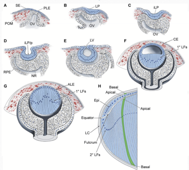
What is the first morphological sign of lens development?
The formation of the lens placode.
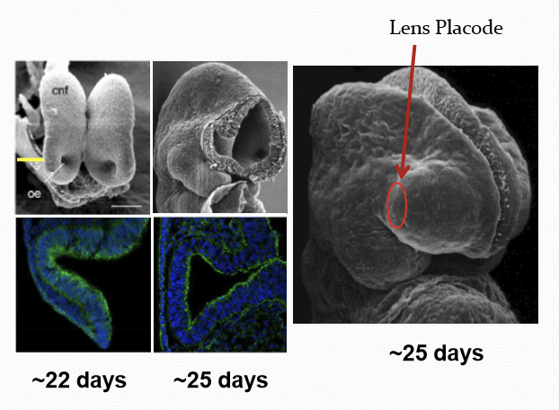
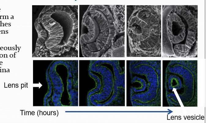
What does the lens placode form into?
It invaginates to form a lens pit and pinches off to from the lens vesicle.

What forms simultaneously during the invagination of the lens placode?
Formation of the optic cup and the future neural retina.
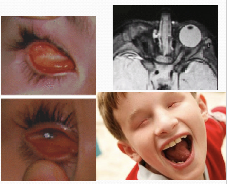
What is Peters Anomaly type II?
When the embryonic lens vesicle fails to separate from the prospective cornea. If forms a lens stalk that is connect to the cornea and persists into the mature eye.
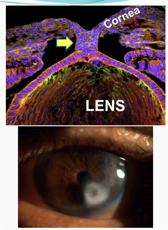
What does the residual stalk of peter's anomaly disrupt?
The anterior segment development.
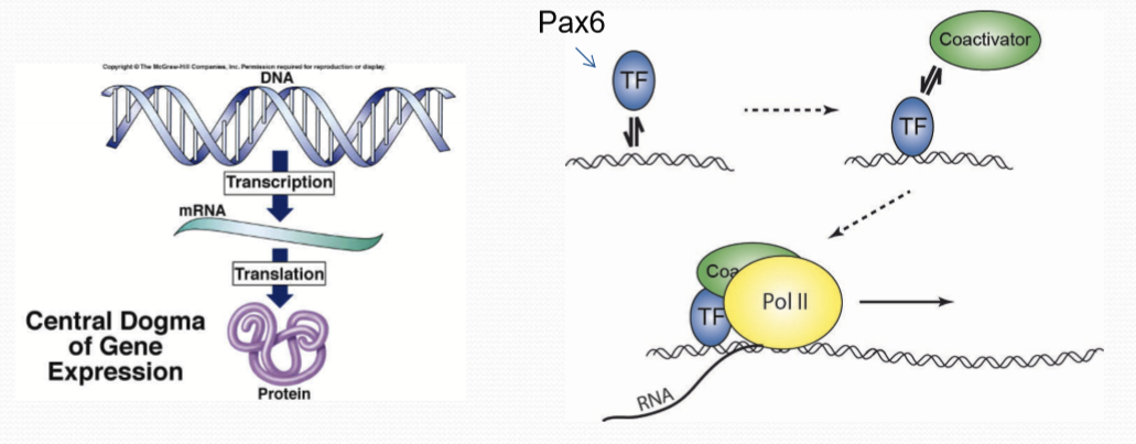
What does PAX6 gene do?
It is the “master control gene” that is required for lens induction, lens fiber differentiation, corneal development, iris formation, retinal development, ciliary body, and lacrimal gland. Disruptions in PAX6 gene will cause severe ocular defects.
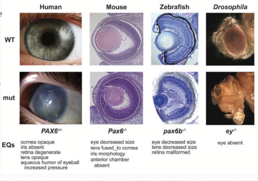
What happens if PAX6 gene is lost?
It can cause the complete loss of an eye (anophthalmia). This is due to the lack of lens formation, which adversely affects the development of additional ocular structures.
What is the role of the lens in influencing eye development?
The lens is believed to secrete signals that influence the development of surrounding ocular structures.
What does PAX6 do?
It is a transcription factor that promotes transcription of specific target genes by binding specific DNA sequences and recruit the transcriptional machinery. It triggers a cascade of pathways that are required for eye development and function.
What occurs after the lens vesicle forms?
Primary lens fibers begin to extend from the posterior lens fibers begin to extend from the posterior cells and obliterate the space within the lens vesicle.
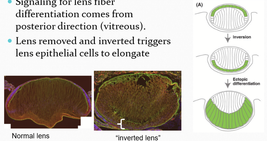
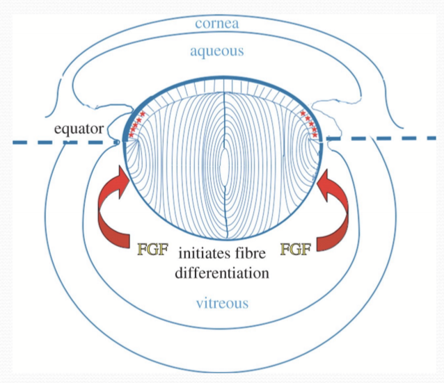
From experimentation, what was found about the lense fibers?
Signaling for lens fiber differentiation comes from the posterior direction (vitreous). As inverting the lens triggers lens epithelial to elongate.
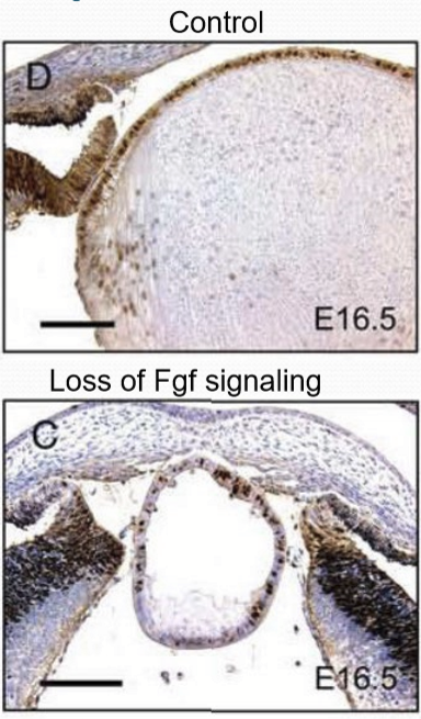
What proteins are required for lens fiber development?
Fgf/Fibroblast growth factor. It is a secreted signaling molecule.
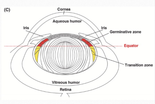
What happens to the secondary lens fibers after they enter the transitional zone?
Differentiate from the lens epithelial cells and begin to elongate. They displace the central primary fibers from their attachments with the posterior lens capsule and lens epithelium.
Where does mitosis of cells occur?
In the germinative zone and continually supplies new cells to the transitional zone.
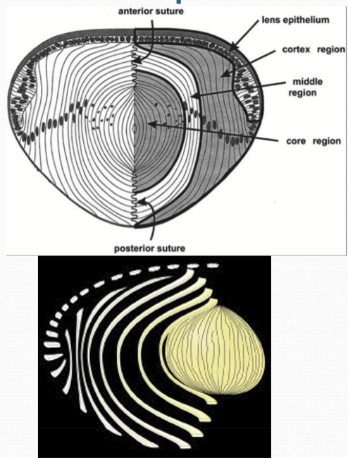
What does the displacement of lens fibers from the lens capsule and lens epithelium allow the secondary fibers to do?
Allows secondary fiber cells on opposite sides of the lens to join together at their apical and basal faces to generate the lens sutures.
What happens to the number of mitotic cells during development?
They decrease in numbers and are found in the perimeter of the lens instead of on the entire anterior side.
What structure forms around the lens soon after it is formed?
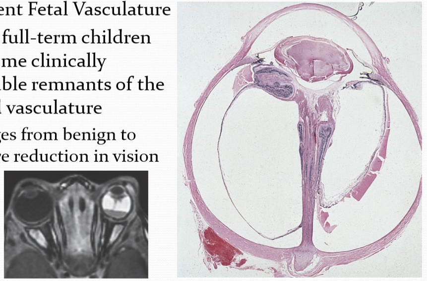
A meshwork of capillaries. Posterior end arises from the hyaloid artery and the anterior side arises from the iris stroma.

When does the lens meshwork get broken down?
During the second trimester. Capillaries regress/breakdown.
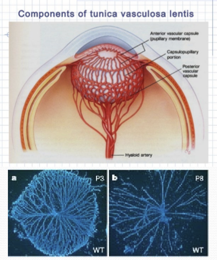
What is persistent fetal vasculature?
When there are clinically detectable remnants of the hyaloid vasculature. Can rang from benign to severe reduction in vision. ~3% of fell-term children have some clinically detectable remnants of the hyaloid vasculature.
Where is the germinative zone located?
The region of the anterior ciliary zonules.
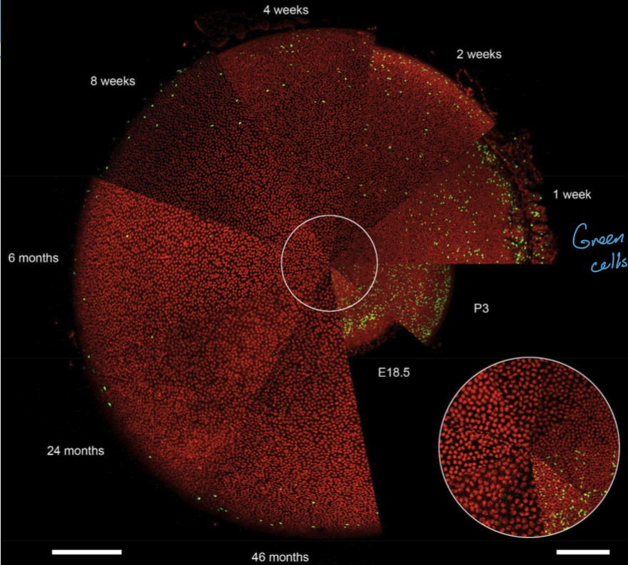
What is the difference between embryonic growth and lengs growth throughout life?
Not much, same mechanisms regulate both. Rate at which growth slows with age. Fgf proteins are still essential in differentiation in mature lens.
Where are younger lens fibers found?
Near the equatorial periphery. Older lens fibers near the lens nucleus.
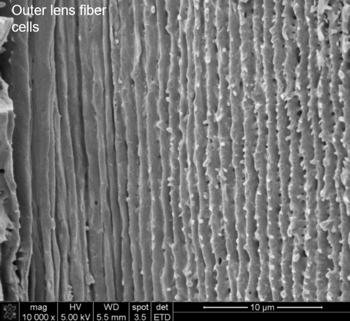
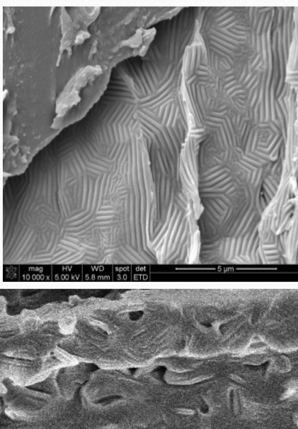
What happens to the lens fibers as they get deeper into the lens?
They gain protrusive structures: Protrusions, paddles, ball-and-sockets. Typically 50-700 micrometers from surface
What is the purpose of the protrusions?
Cell adhesion
transcellular ion/water transport
ball-and-socket have lots of gap-junctions
interlocking protusions have lots of aquaporins
congenital mutations in encoding genes of these proteins causes cataracts
What patterns do the protrusions forms?
Nuclear microridges
Why is adhesion so important?
It helps promote lens transparency.
What does alpha-crystallin do?
Facilitates transparency, refraction of lens. Creates destructive interference of scattered light and prevents protein unfolding. Lower crystallin conc towards surface of lens.
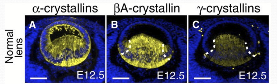
What happens in lens fiber differentiation?
Cell shape altered
increase in cytoplasmic transparency
large increase in crystallin protein expression
organelles and nuclei are degraded
Where does denucleation occur?
Only in erythrocytes, keratinocytes, and lens fibers