8.1 muscle anatomy and contraction
1/40
There's no tags or description
Looks like no tags are added yet.
Name | Mastery | Learn | Test | Matching | Spaced | Call with Kai |
|---|
No analytics yet
Send a link to your students to track their progress
41 Terms
functions of the muscular system
movement of the body, maintenance of posture, respiration, production of body heat, communication, constriction of organs and vessels, contraction of the heart
contractility
ability of a muscle to shorten w force
excitability
capacity of muscle to respond to a stimulus (from our nerves)
extensibility
muscle can be stretched to its normal resting length and beyond to a limited degree
elasticity
ability of muscle to recoil to original resting length after stretched
skeletal muscles
many attached to bones via tendons (connective tissue), appear striated (due to thick and thin filaments), cells are multi-nucleated, under voluntary control
skeletal muscle tissue
responsible for locomotion, facial expressions, posture, respiratory mvmts, other types of body mvmt, voluntary + reflexes but mostly voluntary, fibers (muscle cells) are long, cylindrical, multinucleated, striated appearance
smooth tissue
walls of hollow organs, blood vessels, eye, glands, skin, some functions: propel urine, mix food in digestive tract, dilating/constricting pupils, regulating blood flow, in some locations, autorhythmic, controlled involuntarily by endocrine and autonomic nervous systems, visceral smooth muscle has numerous gap junctions (sheets of smooth muscle function as a unit)
cardiac muscle tissue
heart: major source of mvmt of blood, autorhythmic, controlled involuntarily by endocrine and autonomic nervous systems
a muscle fiber = a __. a __ contains several __. __ are grouped into __ and are also held together and in parallel by __
muscle cell, muscle fiber, myofibrils, muscle cells, fasciculi, connective tissue
sarcolemma
plasma membrane of a muscle fiber (or cell)
sarcoplasm
cytoplasm of a muscle cell
myofibril
contractile elements found in muscle cells (muscle fibers), made of sarcomeres, composed of thick and thin filaments
sarcomere
basic contractile unit of muscle fiber
Z disk
filamentous network of protein, serves as attachment for actin filaments
I bands
btwn thick filaments
A bands
length of thick filaments
H zone
region in A band where actin and myosin do not overlap
M line
middle of H zone; delicate filaments holding myosin in place
tropomyosin
an elongated protein that winds along the groove of the actin double helix
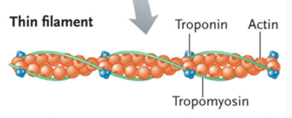
troponin
found between the ends of the tropomyosin molecules in the groove btwn actin strands, composed of 3 subunits: one that binds to actin, a second that binds to tropomyosin, and a third that binds to calcium ions

tropomyosin/troponin complex
regulates the interaction btwn active sites on actin and myosin
myosin ii
myosin motor protein found in skeletal muscle; generates force for muscle contraction, formed from 2 heavy chains, 2 copies each of 2 light chains, head: binds and hydrolyzes ATP and generates force for mvmt, tail: formed from coiled-coil interaction 2 alpha-helices of heavy chains
myosin thick filaments
large, bipolar filaments formed from tail-tail interactions btwn myosin molecules, contain several hundred myosin heads, heads are oriented in opposite directions and project to the outside, thick filament can slide oppositely oriented pairs of actin filaments past each other
what was this one random experiment’s results?
actin filaments glide along slide and so motor activity is contained in the head
which bands will decrease when muscle shortens?
I band and H zone
sliding filament mechanism of muscle contraction
thin filaments slide over thick filaments simultaneously on each side of sarcomeres (note: the thick filaments doesn’t slide), shortens sarcomeres and muscle fibers, produces force that contracts the muscle
motor neurons
stimulate muscle fibers to contract
neuromuscular junction (NMJ)
contact point btwn the axon and muscle
synapse
region where the axon terminal of a neuron rests on an invagination of the sarcolemma
what does the neuromuscular junction consist of?
presynaptic terminal - axon terminal w synaptic vesicles
synaptic cleft - space
postsynaptic membrane (sarcolemma) / motor end-plate
synaptic vesicles in NMJ contain
neurotransmitter: substance released from a presynaptic membrane that diffuses across the synaptic cleft and stimulates (or inhibits) the production of an AP in the postsynaptic membrane
acetylcholine
used to stimulate an AP in skeletal muscle
acetylcholinesterase in NMJ
a degrading enzyme in synaptic cleft, prevents accumulation of ACh
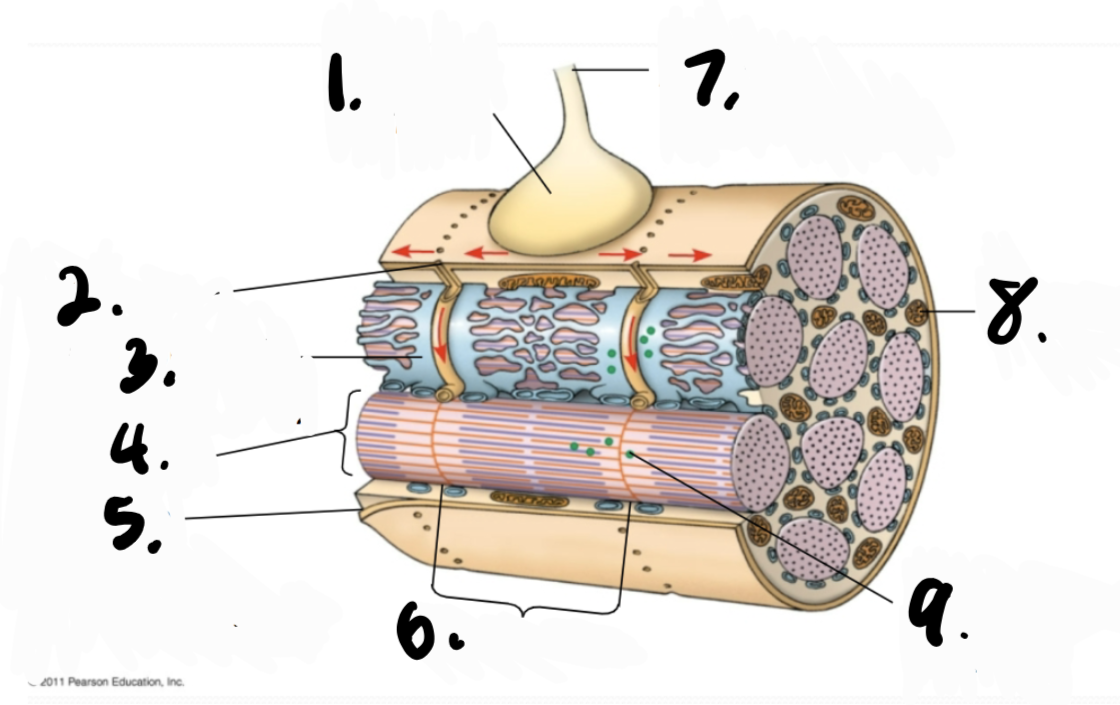
synaptic terminal, T tubule, sarcoplasmic reticulum (SR), myofibril, plasma membrane of muscle fiber, sarcomere, axon of motor neuron, mitochondrion, Ca2+ released from SR
T-tubule
transverse tubule, deep invagination of the sarcolemma, only found in skeletal and cardiac muscle cells, these invaginations allow depolarization of the membrane to quickly penetrate to the interior of the cell
how a muscle contracts
AP arrives at the neuromuscular junction, causing release of acetylcholine → acetylcholine triggers an AP in the muscle fiber that spreads over its plasma membrane, into transverse tubules → stimulates the sarcoplasmic reticulum to release Ca2+ into the cytosol → Ca2+ combines w troponin, inducing a conformational change that moves tropomyosin away from myosin-binding sites on thin (actin) filaments → exposure of sites on actin allows myosin cross bridges to bind and initiate the cross bridge-cycle → myosin heads of thick filaments attach to a thin filament, pull, and release in cyclic reactions powered by ATP hydrolysis
cross bridge cycle
myosin head has ATP bound, not in contact w actin → myosin binding site on actin becomes available → ATP → ADP + P and myosin head attaches to actin and initiates the “power stroke” (bending of the myosin head and mvmt of the actin filament) and releasing ADP, myosin head binds to a new ATP and detaches from actin
system is reset
APs to the muscle stop → Ca2+ is removed from the troponin and actively transported (via Ca2+ pumps) back (from the cytosol) into the sarcoplasmic reticulum → tropomyosin moves to its original location on the actin, blocking myosin binding sites → actin thin filaments slide back to their original position
differences btwn AP stimulation btwn neurons and AP stimulation at the neuromuscular junction
neuron to neuron synapse:
need a lot of synaptic input from a lot of presynaptic cells to get to threshold: a lot of integration, a lot of EPSPs to reach threshold
can have inhibitory input
neuromuscular junction:
one AP in a motor neuron almost always cause an AP in the muscle cell
generally, no inhibitory input. have a large excitatory end plate potential (EPP) that reaches threshold for an AP
since neuromuscular junctions are in the middle of a muscle cell, the AP generated can go out in both directions to cover the entire muscle cell
rigor mortis
few hours after death, Ca2+ leaks into cytoplasm causing myosin head to attach to actin → muscle contraction → muscles stiffen → no ATP (dead), so myosin head cannot be released from actin → in humans, it commences after about 3-4 hrs, reaches max stiffness after 12 hrs, and gradually dissipates until approx 48-60 hrs after death