KAAP630 Test 1
 Call Kai
 Learn
Learn Practice Test
 Spaced Repetition
 Match
Match Flashcards
 Knowt Play
1/92

There's no tags or description
Looks like no tags are added yet.
Name | Mastery | Learn | Test | Matching | Spaced | Call with Kai | Chat |
|---|
No analytics yet
Send a link to your students to track their progress
93 Terms
What are the sections of the adrenal glands?
Adrenal Cortex (outside)
Adrenal Medulla (inside)
What is released from the adrenal cortex (AC)?
corticosteroids (mineralocorticoids and glucocorticoids)
aldosterone (released from zona glomerulosa of AC)
cortisol (released from zona fasciculata and reticularis of AC)
androgenic hormones
released from zona fasciculata and reticularis of AC
What is released from the adrenal medulla?
catecholamines
What are the effects of mineralocorticoids?
Affects electrolytes (minerals) in extracellular fluid
especially Potassium and Sodium
Aldosterone accounts for 90%; cortisol has small contribution
What are the effects of glucocorticoids?
increase blood glucose (cortisol) and influence metabolism
Explain the synthesis, metabolism, and excretion of corticosteroids
LDL-C (low-density-lipoproteins, cholesterol) interact with LDL receptors on surface of cells
Brought into the cell via endosome (endocytosis) and transported to mitochondria
LDL-C cleaved by cholesterol desmolase into pregnenolone
Rate-limiting step
Pregnenolone shuttled out of mitochondria into cytoplasm, then crosses cell plasma membrane
Bound to plasma proteins in blood
metabolized in liver
Excreted in urine
Function of aldosterone
Salt regulation (absorption) and potassium secretion
Regulates sodium-water balance
Regulates blood volume and pressure
Part of RAAS
Two levels of control:
Triggered by high levels of potassium (wants to prevent hyperkalemia)
Triggered by decreased levels of blood volume/decrease in blood pressure; triggers Angiotensin II
Explain the simplified RAAS pathway
Angiotensinogen cleaved by Renin (from kidney) to Angiotensin 1
Angiotensin 1 (vasoconstrictor) converted to Aldosterone and AVP/ADH by Angiotensin-Converting-Enzyme (ACE)
Aldosterone regulates salt and water retention
AVP/ADH regulates water retention and vasoconstriction
Why is Renin released?
Triggered by reduction in blood volume
Reduced blood volume = reduced renal afferent arterial pressure, sensed by juxtaglomerulus (JG) apparatus (part of juxtaglomerulus complex)
Reduced blood volume = reduced glomerular filtration rate (GFR) = lower NaCl concentration in tubular fluid, stimulates JG cells
Sympathetic stimulation of beta receptors on JG cells causes release of Renin
Explain aldosterone stimulation pathway
Aldosterone diffuses into principle cells of kidney
Aldosterone bind to mineralocorticoid receptors (MR), forms hormone-receptor complex (HRC)
Can be blocked by presence of spironolactone (drug)
HRC diffuses into nucleus → production of mRNA
mRNA translated into proteins (Na/K ATPase, ENaC (epithelial sodium channel))
ENaC can be blocked by amiloride (drug)
Takes at least 45 minutes for process to occur, takes hours to see full effects
Aldosterone dysregulation disorders
Hyperaldosteronism (Conn syndrome)
Caused by tumors on adrenal glands
hypertension, hyperkalemia
Secondary hyperaldosteronism
excess aldosterone production due to increased RAAS activation
Tertiary hyperaldosteronism (Bartter and Gitleman syndrome)
Mutations in ion transporters in kidney, excess sodium is lost
stimulates aldosterone in excess via RAAS
Hypoaldosteronism
Addison’s disease: destruction of adrenal glands due to infection, injury, or autoimmune disorder
Renal insufficiency leads to reduced RAAS
Explain cortisol stimulation pathway and its effects
Anterior pituitary releases adrenocorticotropic hormone (ACTH)
ACTH stimulates adrenal cortex; cortisol released
cortisol stimulates:
Gluconeogenesis
Protein mobilization
Fat mobilization (lipolysis)
Stabilizes lysosomes
Relieves stress, excites hypothalamus (state dependent)
PRESENCE OF CORTISOL INHIBITS FURTHER SECRETION OF ACTH
Describe ACTH stimulation pathway
ACTH connects to receptor, makes HRC
Alpha subunit detaches, stimulates cAMP
cAMP stimulates Protein Kinase A
Protein Kinase A stimulates CEH (cholesterol ester-rich droplets) in lipid droplet to convert cholesterol esters into cholesterol
Cholesterol converted to pregnanolone, shuttled into mitochondrion
Pregnanolone converted to 17-OH pregnanolone; 17-OH preg converted to 11-deoxy-cortisol; 11-deoxy-cortisol converted to cortisol
Conversion steps occur in smooth endoplasmic reticulum which works with mitochondria
Cortisol released from mitochondria, diffuses across cell membrane, bound to plasma protein and circulates in blood
Example stimulation path for cortisol
broken bone/stress/burns/exercise → positive stimulation of higher brain centers
higher brain centers → positive stimulation of hypothalamus
hypothalamus releases CRH → stimulates anterior pituitary gland to release ACTH
ACTH → positive stimulation of adrenal cortex
cortisol released from adrenal cortex
Physiological effects of cortisol
Mobilizes tissue amino acids (muscle breakdown)
blocks entry of glucose into tissue (Blocks GLUT 4 transporters)
stimulates gluconeogenesis
antagonizes insulin in liver, stops inhibition of gluconeogenesis
mobilizes free fatty acids (lipolysis)
Decrease growth hormone (GH) and IGF-1; can impair bone and muscle growth
Anti-inflammatory effects of cortisol
stabilizes lysosomal proteins
decreases capillary permeability
decreases WBC mobilization and phagocytosis of damaged cells
suppresses immune system → decline in lymphocyte reproduction
lowers fever by reducing release of interleukin-1 from WBCs
Prednisone (drug) used to treat inflammatory conditions; mimics cortisol
Glucocorticoid dysregulation disorders
Cushing’s disease
excess cortisol production
usually results from tumor, can be ACTH independent or dependent
excess weight gain, round/moon face, fat deposition, hypertension, muscle loss, osteoporosis
Addison’s disease
glucocorticoid deficiency
auto-immune disease, destroys adrenal gland/interferes with steroid hormone synthesis
weakness/fatigue, low BP/hypotension, decreased appetite, weight loss, hyperkalemia
Can be life-threatening
Name the relevant catecholamines and their receptor types
acetylcholine (ACh, nicotinic)
epinephrine (E, adrenergic)
norepinephrine (NE, adrenergic)
Name the two types of adrenergic receptors and their effects once stimulated
alpha
GPCR
vasoconstriction/increased peripheral resistance
reduced mucosal edema (in airway, makes it easier to breathe)
decreased cAMP
inhibition of NT release
autoreceptor feedback regulation
beta
GPCR
increased HR/CO
increased contractility
bronchodilation
vasodilation
reduced inflammatory mediator disease
lipolysis
metabolic regulation
Explain and understand the GPCR pathway

What is the function of the pancreas?
glucose homeostasis, digestion aid
alpha cells secrete glucagon
beta cells secrete insulin
Structure of insulin?

Glucose stimulates insulin release when it passes through the _____ channel of beta-cells in pancreas
GLUT 2
Steps of insulin release
Glucose enters beta cell via GLUT 2 channel
Glucose undergoes glycolysis, generates ATP
ATP closes ATP-sensitive K+ channels
Plasma membrane depolarizes, Ca++ voltage-gated channels open, Ca++ flows in
Influx of Ca++ → insulin vesicles mobilize + fuse with cell membrane
Translocation via GLUT 4 vesicle
Insulin released into circulation
Insulin effect on carbohydrate metabolism
stimulates:
glucose transport in fat tissue and muscle
rate of glycolysis in fat tissue and muscle
glycogen synthesis in fat tissue, muscle, and liver
inhibits:
glycogen breakdown in liver and muscle
rate of glycogenolysis and gluconeogenesis in liver
Insulin effects on lipid metabolism
stimulates:
fatty acid and triacylglycerol synthesis in tissues
uptake of triglycerides from blood into fat tissue and muscle
rate of cholesterol synthesis in liver
inhibits:
lipolysis in fat tissue (lower fatty acid level in plasma)
fatty acid oxidation in muscle and liver
ketogenesis
Insulin effects on protein metabolism
stimulates:
amino acid transport into tissue
protein synthesis in muscle, fat tissue, liver, etc.
inhibits:
protein degradation in muscle
urea formation (urea made when amino acids are broken down)
What are the types of diabetes and what defines them?
Type 1: no insulin
autoimmune, system attacks and destroys insulin/beta cells
Type 2: less insulin and increased insulin resistance
95% of cases, often lifestyle influenced
What are the diagnostic criteria for diabetes?
fasting plasma glucose (FPG) > 126 mg/dL
2 hr plasma glucose > 200 mg/dL during Oral Glucose Tolerance Test (OGTT)
test measures ability of body to store glucose and return glucose to normal after glucose load (ie: eating)
hemoglobin A1C (hemoglobin with glucose attached) > 6.5%
symptoms of hyperglycemia & plasma glucose (p[GI]) > 200 mg/dL
Frequent urination/excretion of large amounts of dilute urine
Excessive thirst
Blurred vision
Fatigue
*Pre-Diabetes: FPG 100-125 mg/dL
What are the treatments for diabetes?
control and monitor blood glucose
exercise
medication
sulfonylureas: close K+ ATP channels in beta cells of pancreas; stimulate insulin release
metformin: inhibits gluconeogenesis; enhances insulin receptor tyrosine kinase activity —> stimulates glucose transport
Diabetes can lead to renal (kidney) failure, cardiovascular disease, autonomic disease, and blindness
Function of glucagon
make glucose/free up glucose
glycogenolysis: breakdown of glycogen into glucose
gluconeogenesis: make glucose from amino acids, glycerol, and lactate
released by alpha cells of pancreas
inhibits:
glycolysis: breaking glucose down to pyruvate
glycogenesis: converting glucose to glycogen for storage
What are normal blood glucose levels in a healthy individual?
4-6 mM of glucose in blood
Describe the pathway for hormonal stimulation of the ovaries
Hypothalamus secretes gonadotropin-releasing hormone (GnRH) → anterior pituitary stimulated to release luteinizing hormone (LH) and follicle-stimulating hormone (FSH)
LH and FSH (through different mechanism) stimulate ovaries → ovaries are stimulated; release estradiol and progesterone
on god just know this slide

What is the purpose of the female reproductive cycle?
Grow 1 follicle
Prepare endometrium for egg implantation
What happens during the follicular/proliferative phase of the female reproductive cell?
FSH and LH make 6-12 primary follicles to grow
Granulosa cells secrete fluid (antrum) that has LOTS of estrogen
Upregulation of FSH and LH receptors → more estrogen made → explosive growth of ovum and follicle
Only 1 lives/out grows the others; other follicles become atretic (degraded)
Remaining follicle released during ovulation
surge in LH
needed for ovulation; causes progesterone release

What happens during the luteal/secretory phase of the female reproductive cell?
after ovulation → LH causes granulosa/thecal cells to become lutein cells = corpus luteum → secretes estrogen & progesterone (and inhibin)
Estradiol (E2) and Progesterone (P4) inhibit further FSH and LH production
Causes proliferation, enlargement, and secretion (goal of fertilization)
If no fertilization, corpus luteum degrades; fall in E2 and P4; FSH and LH begin to rise for next cycle
menstruation begins

Properties of estradiol and progesterone
Derived from cholesterol (steroid hormone)
Bound loosely to plasma proteins
Metabolized and degraded in liver

Functions of estrogen
Cell proliferation and growth
ovaries, fallopian tubes, uterus, breasts
sodium and water retention
chemical similarity to ACTH
temperature regulation

What is the primary function of progesterone?
promote secretory changes in uterine endometrium → prepare for inflammation

What characterizes menopause?
little to no primary follicles left
estrogen levels fall
Rise in FSH and LH
Symptoms:
hot flashes
anxiety/depression
trouble sleeping
vaginal dryness
fatigue
Controversy around treating menopause with HRT
What were the findings of the study done by the Women’s Health Initiative?
use of HRT with estrogen and progesterone led to:
increased risk of stroke
increased risk of heart attack
increased risk of blood clots
increased risk of breast cancer
decreased risk of colorectal cancer
fewer bone fractures
use of HRT with estrogen alone led to:
no difference in heart attack risk
increased risk for stroke
increased risk of blood clots
uncertain effect on breast cancer risk
no difference in colorectal cancer risk
reduced risk of bone fractures
What are the limitations of the Women’s Health Initiative’s study?
Low participant adherence
High dropout rate
Not truly representative sample:
overweight participants
smokers
primarily Caucasian
older participants** (very important)
Why was the age of participants in the WHI study such an issue?
timing hypothesis: the principle that the when of an intervention is as important as the what, because the outcome depends on the physiological state of the system at that moment

What is CEE?
Conjugated Equine Estrogens (CEE): mixture of estrogens derived from pregnant horse urine
used in post-menopausal HRT
What is tE2?
17-B estradiol (transdermal, tE2): biochemical estradiol from body, used in post-menopausal HRT
Preferred HRT method because it’s endogenous to the body and it’s not synthetic
When (roughly) do testosterone levels in men begin to decline after puberty?
around age 70
Explain the pathway for testosterone stimulation
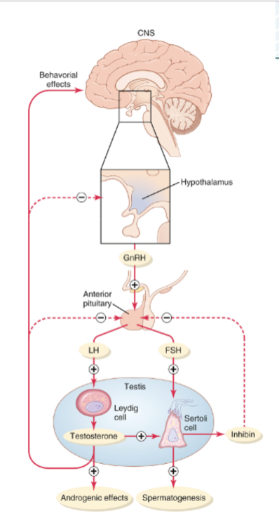
What are the functions of testosterone?
promotes growth of hair, bones, protein formation/muscle, basal metabolic rate
skin health
voice
What are the symptoms of “manopause”?

What are the main functions of the parathyroid gland?
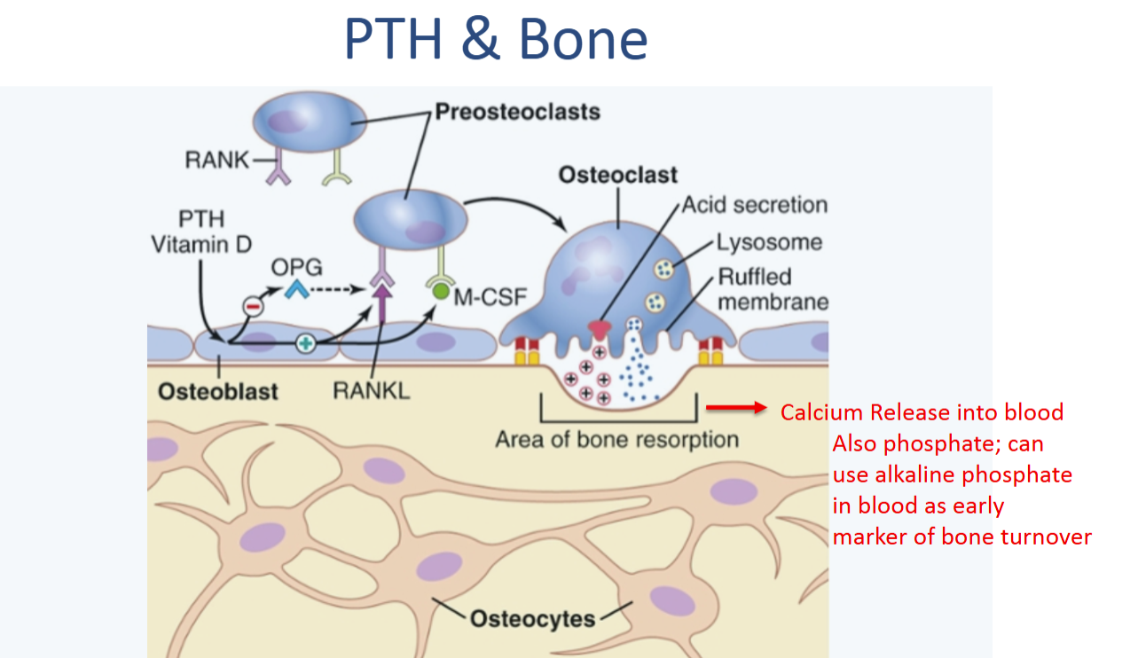
bone resorption (breaking down) and releasing Ca++ into blood
Ca++ reabsorption in the kidneys
Phosphate excretion
Activation of Vitamin D (calcitriol)
What are the characteristics of blood calcium control?
blood calcium is very finely controlled (small amount in blood)
Ca++ needed for:
Muscle contraction, neural transmission, bone development, etc.
Hypercalcemia depresses CNS, hypocalcemia causes tetany
Inverse relationship with inorganic phosphate in blood
Understand this graph for homeostatic regulation of blood calcium


What do high Ca++ blood levels stimulate the parathyroid gland to do?
Ca++ interacts with GPCR → Phospholipase A2 → Arachidonic acid → Leukotrienes
Leukotrienes degrade PTH and inhibit its release

What do low Ca++ blood levels stimulate the parathyroid gland to do?
Blood Ca++ doesn’t interact much with GPCR, so GPCR relaxes
GPCR relaxation allows PTH release
What is a biphasic response (in terms of PTH)?
pre-formed PTH can be released to acute changes in Ca++ in blood
Chronic low Ca++ → increased PTH synthesis (long-term)
What is Phosphate’s relationship with Calcium the in blood?
Positive correlation
Phosphate increases PTH by removing inhibitory effect stopping formation of Arachidonic acid from Phospholipase
In renal disease, elevated Phosphate can → hyperparathyroidism and bone loss
Know this diagram for how PTH releases calcium from the bone
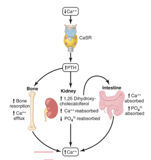
Understand the target organs and effects of PTH

What does PTH do to the kidneys?
Increase Ca++ reabsorption by increasing # of Ca++ channels in membrane to draw more Ca++ back in
Excrete phosphate and reduce intake (reduced gene expression of Na+/PO4 cotransporter)
Activation of enzyme 1-alpha-hydroxylase which activates Vitamin D (calciferol → calcitriol)

How does active Vitamin D (calcitriol) impact blood Ca++?
function: Ca++ reabsorption in gut; deposition and resorption of bone
How:
Calcitriol binds Vitamin D receptors (VDRs) on nuclei of gut cells (enterocytes) → stimulates transcription of Ca++ transport proteins (Calbindin, TRPV6, etc.)

What does PTH do to Vitamin D?
PTH causes conversion of calciferol (inactive Vitamin D) to calcitriol (active Vitamin D) in proximal tubule of kidneys
What is CaSR?
GPCR on parathyroid chief cells, kidney tubules, bone cells, etc.
monitors extracellular Ca++ concentrations
high Ca++ concentration → activates CaSR → fall in expression of hormones/mechanisms that increase Ca++ concentration in blood (ie: PTH, Vitamin D)
True or false: hypoparathyroidism is more common than hyperparathyroidism
False
What are the causes, symptoms, treatments, and complications of hyperparathyroidism?
causes:
adenoma (tumor of parathyroid gland); causes overproduction of PTH
kidney disease
symptoms:
high blood Ca++
bone loss/pain/fracture
increased urination (more Ca++ filtered and reabsorbed)
kidney stones
muscles weakness/twitches
heart palpitations
treatments: surgery to remove adenoma or medication
Complication: “hungry bones”; post-op, bones can overcorrect due to the sudden shift in metabolism → hypocalcemia as bones take in too much Ca++ from blood
What does hypoparathyroidism do to levels of Ca++ and Phosphate in blood?
decreases Ca++, raises Phosphate (less excreted)
What are the alternative names for the pituitary gland, the anterior pituitary gland, and the posterior pituitary gland?
pituitary gland: hypophysis
anterior pituitary gland: adenohypophysis
communicates with hypothalamus via hypothalamic-hypophysial portal vessels
posterior pituitary gland: neurohypophysis
neural tissue extending down from hypothalamus
Describe the basic stimulation pathway for the anterior pituitary gland
hypothalamic stimulating or releasing hormones → median eminence → hypothalamic-hypophysial portal vessels system → anterior pituitary

Name the hypothalamic hormones that stimulate the anterior pituitary gland and their effects
thyrotropin-releasing hormone (TRH) → causes release of thyroid-stimulating hormone (TSH)
corticotropin-releasing hormone (CRH) → causes release of adrenocorticotropic hormone (ACTH)
growth hormone-releasing hormone (GHRH) → causes release of growth hormone (GH) and growth hormone inhibitory hormone (GHIH)/somatostatin
gonadotropin-releasing hormone (GnRH) → causes release of two gonadotropic hormones (FSH, LH)
prolactin-inhibiting hormone (PIH) → causes inhibition of prolactin secretion (lactation hormone)
Stimulation pathways of the hypothalamic hormones
Phospholipase C = PLC
Adenyl Cyclase = AC

What are the functions and effects of growth hormone (GH)?
causes growth in tissues
increases size of cells, mitosis, cell differentiation
Target organs:
Skeleton/bones: increase amino acid uptake, protein and collagen synthesis, chondrocyte (cartilage cell) proliferation, longitudinal growth of bones
Adipose tissue: increases lipolysis → raises free fatty acids in blood, lowers fat storage
Muscle: increases amino acid uptake, protein synthesis → bigger muscles
Liver: increases IGF secretion, IGFBP synthesis, and glucose output → rise in blood sugar
Describe the stimulation pathway from GHRH to GH
GHRH → GPCR → Adenylate cyclase/cAMP → Protein kinase A → GH
True or false: IGF-1 from the liver can inhibit GH; this is an example of negative feedback
True
True or False: IGF-1 is necessary for linear growth
True
Fill in the blanks
Most GHs in circulation are bound to ____________ ______. GH binds to _______ _______ ________ and activate downstream signaling proteins for target-organ-specific effects.
GH-binding proteins
surface cytokine receptors
What are the metabolic effects of GH?
increase rate of protein synthesis
enhances transport of AA through the cell
increased transcription
increased translation/protein synthesis
decreased protein breakdown in cell
enhances fat utilization for energy (makes body more dependent on fat energy by raising adipose tissue insulin resistance)
decreases rate of glucose utilization (protects blood glucose for brain)
GH disorders
dwarfism: too little GH
giantism/acromegaly: too much GH
What hormones are released by the thyroid gland?
Calcitonin (reduce blood Ca++)
T3
Less common than T4, but more potent
T4
More common than T3, but less potent
T4 → T3 in tissues
What makes up the thyroid?
follicles lined with cuboidal epithelial cells that secrete colloid
C cells that secrete calcitonin
What is colloid?
secreted by cuboidal epithelial cells in follicle
rich in thyroglobulin, which has a lot of thyroid hormones
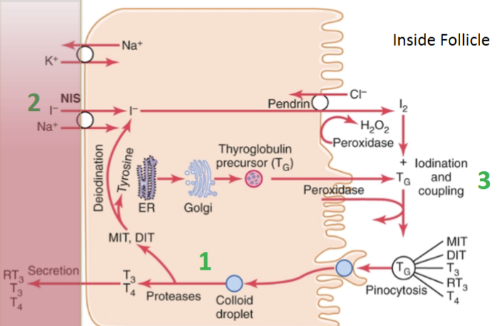
Describe the production of T3 and T4 in a thyroid follicle, beginning with Iodide
iodide is co-transported into cuboidal cell with sodium (Na+); ‘iodine trapping’
Pendrin (counter transport molecule) transports iodide into the follicle
Thyroglobulin secreted by ER and Golgi into follicle
Iodide oxidized to by iodine by thyroid peroxidase (TPO)
Iodide → iodine by TPO lets TPO attach iodine to the tyrosine AAs on thyroglobulin
Iodine-tyrosine AAs coupled → thyroid hormones
Colloid droplet taken into cuboidal cell via endocytosis
Proteases cleave thyroglobulin → T3 and T4
Thyroid hormones stored for months until secreted into blood

How does TSH (from anterior pituitary gland) increase T3 and T4?
increases:
TG cleavage
activity of iodine pump
iodination
number of cells
cell size and secretory activity
stimulates cells via adenylate cyclase/cAMP (2nd messenger system)
Describe T3 and T4 transport and activity
circulate bound to plasma proteins; released slowly and used slowly to and by the target cells
T4 → T3 via iodinase before entering nucleus
T3 acts on thyroid hormone receptor in nucleus
Upregulation of gene expression → mRNA transcription → increases protein synthesis
Revs up body’s functional activity (higher cardiac output, increased glycogenesis, etc.)
What are the effects of thyroid hormone?
bone growth and development
increases cardiac output (CO)
increases lipolysis
cholesterol metabolism in the liver
axon myelination in brain
stimulates GH; inhibits TSH (negative feedback)
True or false: disorders of the thyroid do not involve resistance to TH at the level of the tissues
False
What are hyperthyroidism and hypothyroidism?
hypothyroidism: usually disease of the thyroid gland; low metabolic state (→ weight gain), low hormone levels
thyroid gland can be enlarged (goiter caused by too little iodine)
myxedema: swelling of face/eyes; bags under eyes
atherosclerosis: lack of TH → buildup of cholesterol (bc TH stimulates cholesterol metabolism in liver)
Hashimoto’s: autoimmune; immune system destroys the thyroid gland
treated with TH
hyperthyroidism: excessive thyroid function; increased BMR, cardiac and ANS dysfunction
Grave’s disease (autoimmune): immune system attacks the thyroid gland → too much TH produced → hyperthyroidism in blood
characterized by bulging eyes
What can cause goiter?
hyperthyroidism
hypothyroidism
too little iodine
tumors
autoimmune condition
What hormones are released from the posterior pituitary gland (hypophysis)?
Oxytocin: stimulate smooth muscle contraction
childbirth, lactation
pitocin (synthetic oxytocin) given to induce labor
ADH/AVP: reduce body water loss to maintain plasma volume
reabsorbs water in the kidneys (acts on V2 receptors) via tubules; water moved to capillaries → body fluid maintained
main trigger: osmolality of blood, NOT LOW BLOOD VOLUME but that does still trigger it
hormones are secreted from nerve endings in the hypothalamus via exocytosis into adjacent capillaries
Describe the mechanism of activation, function, and effect of ADH/AVP
Osmoreceptors in hypothalamus sense blood osmolality and water concentration
high osmolality/low water concentration → osmoreceptors shrink → neural reflex in hypothalamus → ADH released
ADH released → kidneys retain water
If osmolality normal but plasma volume low (ie: in cases of hemorrhage) → stretch receptors in heart (aorta and carotid arteries) initiate reflex → ADH released and vasoconstriction is triggered
Understand the ADH stimulation pathway in the collecting duct of the kidneys
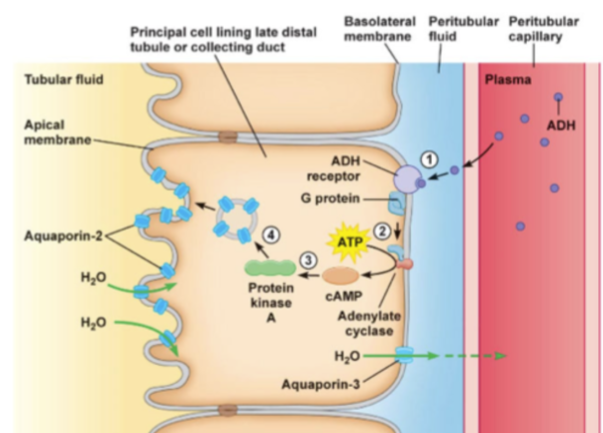
Understand the ADH/AVP stimulation pathway in response to normal osmolality and low plasma volume

Know PLC (phospholipase-C) vs AC (adenylate cyclase)

ADH/AVP disorders
hyper: causes body to hold too much fluid
can happen in heart failure or nephrotic syndrome
hypo: causes excess excretion
diabetes insipidus: caused by ADH/AVP deficiency instead of insulin
neurogenic: most common; AVP can’t be released due to trauma, inflammation, cancer, etc.
nephrotic/renal: mutations in receptors or aquaporin-2 channels (AQP2) → water can’t be reabsorbed