Abdomen and GIT imaging
 Call Kai
 Learn
Learn Practice Test
 Spaced Repetition
 Match
Match Flashcards
 Knowt Play
1/31

There's no tags or description
Looks like no tags are added yet.
Name | Mastery | Learn | Test | Matching | Spaced | Call with Kai | Chat |
|---|
No analytics yet
Send a link to your students to track their progress
32 Terms
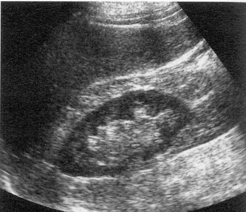
What organs are show in this ultrasound?
Liver and kidney
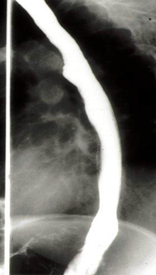
Esophageal compression
*smooth-no problem
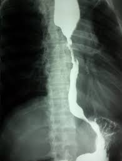
Diagnosis
Ca esophagus -cancer
*shouldering
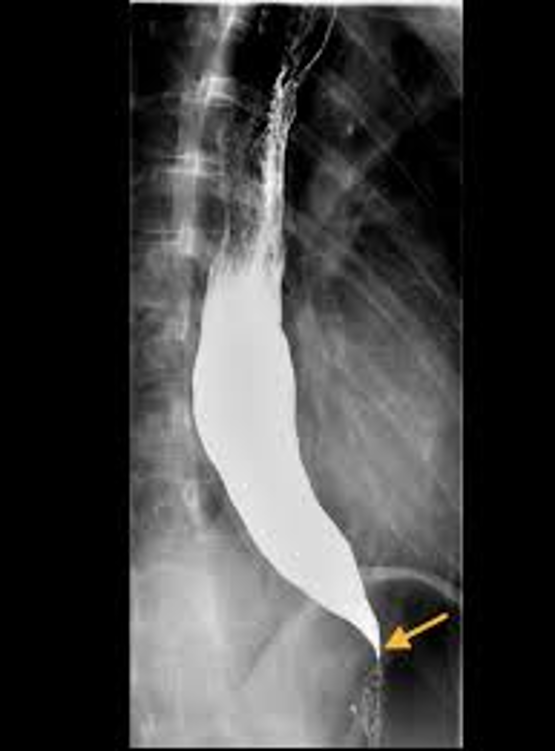
diagnosis + sign
Achalasia of the cardia
Rat tail / Bird peak
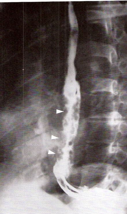
diagnosis
Varices
*think varicose veins, but in esophagus
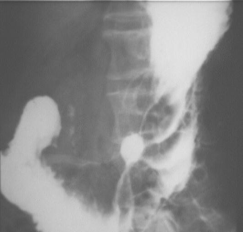
Ulcer-malignant
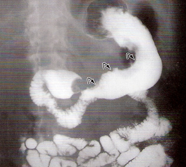
Gastric wall thickening (with irregular outlines indicated gastric mass)
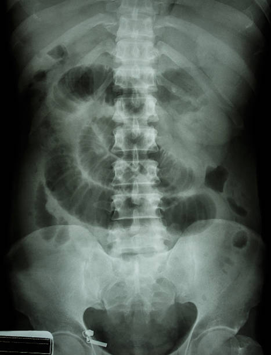
Small bowel obstruction
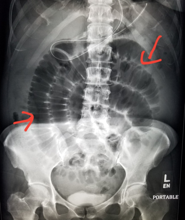
Small bowel obstruction
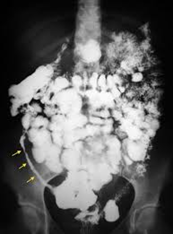
multiple strictures with long segment of stricture at terminal ileum in Crhons disease
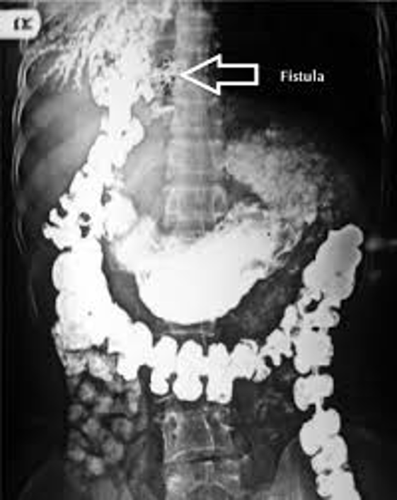
fistula formation and reflux of contrast to biliary system
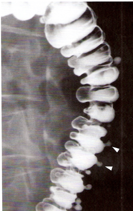
Diverticulosis (multiple contrast filled small sacs)
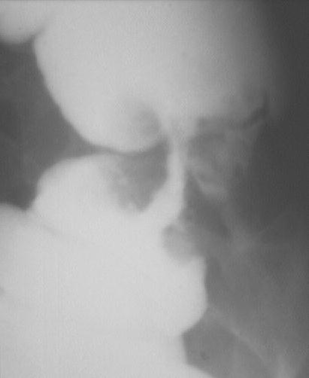
Ca colon
apple core
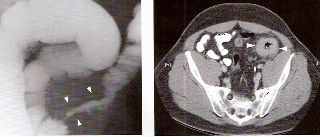
Circumferential wall thickening of colon in CT and long stricture on contrast enema
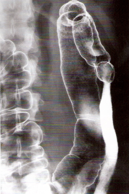
Ulcerative colitis
*lead pipe appearance due to loss of haustral pattern
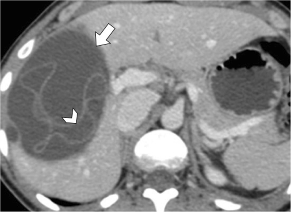
Hydatid cyst
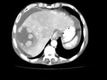
Diagnosis + what phase?
Hepatic hemangioma
Arterial phase
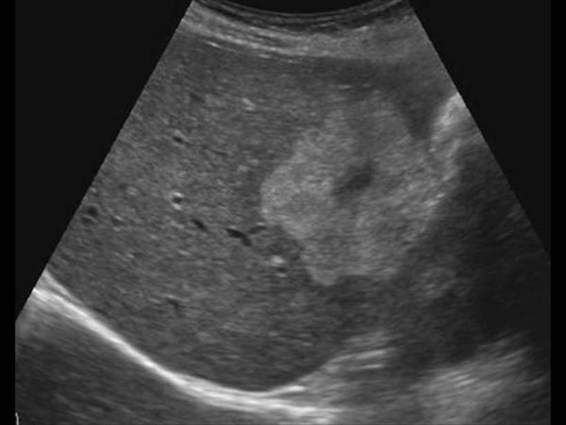
Echogenic focal lesion mostly hemangioma
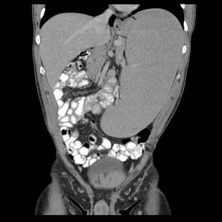
Splenomegaly
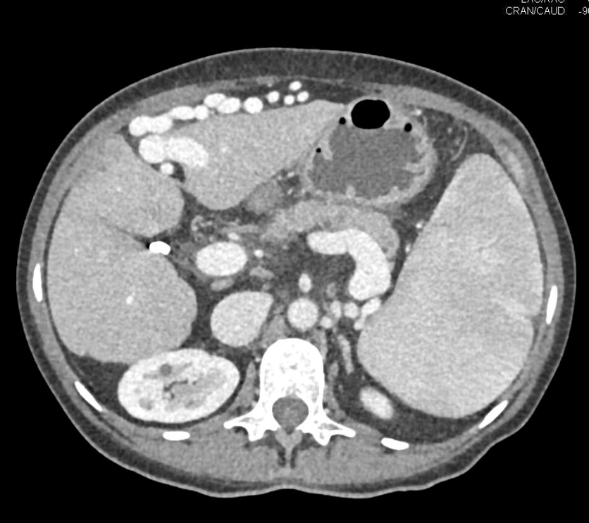
Portal hypertension (bulky spleen, dilated portal, splenic veins and collaterals)
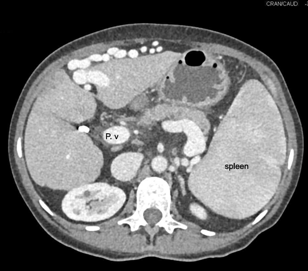
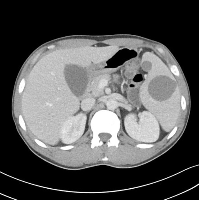
what is in the spleen?
Cyst
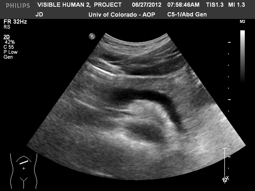
Pancreas
The pancreas is best seen through what window?
Fluid filled window, like stomach
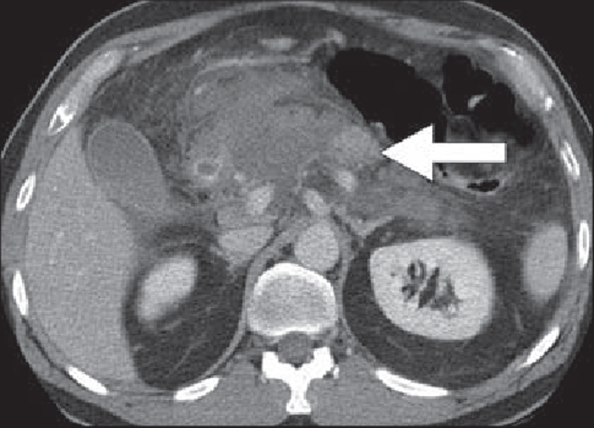
Pancreatitis
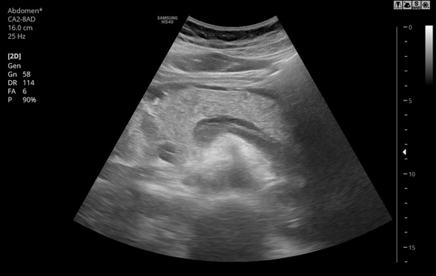
Pancreatitis
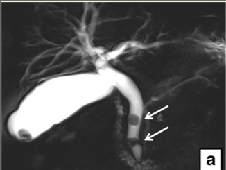
GB and CBD stones
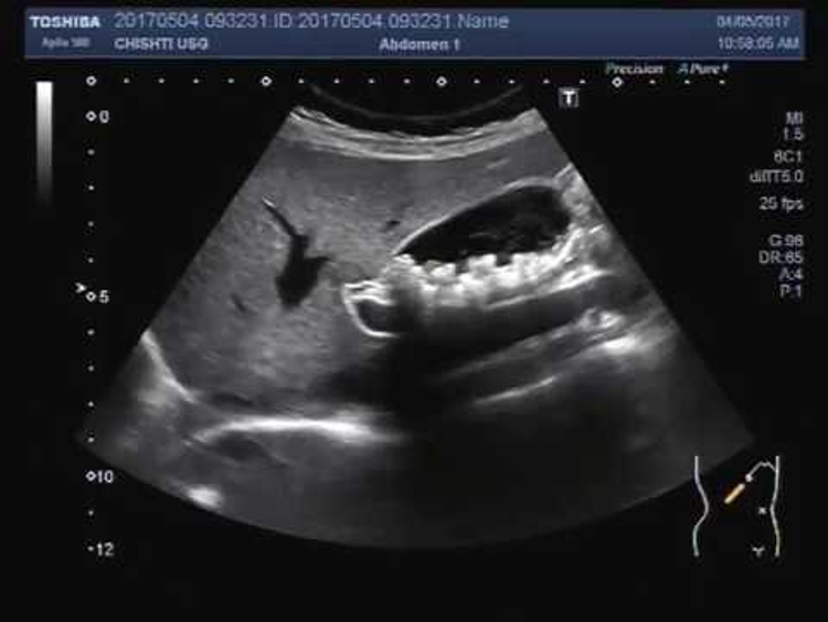
Multiple GB stones
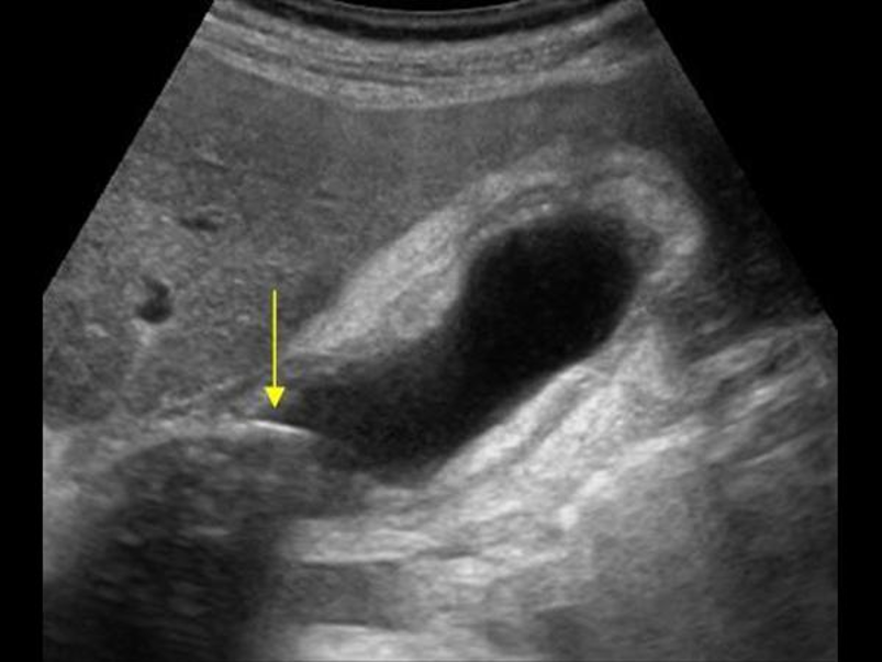
A calcular cholecystitis
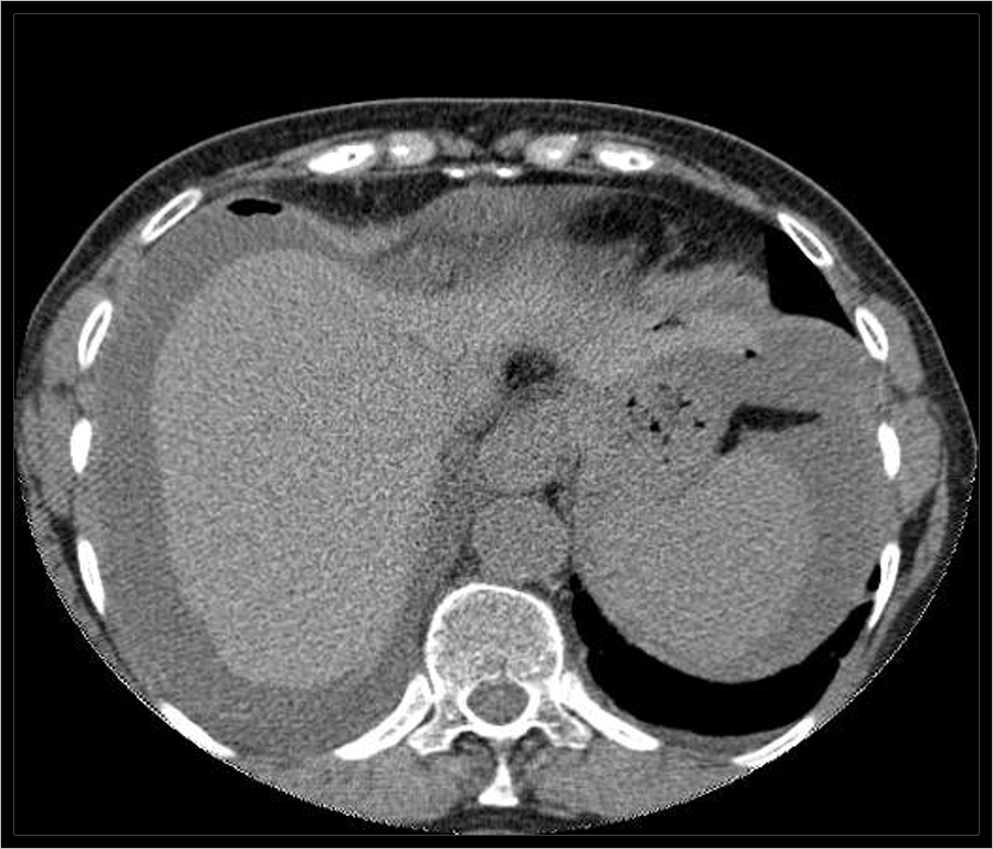
Peritoneum (ascites with small focus of free air)
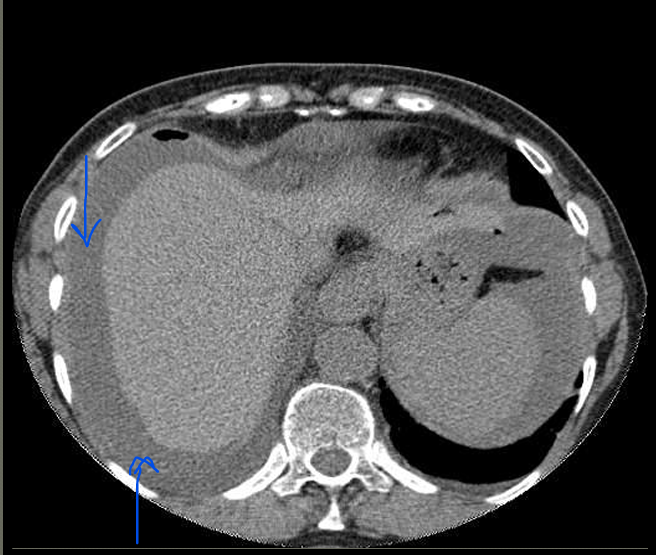
Peritoneum (ascites with small focus of free air)
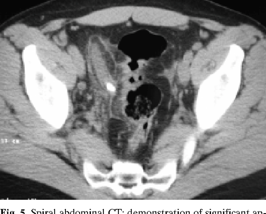
Fecaleith
stone within the appendix