3 Structure of Biomolecules
 Call Kai
 Learn
Learn Practice Test
 Spaced Repetition
 Match
Match Flashcards
 Knowt Play
1/63

There's no tags or description
Looks like no tags are added yet.
Name | Mastery | Learn | Test | Matching | Spaced | Call with Kai | Chat |
|---|
No analytics yet
Send a link to your students to track their progress
64 Terms
Amino acid parts
an alpha carbon with an amino group, COO- (carboxylic acid), hydrogen atom, and an R group.
proteins only contain (isomer)
L isomers
pH vs pKa amino acids
if the pH is above the pKa, then the protein is most likely to be in its deprotonated form. if the pH is below the regular pKa, then the protein is more in the protonated form. The higher usual pKas are due to R groups.
protein primary structure
amino acids linked by peptide bonds
a residue
a single amino acid unit in a polypeptide
peptide bond formation involved
the loss of a watermolecule through dehydration synthesis and the a-carboxy group of one amino acid links to the a-amino group of another amino acid
C and N terminus
C terminus is carboxyl N terminus is amino end
oligopeptides
polypeptide chains made of small numbers of amino acids
1 dalton =
1amu (for protein molecular mass)
fatty acids
part of lipids, hydrophobic, long hydrocarbon chains that end with carboxylic acid groups
Fatty acid names
derived from the parent hydrocarbon with oic at end. like decanoic acid (10:1). The first number is the number of carbon atoms total, second is the number of double bonds.
Naming fatty acid carbon atoms (2 ways)
omega (ω) carbon way
The methyl carbon end, that carbon is called the omega ω carbon, also carbon 1.
Number down from there, with the double bond being the ω-first carbon of the double bond double bond. For naming, it is omega-first carbon of double bond.
cis/trans way
the carbon of the carboxylic acid is the carbon 1.
number down from there, with carbon 2 alpha and 3 beta (organic chem numbering system). Then for naming, denote cis or trans, with a delta Δ symbol and the first carbon from the double bond as a superscript.

number of carbons in fatty acids
fatty acid chains contain an even number of carbon atoms between 14 and 24 - 16 and 18 are the most common.
Three common types of membrane lipids in biological membranes
phospholipids, glycolipids, and cholesterol
phospholipid components
one or more fatty acid tails (hydrophobic), glycerol (platform fatty acids are attatched), phosphate group (charged group), and an alcohol attatched to the phosphate (hydrophilic side, gives up H ion easily).
phosphoglycerides vs sphingophospholipids
both are a type of phospholipid, phosphoglycerides have a glycerol backbone with sphingophospholipids have a sphingosine backbone.
Common phosphoglyceride found in membranes
phosphatidylcholine
Common sphingophospholipid found in membranes
sphingomyelin, found extensively in myelin sheaths around neural axons.
Cholesterol structure
Big, blocky structures. a steroid built from 4 linked hydrocarbon rings. Contains a linked hydrocarbon tail at one end, -OH group at other end.
cholesterol interactions and orientation
-OH group interacts with phospholipid head groups, oriented parallel to fatty acid chains of phospholipids in membranes.
amphipathic
a molecule that has both a hydrophobic and hydrophilic region
moiety
part of portion
phospholipid moietys
head hydrophilic, tail hydrophobic
micelle
a globular structure with the polar head groups on the outside surface and hydrocarbon tails inside. (phospholipid bilayer is NOT a micelle). Usually single tailed salts of fatty acids do.
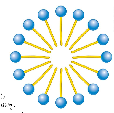
How are lipid bilyers formed
Spontaneously, stabilized by non covalent interactions.
lipid bilayer - biological consequences of hydrophobic interactons
Bilayers ted to close on themselves so there are no edges with exposed hydrocarbon chains (forms compartments), self-sealing. A hole is very unfavorable.
What passes through lipid bilayer
small molecules that are nonpolar pass well, and water.
Permeability coefficients for lipid bilayer
Denoted by P. Water is around 10^-2. going down to smaller P is Na+ (charged ions that are hard to go across), glucose is around the middle.
Most membrane processes are carried out by
Proteins. they transport molecules and information across.
integral membrane proteins, and how to remove/extract
Stuck inside the lipid bilayer. Interact extensively with the hydrocarbon chains of membrane lipids. They have a hydrophobic section for this (and hydrophilic section!) They are released by agents that compete for nonpolar interactioons. Difficult to extract because deeply embedded, chemicals needed.

Peripheral membrane proteins, and how to remove/extract
Bound to membranes mainly, sit on the surfaces and don’t penetrate deep into core. Primarily bound through electrostatic and hydrogen bond interactions with the head groups of lipids. Hydrophilic. Disrupted by using salts of pH, or shaking loose. Easier to remove than integral.

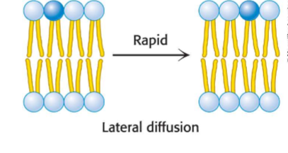
lateral diffusion
a process by which lipids and membrane proteins are constantly in lateral motion (moving side to side throughout a membrane).
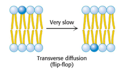
transverse diffusion
movement of lipids and membrane proteins across to other end of membrane (flip-flop). Much more difficult to do. Slow process compared to lateral diffusion.
FRAP
way to visuzalize lateral diffusion. Fluorescence recovery after photobleaching. you bleach an area, and see how long it takes for the bleached spot to recover, showing the amount of lateral diffusion occuring. The recovery is the increase in fluoresence after photobleaching.

Tm (melting temperature) of a fluid membrane.
The transition from rigid to fluid state takes place above the Tm. Depends on the degree of unsaturation, saturated fatty acids favor rigid state. unsaturated (with a double bond) interfered with highly ordered packing. Think about it, because of the kink in the bond, they have more room to wiggle around and move, increasing fluidity, however it causes weak intermolecular attraction and inability to pack tightly. Unsaturated bonds lower the Tm necessary to be fluidlike.

Cholesterol and fatty acid tight packing
Disrupts this because it is a 4-ring bulky steroid. Disrupts the regular interactions between the chains, helps maintain proper fluidity in animal membranes.
current plasma membrane model
fluid mosiac model
lipid rafts
membrane domains having reduced fluidity. cholesterol forming complexes with lipids that contain the sphingosine backbone.
monosaccharide and structure.
the basic building block for carbs. empirical formula (CH2O)n. contain an aldehyde or ketone with 2+ hydroxyl group.
Epimers
sugars that are diasteromers differing in configuration only at a single asymmetric center.
most monosaccharides exist as
interchanging cyclic forms
identify pyran and furan

anomer
a diasteriomeric form of sugars that forms when a cyclic hemiacetal is formed (ring created) and an additional symmetric center is created.
d fructose
rapidly interchanges between four distinct ring structures. pyranose form predominates due to reduced steric hinderance.
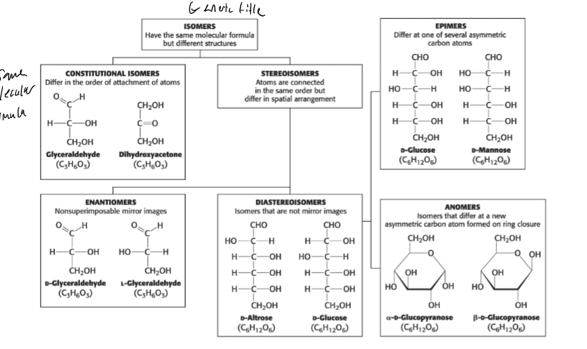
ISOMER TYPES (6)
See chart. know this.
pyranose and furanose ring conformations
chair or boat.
Beta - D - glucose conformation
the chair form predominates because all axial positions are occupied by H, the boat form is disfavored because it is sterically hindered.
glycosidic linkage
monosaccharides are joined to alcohols and amines. O and N type.
O-linkage: covalent linkage between the oxygen atom of alcohol
N-linkage: covalent linkage formed between the nitrogen atom of an amine.
oligosaccharides
sugars that contain two or more monosaccharides linked by o-glycosidic bonds.
sucrose
common disaccharide. glucose + fructose. table sugar.
lactose
milk sugar, galactose +glucose.
maltose
disaccharide resulting from the hydrolysis of large oligosaccharides that consists of two linked glucose.
glycogen
most common polymer in animal cells, storage form of glucose. hydrolyzed by alpha amylase.
starch
polymer for nutrition in plants. amylose and amylopectin. amylose is unbranched with no 1-6 linkage, amylopectin is branched.
cellulose
main structural polysaccharide of plants. B configuration of linkage that allows cellulose to form long straight chains for structure. Interact together with hydrogen bonds.
insoluble fiber in diet
while we cant digest it, they increase the rate digestion products pass through the large intestine. softens stools.
Soluble fiber in diet
slows the movements of food through the digestive tract (harder to break down).
RNA vs DNA
ribose is the sugar in RNA, deoxyribose is the sugar in DNA. Ribose has an extra 2’ OH group that increases ability to be hydrolyzed.

backbone of DNA and RNA
sugars linked by phosphodiester bridges with a negative charge.
Purines and Pyrimidines (and attatchment)
The nitrogenous bases of DNA and RNA. Attatched to the 1’ carbon atom of sugar. Purines are adenineand guanine with two rings. pyrimidies are thymine and cytosine with one ring. RNA has uracil instead of thymine.
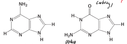
identify
adenine on left, guanine on right. Guanine has an extra carbonyl.

identify
Cytosine, uracil, and thymine left to right. Pyrimidines have one ring. cytosine only has one carbonyl, uracil doesn’t have an extra methyl.
nucleoside
the unit consisting of a base bonded to a sugar (no phosphate group).
nucleoside triphosphates
has three phosphate groups. high energy bonds are between the phosphate groups that can provide energy.