N230 Exam #3 Study Guide
 Call Kai
 Learn
Learn Practice Test
 Spaced Repetition
 Match
Match Flashcards
 Knowt Play
1/23

There's no tags or description
Looks like no tags are added yet.
Name | Mastery | Learn | Test | Matching | Spaced | Call with Kai | Chat |
|---|
No analytics yet
Send a link to your students to track their progress
24 Terms
Postpartum Infection (also known as Puerperal Infection)
any clinical infection of the genital tract that occurs within 28 days after miscarriage, induced abortion, or birth
defined as presence of a fever of 100.4 degrees fahrenheit (38 degrees celsius) in the first 10 postpartum days (not including the first 24 hours after birth)
Endometritis (Endomyometritis) → Postpartum Infection
most common postpartum infection
infection of the lining of the uterus which causes fundal tenderness and fever
caused by prolonged rupture of membranes or manual introduction of bacteria
Not usually cultured, just treated
managed with IV broad spectrum antibiotic therapy like gentamicin and clindamycin → treat for 24 hours afebrile
Wound Infections → Postpartum Infection
develop after mothers are discharged home
rates of wound infection after cesarean birth are 3-5%
cellulitis/abscess of wound, laceration
Urinary Tract Infections → Postpartum Infection
dysuria, burning, frequency, suprapubic pain
get urinalysis
foley, straight cath
occur in 2-4% of postpartum women
Mastitis → Postpartum Infection
usually staph aureus which is treated with antibiotics such as dicloxacillin or cephalexin
causes fever, flu-like symptoms
Postpartum Hemorrhage Definition
cumulative blood loss of greater than or equal to 1,000 mL
bleeding associated with signs/symptoms of hypovolemia within 24 hours of birth
leading cause of maternal morbidity and mortality
often unrecognized until mother has profound symptoms → early, acute or primary PPH occurs within 24 hours of birth
late or secondary PPH occurs more than 24 hours but less than 6 weeks after the birth
PPH Signs and Symptoms
lightheadedness
pallor
diaphoresis
altered mental status
tachycardia
hypotension
restlessness
tachypnea
Causes of PPH
Tone: uterine atony (marked hypotonia of uterus) is most common
blood supply to the uterus to supply the placenta is cut off by myometrium contractions
risks: high parity, polyhydramnios (excessive amniotic fluid surrounding the fetus), macrosomia (large baby), uterine fatigue due to long labor, tocolytic drugs (used to stop or slow contractions)
Trauma: lacerations of the genital tract (uterus, cervix, vagina, perineum) and hematomas
Tissue: retained products of the placenta or a placental anomaly
Thrombin: when there is no identifiable cause, think coagulopathy.
TXA is first line therapy
Von Willebrand is the most common inherited bledding disorder due to a missing clotting factor.
idiopathic or immune thrombocytopenic purpura (ITP)
Subinvolution of the Uterus: causes late postpartum bleeding with signs and symptoms of prolonged lochial discharge, irregular or excessive bleeding, and sometimes hemorrhage
Inversion of the Uterus: potentially life threatening but rare; occurs in 1 in 2000 to 1 in 25000 births.
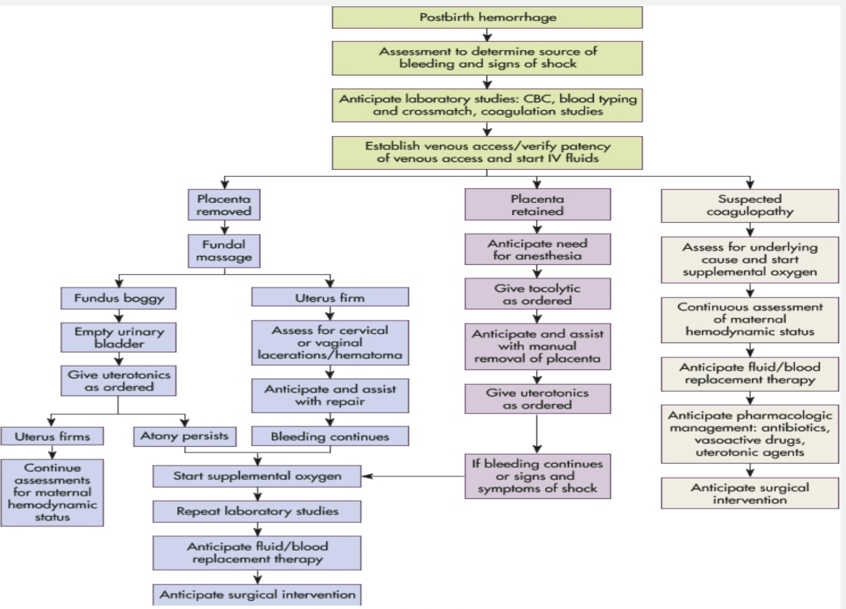
Management of PPH
uterine massage → call code and get help → hemorrhage cart → administer IV fluids and uterotonic medications (pitocin, methergine, hemabate, misoprostol) and TXA (antifibrinolytic) → empty bladder → physical pressure (jada) → surgery
Breastfeeding Positions
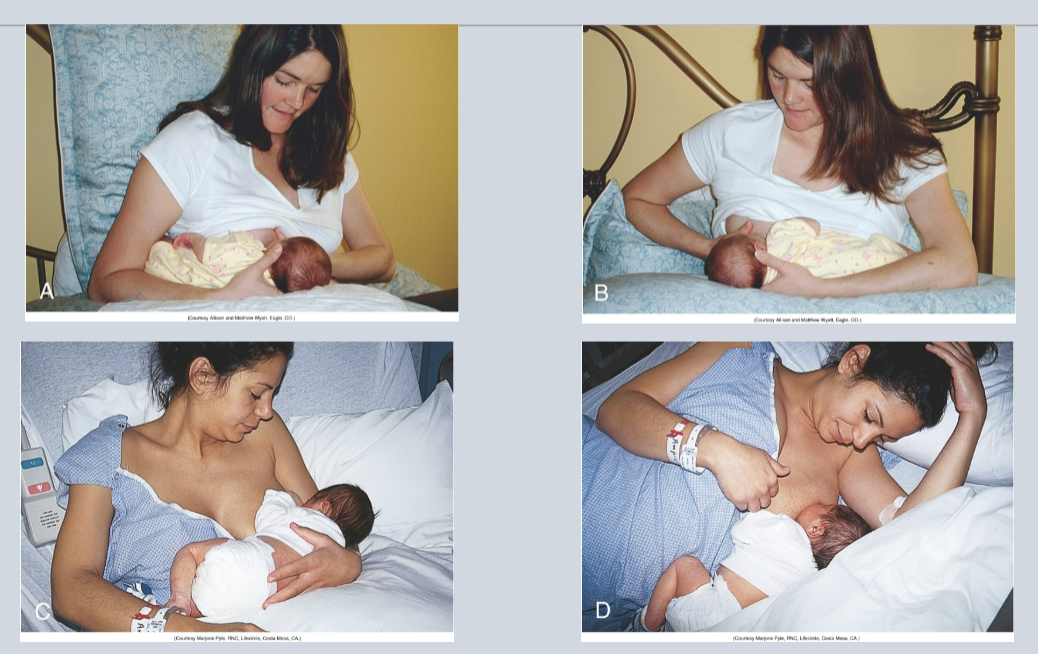
football or clutch hold (under the arm) → recommended for early feedings and is particularly helpful for mothers who gave birth by cesarean section and late preterm and early term infants to avoid flexing the head, which can impede breathing and cause positional apnea
across the lap (cross-cradle or modified cradle)
cradle: the most common breastfeeding position for infants who have learned to latch easily and feed effectively
side-lying → allows the mother to rest while breastfeeding and is recommended for women with perineal pain and swelling
semireclining position: mother is more relaxed, nipple pain is reduced or eliminated, more freedom of movement to use hands, baby can use inborn reflexes to latch and feed effectively
Engorgement
condition where the breasts become overfilled with milk, causing them to feel hard, warm, enlarged, shiny, and painful
normal engorgement is not unusual in the first 24-48 hours after the breasts fill with milk
occurs around 3 days after birth and with appropriate treatment resolves within 24 hours
all postpartum women are at risk, not just those who are breastfeeding
management for all women: icepacks and cabbage leaves, well-fitted support bra, anti-inflammatory medications
management for breastfeeding women: feed frequently, hand expression or breast pump to reduce engorgement and promote comfort
management for formula feeding women: do NOT express breast milk as it can stimulate milk production and worsen engorgement
Newborn Jaundice
jaundice in newborns differs from adults because newborns produce bilirubin 2-2.5 times that of an adult. it occurs when bilirubin levels exceed 5mg/dL and is observed in 60% of term infants and 80% of preterm infants in the first week after birth
Physiologic Jaundice
the most common type and is not associated with any pathologic process
occurs due to the newborn’s immature liver function
appears after 24 hours of age and peaks between 2-4 days and gradually decreases between days 5-7
Breastfeeding Related-Jaundice
Early Onset
begins at 2-4 days of age
related to poor latch or inadequate milk supply resulting in decreased caloric and fluid intake
Increased enterohepatic circulation from delayed meconium passage
Late Onset
occurs in up to 30% of breastfed infants
bilirubin continues to rise while formula-fed infants’ levels around day 5
peaks between 5-15 and may persist beyond 1 month, lasting up to 3 months
safe condition - breastfeeding should NOT be discontinued
Pathologic Jaundice
warning signs requiring investigation
jaundice appearing within 24 hours after birth
total serum bilirubin >15-17mg/dL in newborns
TSB increasing >5mg/dL during the newborn period
risk factors include preeclampsia, maternal diabetes, ABO/Rh incompatibility, prematurity, delayed meconium passage, G6PD deficiency, infections, and birth trauma
Jaundice Treatment
phototherapy (UVB light)
absorbed through skin, causes photochemical reaction with bilirubin facilitating dissolution of it of excretion
overhead lights, blanket
Fetal Loss Communication
Communicating With Bereaved Parents After Fetal Loss
Effective communication with families experiencing perinatal loss requires intentional presence, active listening, and avoiding common pitfalls.
Core Principles
Validate and Listen:
Acknowledge the loss with a simple but sincere "I am sorry"
Encourage parents to tell their stories and listen with care
Women who had a stillbirth or whose baby died may have a particularly strong need to share details about the birth
Ask the father or partner directly about their views and feelings, as nurses often focus primarily on the mother
Therapeutic Presence:
Have a presence of self and willingness to be alongside, quietly supporting the bereaved person
Lean forward, nod, and use phrases like "Tell me more" to encourage sharing
Sitting through silence can be therapeutic - it gives parents time to collect thoughts and process emotions
What to Avoid
Resist unhelpful responses:
Don't give advice or use clichés
Don't try to reduce their pain with quick fixes - the initial impulse to say or do something to stop crying can actually stifle further expression of emotion
Be cautious with touch:
Assess before using touch as a therapeutic technique
For some it's meaningful; for others it's an invasion of privacy or contrary to cultural/religious beliefs
Practical Considerations
Time allocation:
Providing adequate care for grieving families is time-consuming
Nurses may need adjusted shift assignments to give adequate attention without being rushed
Empowering decisions:
Parents should make decisions about when and by whom nursery items are stored or given away
Well-meaning relatives who rush to dismantle the nursery without involving parents can compound their grief
Address guilt:
Women are particularly vulnerable to feelings of guilt and may wonder what they did to cause the loss
Neonatal Heat Loss
Newborns, especially preterm and low birth weight (LBW) infants, are highly vulnerable to heat loss due to several physiological factors.
Why Newborns Lose Heat
Preterm infants face additional challenges:
Smaller muscle mass and fewer brown fat deposits for heat production
Lack of insulating subcutaneous fat
Poor reflex control of skin capillaries
Thin skin in very low birth weight (VLBW) and extremely low birth weight (ELBW) infants
In VLBW and ELBW infants, heat loss from radiation, evaporation, and transepidermal water loss is three to five times greater than in larger infants.
Four Mechanisms of Heat Loss
1. Radiation:
Heat loss to cooler objects not in direct contact with the infant
Example: Crib placed near a cold window or air conditioning unit cools the crib walls, which then cools the baby's body
Prevention: Place cribs away from exterior walls, windows, and ventilating units
2. Conduction:
Heat loss through direct contact with a cooler solid object
Prevention: Place infant on padded, covered surfaces; use clothes and blankets for insulation
3. Convection:
Heat loss aided by surrounding air currents
Example: Placing infant in direct airflow from a fan or air conditioning vent
Prevention: Transport in cribs with solid sides to reduce airflow
4. Evaporation:
Heat loss through moisture evaporating from skin
Particularly significant in preterm infants with increased transepidermal water loss 2
Consequences of Cold Stress
For every 1°C temperature decrease from normal, mortality risk increases by 28%.
Cold stress produces 2:
Hypoxia
Pulmonary vasoconstriction
Metabolic acidosis
Hypoglycemia
Prevention Strategies
Skin-to-skin contact with parent immediately after birth if medically stable
Prewarmed incubator
Head coverings (fabric-insulated or wool caps)
Heated gel mattress with radiant heat in delivery room
Plastic wrap or heated humidified gas in delivery room
APGAR
Neonatal Head to Toe Assessment Findings
General Appearance
Posture: Term newborns exhibit complete flexion - head flexed with chin on chest, arms flexed with hands clenched, legs flexed at knees and hips with thighs on abdomen, and feet dorsiflexed. The vertebral column is also flexed.
Behavior: Assess alertness, drowsiness, and irritability, which may reflect neurologic problems.
Neurologic System
Reflexes to assess:
Grasp reflex: Touching palms or soles near digit base causes flexion/grasping
Babinski reflex: Stroking outer sole upward causes big toe dorsiflex and other toes hyperextend
Mass reflexes should be tested at exam end as they may disturb the infant
Document: Absence, asymmetry, persistence, or weakness of reflexes.
Extremities
Digits:
Count fingers and toes
Note polydactyly (extra digits) or syndactyly (fused digits)
Partial syndactyly between 2nd and 3rd toes is a common normal variation
Nails:
Should be pink (slight blueness in acrocyanosis is normal)
Yellowing may indicate intrauterine distress, postterm birth, or hemolytic disease
Short/absent nails: preterm; Long nails: postterm
Palmar creases:
Should have usual creases
Transverse palmar crease (simian crease) suggests Down syndrome but may be normal
Sole creases:
Full-term: creases cover entire sole
Postterm: deep creases
Preterm: partial or absent creases
Soles are flat with prominent fat pads
Range of motion:
Should demonstrate full ROM in elbow, hip, shoulder, and knee joints
Movements should be symmetric, smooth, and unrestricted
Absence of arm movement signals potential birth injury (Klumpke or Erb-Duchenne palsy)
Asymmetric Moro reflex warrants upper extremity evaluation
Muscle tone:
Extension of flexed extremity meets resistance and returns to flexed position when released
Hypotonia suggests hypoxia, neurologic abnormalities, or Down syndrome
Asymmetry may indicate paralysis from CNS damage
Failure to move lower limbs suggests spinal cord lesion
Neonatal Hypoglycemia
Hypoglycemia is a blood glucose concentration inadequate to support neurologic, organ, and tissue function . It's particularly common in infants of diabetic mothers (IDMs), especially those who are macrosomic or large-for-gestational-age.
Diagnostic Thresholds
Symptomatic infants - treat when glucose is <40 mg/dL with a goal to maintain prefeeding levels ≥45 mg/dL.
Asymptomatic infants - treat when glucose is:
<25 mg/dL if <4 hours of life
<35 mg/dL between 4-24 hours of life
General guidelines: Hypoglycemia is defined as glucose <40 mg/dL in the first 4 hours and <45 mg/dL once 4-24 hours of life.
Pathophysiology in IDMs
In utero: High maternal glucose crosses the placenta, stimulating fetal pancreatic islet cell hypertrophy and hyperplasia, leading to hyperinsulinism. This causes excessive fetal growth and fat deposition (macrosomia).
At birth: Umbilical cord clamping abruptly removes the maternal glucose supply. The infant's continued high insulin production rapidly depletes circulating glucose, creating hypoglycemia within 0.5-4 hours (most commonly 1-6 hours) after birth.
High insulin levels also inhibit gluconeogenesis and glycogenolysis, further contributing to hypoglycemia.
Clinical Presentation
Signs include:
Jitteriness
Apnea
Tachypnea
Hypotonia
Decreased activity
Cyanosis
Seizures (in severe cases)
Many infants remain asymptomatic. Asymptomatic hypoglycemia is concerning and associated with increased risk of poor executive function and visual motor function.
Management
Feeding:
Begin breast milk or formula within the first hour if cardiorespiratory status is stable
Avoid oral glucose - it can trigger massive insulin release causing rebound hypoglycemia
IV therapy:
10% dextrose and water (IV) for IDMs with poorly controlled maternal diabetes
Goal: maintain serum glucose >45 mg/dL
Monitoring:
Frequent blood glucose checks for first 2-4 days
Use heel-stick glucometers for screening, confirm with laboratory testing
Neonatal Infections
Sepsis (septicemia) is a generalized bacterial infection in the bloodstream. Neonates are highly susceptible due to diminished immune function - impaired phagocytosis, delayed chemotactic response, minimal/absent IgA and IgM, and decreased complement levels.
Key challenge: Infants show no local inflammatory reaction at the infection portal, and symptoms are vague and nonspecific, delaying diagnosis and treatment. Therefore, maintain high suspicion for infection and begin antibiotics promptly when concerned.
Early-Onset Sepsis (EOS)
Timing: Less than 3 days after birth
Acquisition: Perinatal period through:
Direct contact with maternal GI/GU tract organisms
Aspiration of infected amniotic fluid
Transplacental transfer from maternal bloodstream
Common pathogens:
Term infants: Group B streptococcus (GBS) - most common
Preterm infants: Escherichia coli
Others: Viridans streptococci, Listeria monocytogenes, enteroviruses, coagulase-negative Staphylococcus
Vaginal pathogens: Syphilis, gonococci, Candida albicans, HSV type II, Chlamydia
Risk factors:
Prolonged rupture of membranes
Preterm labor (<37 weeks)
Maternal intrauterine infection
Maternal GBS colonization
Current rates: Despite maternal screening/prophylaxis, early-onset GBS infection remains at approximately 0.25 per 1000 live births.
Late-Onset Sepsis (LOS)
Timing: After 72 hours up to 30 days of life
Sources: Maternally derived or health care-acquired infection
Common pathogens: Coagulase-negative staphylococci, Klebsiella, Enterobacter, E. coli, Candida species
Risk factors:
Prematurity (most significant)
Invasive procedures (intubation, catheterization, surgery)
Infection entry through umbilical stump, skin, mucous membranes, or internal systems
Treatment
Empiric antibiotic therapy initiated before lab confirmation:
Early-onset sepsis: Ampicillin + aminoglycoside
Late-onset sepsis: Vancomycin + aminoglycoside
Narrow therapy to confirmed pathogen ASAP due to antibiotic-resistance concerns
Supportive care:
Oxygen administration (if respiratory distress/hypoxia)
Fluid regulation and electrolyte/acid-base correction
Temporary NPO status (if septic ileus/NEC concern)
Blood product transfusions (RBCs, platelets, FFP, cryoprecipitate)
Vital sign monitoring and thermal regulation
Neonatal Skin Findings
Normal Skin Findings
Color changes:
At birth: Skin may appear grayish-white with vernix caseosa (cheeselike mixture of sebum and desquamating cells) absorbed within 24-28 hours
By day 2-3: Skin turns to natural tone, becomes drier and flakier
Lanugo: Fine, downy hair on forehead, cheeks, shoulders, and back
Benign Birthmarks
Slate grey nevi (previously called Mongolian spots/congenital dermal melanocytosis) and telangiectatic nevi require no therapy, only parental reassurance
Café-au-lait spots: Multiple light brown spots may indicate:
Neurofibromatosis (autosomal dominant)
McCune-Albright syndrome
Warrants further examination of child and family members
Erythema Toxicum Neonatorum
Description: Benign, self-limiting rash also called "flea-bite dermatitis" or "newborn rash"
Timing: Appears within first 2 days (up to day 10)
Appearance:
Firm, 1-3 mm pale yellow or white papules/pustules on erythematous base
Resembles flea bites
May progress from discrete macular erythematous rash to papules/vesicles/pustules
Distribution: Face, proximal extremities, trunk, buttocks - anywhere except palms and soles (no pilosebaceous follicles there)
Characteristics:
More obvious during crying
No systemic manifestations
Heals without pigmentation changes
Lasts 5-7 days (resolves within weeks)
Diagnosis: Pustule smear shows numerous eosinophils, relative absence of neutrophils
Management:
No treatment necessary
Reassure parents of benign, transient nature
Avoid topical treatments - may exacerbate condition
Birth Trauma-Related Findings
Soft tissue injuries from delivery:
Erythema, ecchymoses, petechiae
Abrasions, lacerations, edema
Localized discoloration over presenting/dependent parts
Face presentation: bruising over face
Breech presentation: bruising/swelling over buttocks and genitalia
Tight nuchal cord: ecchymosis and petechiae over head/upper trunk/face
Benign if: Petechiae disappear within 2 days with no new lesions
Forceps injury: Linear configuration across both sides of face
Newborn Vital Signs
Heart Rate
Normal range: 120-160 beats/min
Variations:
During deep sleep: 80-100 beats/min
During crying: Up to 180 beats/min or higher
At birth: Rates close to 200 beats/min may occur in neonates, decreasing rapidly to ~120 beats/min within hours
Assessment:
Point of maximal impulse (PMI): Fourth to fifth intercostal space, medial to left midclavicular line
Often visible and easily palpable due to thin chest wall
Reevaluate if heart rate is >160 or <100 beats/min within 30 minutes to 1 hour
Abnormal findings:
Sustained tachycardia: May indicate infection or cardiac rhythm disturbance (e.g., paroxysmal atrial contractions)
Irregular heart rate/sinus dysrhythmia: Common in first few hours but may need evaluation thereafter
Respiratory Rate
Normal range: 40-60 breaths/min
Rate up to 80 breaths/min may be noted
Assessment: Count for 1 full minute
Variations:
Infants delivered by cesarean section may have more rapid respiratory rate than vaginal deliveries
Blood Pressure
Normal ranges:
Systolic: 60-96 mm Hg
Diastolic: 30-62 mm Hg
Alternative reference:
Day 1: Average 65/45 mm Hg
Day 4: Average 70/46 mm Hg
Assessment method:
Electronic sphygmomanometer with Doppler or oscillometric technique (e.g., Dinamap)
Cuff width-to-arm ratio: 0.45-0.70 (approximately half to three-quarters)
Best measured when infant is quiet or sleeping
Compare upper and lower extremities - should be equal
Abnormal findings:
Sustained hypertension: May indicate thrombosis (umbilical artery catheter), coarctation of aorta, renal disorders, congenital adrenal hyperplasia, or CNS disease
Additional Screening
Pulse oximetry: Recommended for all newborns after 24 hours of age to screen for critical congenital heart disease (CCHD)