DMI 131 (THORACIC)
 Call Kai
 Learn
Learn Practice Test
 Spaced Repetition
 Match
Match Flashcards
 Knowt Play
1/56

There's no tags or description
Looks like no tags are added yet.
Name | Mastery | Learn | Test | Matching | Spaced | Call with Kai | Chat |
|---|
No analytics yet
Send a link to your students to track their progress
57 Terms
Bony thorax
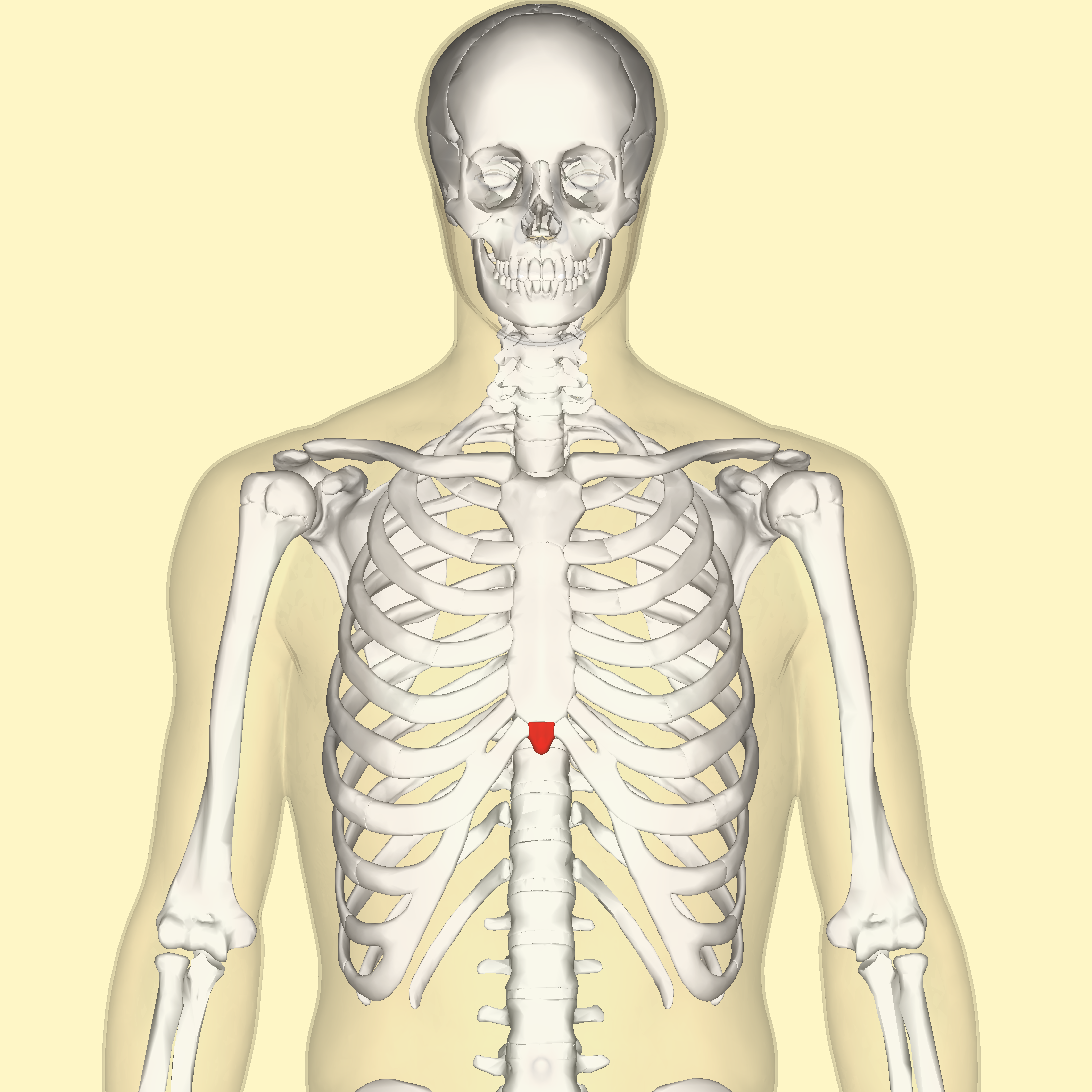
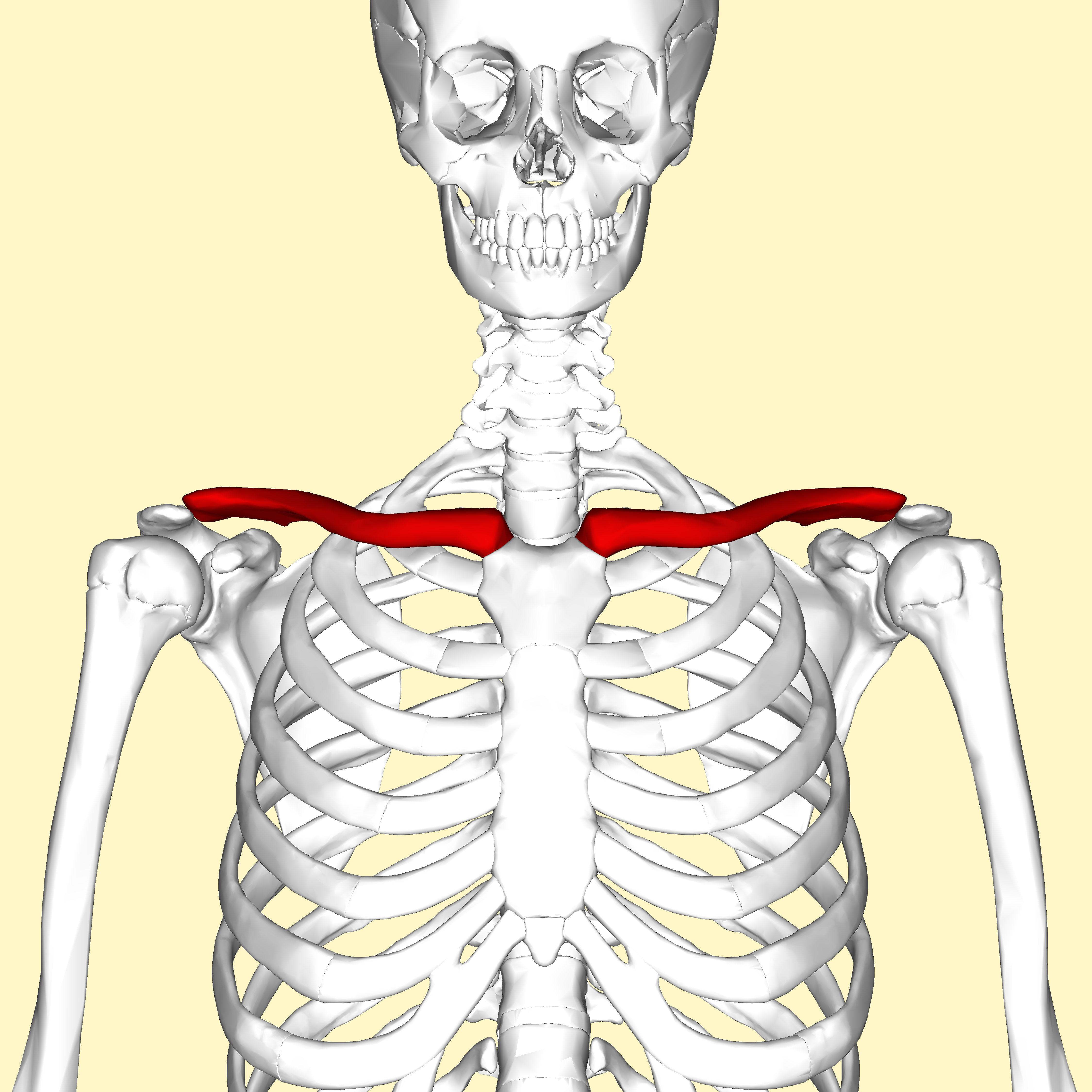
Consists of the Sternum (breastbone), Manubrium, body, Xiphoid Process, Clavicles (collarbones), Scapula ( shoulder blades), 12 pairs of ribs, 12 thoracic vertebrae

Manubrium

Sternum
breast bone located in the center of the chest. Composed of three pieces: Manubrium, body, and xiphoid process

Xiphoid process (T9-T10)
pointy bone projection at the end of the sternum ( also known as the xiphoid tip)
Clavicles ( collarbones)
Scapula (shoulder blades)

12 pairs of ribs

Thoracic vertebrae
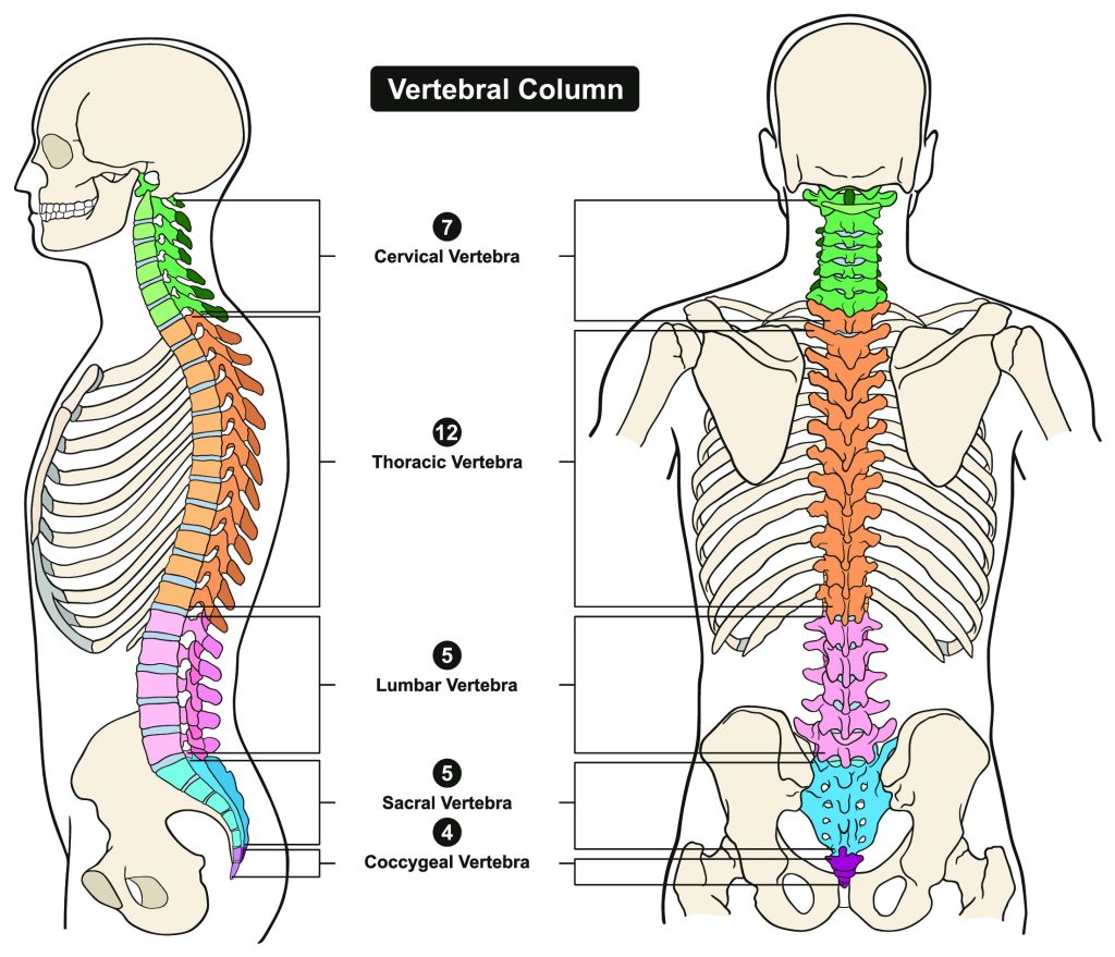

Vertebrae Prominens ( C7)
7th cervical vertebra. Bony bump at the base of the back of the neck that is felt when you bend your head forward


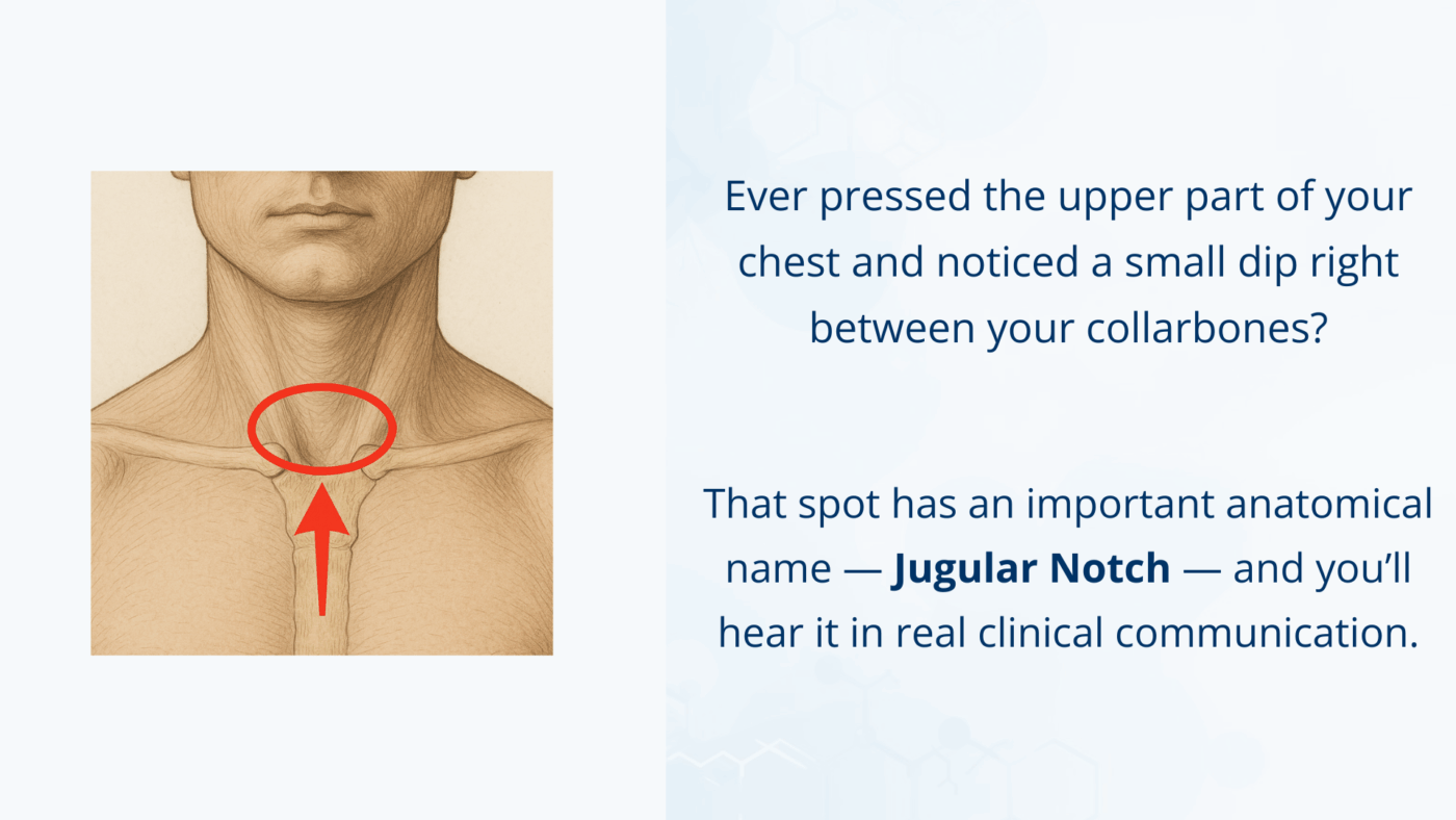
Jugular notch ( T2-T3)
Located on the manubrium, right in the middle between your clavicles. Can feel it at the base of your neck.
Sternal Angle ( T4-T5)
body meets manubrium

Inferior rib angle (L2-L3)
Lowest costal margin, below sternum

Apex
rounded upper area above the clavicles

Costophrenic angle
outermost corner of each lung

Hilum
central area where bronchi, blood vessels, lymph vessels, & nerves enter. ( theres a left and right)

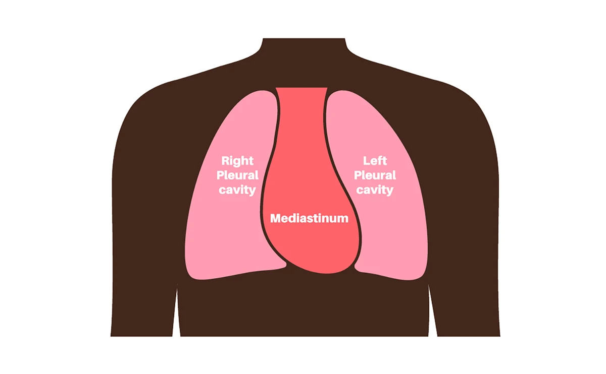
Mediastinum
Middle portion of the thoracic cavity between the lungs. Four structures here: thymus gland, heart and great vessels, trachea ( anterior), esophagus ( anterior)
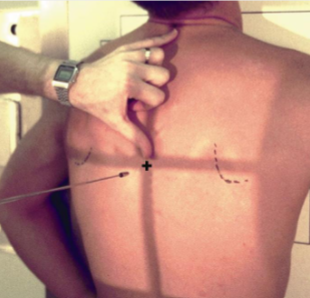
T7
approximately at the level of the inferior angle (bottom tip) of the scapula when the arms are down

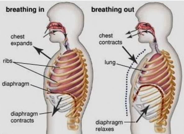
Why inspirate? ( breathe in)
Fills lungs with air, making them bigger/longer, pushes diaphragm downward, letting you see lungs more clearly
Hemidiaphragm

Carina

Sternoclavicular junction
where your Sternum meets your clavicular (collarbone)

Spinous process
one on each vertebrae

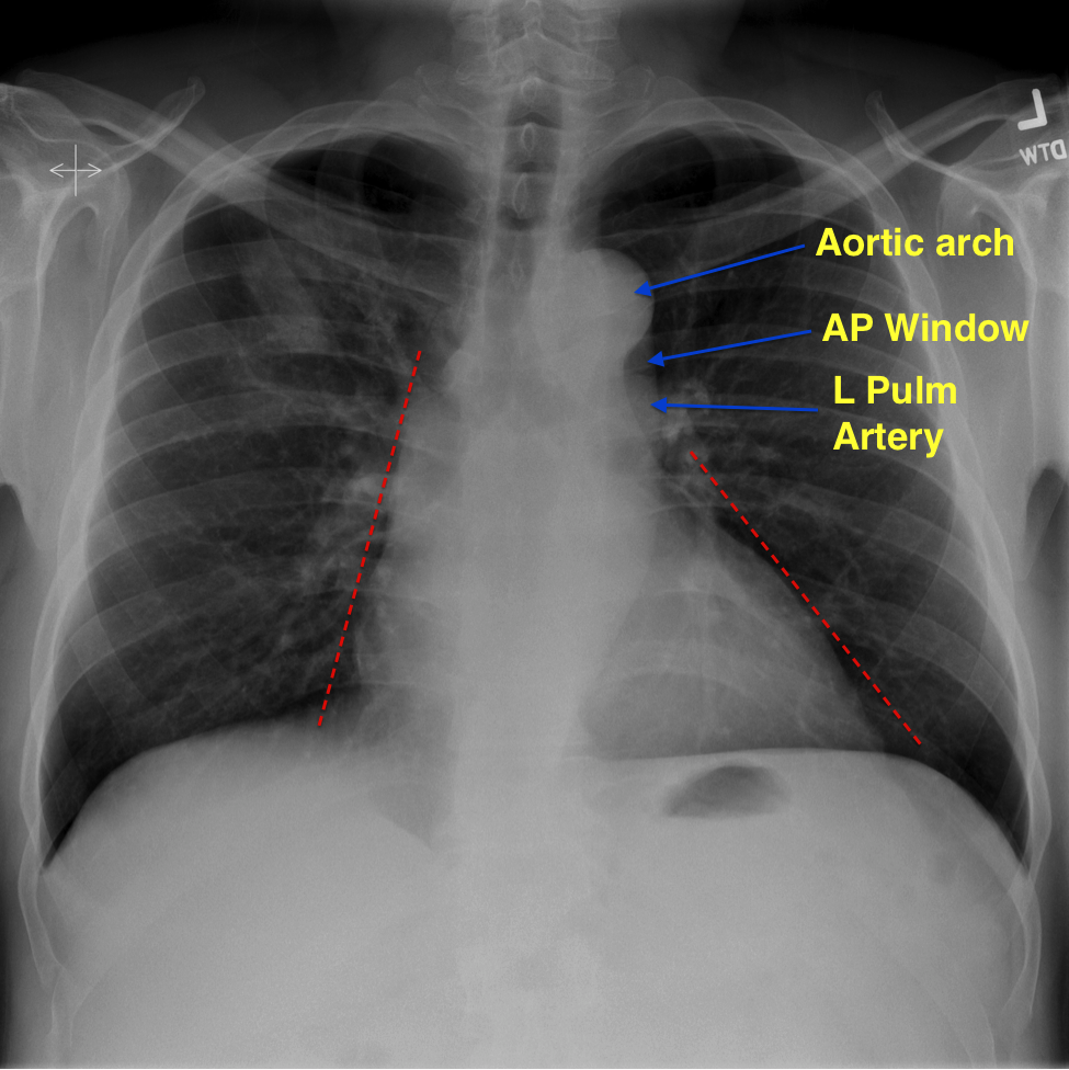
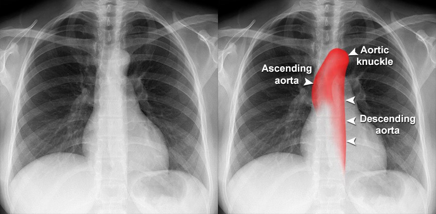
Aortic Arch
located on the left side
Bronchi
branches off trachea

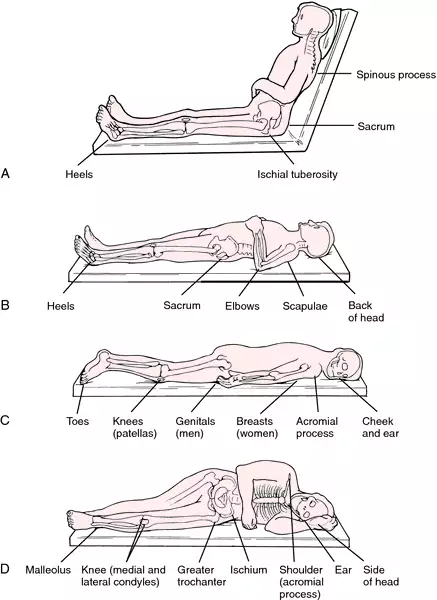
Lateral Decubitis
Patient is lying on side. Looking for air filled/ fluid filled lungs. Use horizontal beams.
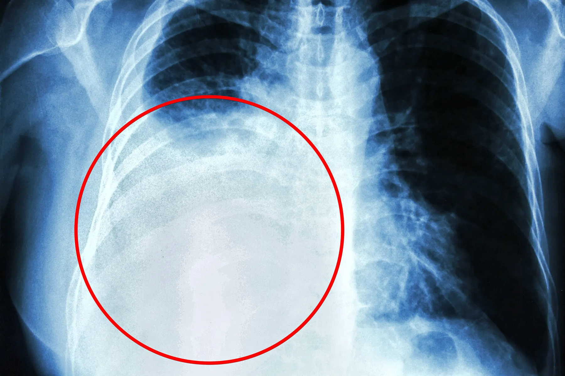
Pleural effusion
Fluid in lungs. Costophrenic angles appear blunt, not sharp
Trachea
air filled b/c of inspiration

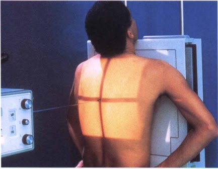
Technique for PA chest
kV: 110-125. mA:200. Time (sec): 0.02-0.04 mAs: 4-8
Technique for LATERAL chest
kV: 110-125. mA:200. Time (sec): 0.08-0.12. mAs: 16-24

Technique AP chest
kV: 75-85. mA:200. Time(sec): 0.02-0.03. mAs: 4-6

CHEST CONTRAST
Long scale, short wavelength, low contrast. High kVp, low mAs, b/c of soft tissues, air-filled lungs, and bone. Want enough penetration and a long scale of contrast ( shades of gray). Low mAs to reduce pt exposure

Hypersthenic
Very wide/ broad, short torso. Broad and short chest/ lungs. IR crosswise landscape to not cut off anatomy

Sthenic
Average build, IR lengthwise/ portrait

Hyposthenic
Between average and skinny, longer, narrower chest/ lungs, IR lengthwise/portrait

Asthenic
Very slender, long torso. IR lengthwise/portrait

Decubitis
Patient lying down AND horizontal x ray beam. Needed when trying to demonstrate air/fluid. Air rises, fluid sinks. Named for the side that is DOWN

SID and Chest
72 inches. The reason is magnification. Long SID lowers magnification, making the heart look closer to its true size ( more accurate)
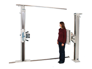
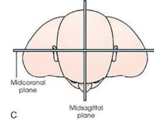
MCP (Midcoronal plane) PA CHEST
Runs left to right. Parallel to IR

MCP ( Midcoronal plane) LATERAL CHEST
Runs left to right. Perpendicular to IR because patient turns 90*

MSP ( Midsagittal plane) PA CHEST
Runs front to back. Perpendicular to IR
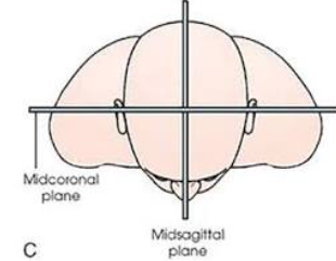
MSP ( midsagittal plane) LATERAL CHEST
Runs front to back. Parallel to IR because patient is rotated 90*
Primary ( main stem bronchus)
Right and left. The right is shorter and wider; the left is narrower but longer.

Thymus Gland
Located behind the sternum, above and anterior to the heart. Bigger in infants and shrinks over time. Can be seen on PEDIATRICS chest radiograph

Heart
Posterior to the sternum and anterior to T5-T8


“ Great Vessels”- Superior Vena Cava
Located in the mediastinum.


“ Great Vessels”- Inferior Vena Cava
Located in the mediastinum. ( can be seen laterally)

“ Great Vessels”- Aorta
Extends from the heart to L4. The thoracic aorta above the diaphragm. Located in the mediastinum.

“ Great Vessels”- Pulmonary Arteries
Locate in the mediastinum


“ Great Vessels”- Pulmonary Veins
Have a right and a left. Located in the mediastinum


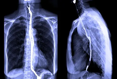
Esophagus
Posterior ( behind) the trachea

The Lungs
Right and left. The right lung has 3 lobes ( superior, middle and inferior); the left lung has 2 ( superior, and inferior)


Pharynx
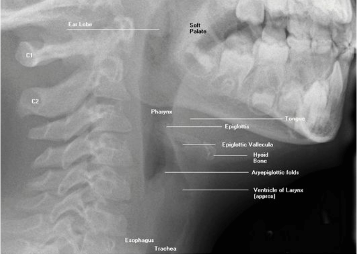
Posterior area between the nose and mouth ( superiorly) and larynx and esophagus ( inferiorly). Serves as passage for food and air. Divided into 3 parts: Naso, oro and laryngopharynx. ( BEST SEEN LATERAL)
AP LORDOTIC
Erect, one foot from Bucky, leaned back. Shoulders, neck, and back of head against IR. Entire lungs, clavicles above the apices, and distorted ribs displayed. SID 72” Lengthwise, 110-125 kV, 200mA, 0.02-0.04 sec, 4-8 mAs


AP UPPER AIRWAY
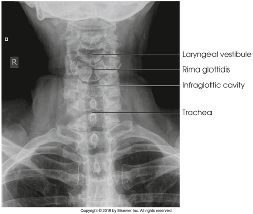
Erect, raised chin. Center at T1-2, 1 inch above jugular notch. SID 40” 75-85 kV, 100mA, 0.10-0.12 sec, 10-12 mAs. Slow, deep inspirations

LATERAL UPPER AIRWAY
Erect left/right lateral. Chin elevated, arms behind back. Center C6-7 and MSP. SID 72” 75-85 kV, 100mA, 0.03-0.04 sec, 3-4 mAs. Slow, deep inspirations

PORTABLES CHEST