Restrictive Lung Disease
 Call Kai
 Learn
Learn Practice Test
 Spaced Repetition
 Match
Match Flashcards
 Knowt Play
1/120

There's no tags or description
Looks like no tags are added yet.
Name | Mastery | Learn | Test | Matching | Spaced | Call with Kai | Chat |
|---|
No analytics yet
Send a link to your students to track their progress
121 Terms
PFT
restrictive lung diseases are diagnosed only by...
FEV1/FVC ratio
in obstructive lung disease, ____ is decreased
restrictive lung diseases
characterized by reduced lung volumes, either because of an alteration in lung parenchyma or because of a disease of the pleura, chest wall, or neuromuscular apparatus.
asthma and COPD
obstructive lung diseases include...
TLC
measures of expiratory airflow are preserved and airway resistance is normal and the FEV1/FVC ratio is normal or increased.
Restrictive lung diseases are associated with a decreased _____ on PFTs
parenchymal lung disease
(intrinsic cause of lung disease)
if caused by _________, restrictive lung disorder are accompanied by reduced gas transfer (reduced diffusion capacity, DCLO), which may be marked clinically by desaturation after exercise.
all lung volumes
In cases of intrinsic lung disease, the physiological effects of diffuse parenchymal disorders reduce which lung volumes?
Restrictive lung disease
In an obstructive pattern, the expiratory airflow is reduced out of proportion to the lung volume. This is why the FEV1/VC is abnormally low in obstructive pattern.
in ________, expiratory airflow is reduced in proportion to lung volume.
in other words, the FEV1/FVC ratio is unaffected, because the two values are reduced proportionately.
- interstitial lung diseases (ILD)
- extrinsic disorders or extrapulmonary diseases
Restrictive lung disease may be divided into two groups based on anatomical structures...
Interstitial lung diseases (ILD)
group of restrictive lung disease causing inflammation of scarring of the lung tissue and/or vasculature and/or result in filling of the air spaces with exudate and debris.
pneumonitis
filling of the air spaces with exudate and debris in ILD.
- smoking related
- hypersensitivity related
- exposure related (asbestosis, silicosis)
- drug induced (amiodarone)
- radiation to chest (breast cancer patients)
known causes of ILD
- idiopathic pulmonary fibrosis
- sarcoidosis
- acute interstitial pneumonia
unknown causes of ILD
Amiordarone
anti-arrhythmic medications that can cause pulmonary fibrosis (ILD)
smoking
cause of ILD that is also a risk for Obstructive lung disease (COPD)
Extrinsic disorders or extrapulmonary diseases
causes of restrictive lung disease that originate in the chest wall, pleura, and respiratory muscles that are needed to function normally for effective ventilation. Diseases of these structures result in lung restriction and impaired ventilatory function.
dyspnea and dry cough over months (>6 months)
Patients with restrictive lung disease, present with nonspecific symptoms, ______ are the most common presenting symptoms.
infectious and Heart Failure
If dyspnea and cough have only been present for days to months, _____ etiologies are more likely than restrictive lung disease.
Interstitial Lung Diseases (ILD)
if infectious and Heart failure etiologies are excluded or empiric treatment is unsuccessful in patients with dyspnea and cough, then ______ should be considered.
- GERD
- Asthma
- Post Nasal Drip
three most common causes of chronic cough
- timing of symptoms
- smoking
- exposure history
- Rheumatology ROS
- FH of autoimmune and lung disease
- medications and past medications
A careful history for patients with restrictive lung disease should include...
CXR
initial imaging for patients with restrictive lung disease which can be normal in up to 20% of patients.
20
___% of patients with restrictive lung disease have a normal CXR
Diffuse Parenchymal Lung Disease (DPLD)
the infiltrates are in a diffused/reticular pattern. Unlike pneumonia in which the infiltrates are focal.
Typical CXR finding of patients with restrictive lung disease is _________. There are increased interstitial reticular and nodular infiltrates in different patterns of distributions.
High-resolution CT of the chest
imaging that should follow CXR in patients with suspected Restrictive lung disease.
Biopsy
gold standard for diagnosis of ILD
PFT
should be performed in patients with chronic symptoms of restrictive lung disease in order to exclude obstructive pattern as the primary cause, but also to rule out obstructive/restrictive pattern.
inflammation due to restrictive disease may lead to bronchoconstriction --> obstructive pattern
Explain how mixed pattern can develop in a patient with a restrictive lung disease?
- progressive exertion dyspnea
- cough (usually dry)
- hemoptysis
- +/- wheezing
symptoms of ILD
progressive exertional dyspnea
predominant symptom of ILD
hemoptysis (or grossly bloody sputum)
symptom of ILD that occurs in patients with diffuse alveolar hemorrhage syndromes and vasculitis.
wheezing
an uncommon symptom in pure ILD but can be present in obstructive/restrictive cases
ILD
Velcro crackles are common in most patients with _______
bronchiolitis
inspiratory squeaks or scattered, late inspiratory high-pitched rhonchi are frequently heard in patients with _______
cyanosis
cutaneous change seen in patients with ILD that usually occurs with exertion. Uncommon at rest and is a sign of advanced disease.
- Velcro crackles
- Cyanosis
- Digital clubbing
physical exam findings associated with ILD
search for the cause
After you have determined the patient has restrictive lung disease, your next step is to...
sarcoidosis
erythema nodosum in patient with restrictive lung pattern suggests _______

connective-tissue diseases or drug-induced lung diseases
a maculopapular rash in patient with restrictive lung pattern suggests _______
connective-tissue diseases
Reynaud phenomenon in patient with restrictive lung pattern suggests _______
scleroderma
telangiectasia in patient with restrictive lung pattern suggests _______
systemic sarcoidosis
peripheral lymphadenopathy, salivary gland enlargement, and hepatosplenomegaly in patient with restrictive lung pattern suggests _______
sarcoidosis and ankylosing spondylitis
uveitis in patient with restrictive lung pattern suggests _______
uveitis causes pain with pupil movement (dilating/constricting to light)
systemic vasculitis
purpura in patient with restrictive lung pattern suggests _______
renal involement
hematuria and anasarca in patient with restrictive lung pattern suggests _______
cor pulmonale
occurs in the late stages of pulmonary fibrosis or advanced kyphoscoliosis
- grossly visible kyphoscoliosis and obesity
- accessory muscle usage
- rapid shallow breathing
extrapulmonary findings in patient with extrinsic disorders (restrictive lung disease)
Idiopathic Pulmonary Fibrosis (IPF or Usual Interstitial Fibrosis)
most common idiopathic interstitial pneumonia. Mean age of onset is 62 years old and can be familial.
Autosomal dominant
Idiopathic pulmonary fibrosis can be familial likely following a ______ pattern with variable penetrance.
MUC5B
IPF is associated with the _____ allele with familial and sporadic forms
smoking
IPF is frequently associated with a history of _______
patients may present with a mixed pattern on PFTs because of this exposure.
IPF
some other causes of intrinsic lung disease have systemic effects/manifestations. IPF usually just limits itself to the lungs.
cause of intrinsic restrictive lung disease that is usually limited to the lungs. There is fibrosis of interstitial spaces with zones of normal lung
3-5 years
the prognosis of IPF is poor, average survival is ______. There is some variability in the course: some progress slowly or stepwise while others rapid decline leading to death.
gradual onset of exertional dyspnea and dry cough
over months to years
presenting symptoms of Idiopathic pulmonary fibrosis
Idiopathic pulmonary fibrosis
PE reveals dry inspiratory crackles at the bases of the lungs, 50% of patients have clubbing. More severe diseases might have pulmonary HTN and signs of right sided heart failure.
Diagnosis of exclusion
you must exclude all other causes of restrictive lung disease
- Restrictive pattern
- decreased DCLO (may be isolated finding)
PFTs of patient with Idiopathic Pulmonary Fibrosis would display...
Idiopathic pulmonary fibrosis
CXR revealing basal-predominant reticular infiltrates in a patient with restrictive pattern on PFTs is indicative...
Idiopathic pulmonary fibrosis
High resolution CT displays basal and peripheral dominant septal line thickening and traction bronchiectasis and honeycomb changes. (This pattern is typically seen with other ILD diseases)
IPF
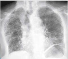
IPF

- primarily supportive (optimize fitness and oxygenation)
- Nintedanib and Pirfenidone to slow fibrosis
- lung transplant
Treatment overview for IPF...
nintedanib
tyrosine kinase inhibitor that blocks activation of fibroblasts in patients with IPF
Pirfenidone
regulates transforming TGF-beta and TNF-alpha activity in patients with IPF
Nintedanib and Pirfenidone
immunosuppressant drugs that slow the decline of IPF on imaging, but do not improve symptoms or quality of life in the patient. The drugs are likely more useful in the disease process, but once fibrosis has occurred it is not reversed
anti-inflammatories (steroids) and immune modulators (azathioprine)
because there is NO inflammation
drugs that are NOT indicated in IPF treatment
lung transplant
only treatment for IPF that prolongs life
mechanical ventilation
patient will never have capacity to be removed from ventilator
_____ should be avoided in IPF patients in acute respiratory failure.
Sarcoidosis
multisystem granulomatous disease of unknown cause. More than 90% of patients have pulmonary involvement.
incidental findings on CXR
many patients are asympatomatic
How is sarcoidosis typically discovered?
African Americans and women
Sarcoidosis in the US is more common among _________. There is some familial clustering.
No, spontaneous regression is common
Is pulmonary sarcoidosis typically a long term disease process?
biopsy is usually needed
diagnosis of exclusion otherwise
Diagnosis of sarcoidosis is made by...
HLA
sarcoidosis involves _____-mediated processing of unknown antigens by macrophages leading to abnormal activation of T cells. This causes organization of inflammatory cells into granulomas. Fibroblasts and collagen encase granulomas and can lead to fibrosis
T cell
in sarcoidosis, HLA-mediated processing of unknown antigens by macrophages causes abnormal activation of _______
granulomas
in sarcoidosis, abnormal activation of T cells lead to organization of inflammatory cells into _____
fibroblasts and collagen
in sarcoidosis, _____ encase granulomas and can lead to fibrosis.
Stage I
stage of sarcoidosis that involves Hilar lymphadenopathy, but lung parenchyma is normal.
90% spontaneous resolution without treatment
Clinical course of stage I sarcoidosis
Stage II
stage of sarcoidosis that involves Hilar lymphadenopathy with abnormal lung parenchyma.
20% spontaneous improvement without treatment. Most patients need to be treated with steroids.
clinical course for stages II and III of sarcoidosis
Stage III
stage of sarcoidosis in which there is no lymphadenopathy, but there is abnormal lung parenchyma
Stage IV
stage of sarcoidosis in which there are parenchymal changes with fibrosis and architectural distortion.
2 years
the progression into the progressive phase of sarcoidosis typically occurs in about ______
Pulmonary Sarcoidosis

Sarcoidosis
CXR displaying bilateral hilar and mediastinal lymphadenopathy (>2cm) is indicative of
- dry cough and dyspnea
- night sweats
- weight loss, fatigue
- Lofgren syndrome
symptoms of sarcoidosis can be insidious or acute, they include...
Lofgren syndrome
involves erythema nodosum, arthritis, bilateral hilar adenopathy, +/- fever, uveitis. Associated with sarcoidosis
Sarcoidosis
CT with upper lobe fibrosis, nodules and ground glass opacities, mediastinal and hilar lymphadenopathy, cystic changes, thickening of the pleural space, and bronchiectasis is indicative of _______
noncaseating granulomas
lung biopsy of patient with sarcoidosis would display ______
beryllium
occupational exposure that is indistinguishable from Sarcoidosis. It is only ruled in/out based on the patients history
ACE
___ levels are elevated in 60% of patients with sarcoidosis. This lab value is not specific or sensitive, but can contribute to your diagnosis.
GLucocorticoids
mainstay therapy of sarcoidosis that are usually reserved patients with systemic symptoms.
short term
studies show that glucocorticoids showed benefit in ____ symptoms in patients with sarcoidosis
immunsuppressive
____ therapies have been tried for patients with Sarcoidosis if glucocorticoid therapy has failed.
topical steroids
If sarcoidosis only affects the anterior eye, localized skin, or causes cough, treatment should include _______
taper to
if patients with sarcoidosis require systemic glucocorticoid therapy, what should your goal be in the next 6 months?
Methotrexate, hydroxychloroquine, azathioprine
If glucocorticoid dose can not be tapered to
Pneumoconiosis
chronic fibrotic lung diseases due to inhalation of inorganic dusts
Silicosis
pneumoconiosis caused by inhalation of silica dust that occurs in mines and sandblasting workers.