APHY 102 M1-2 Study Guide Questions Flashcards
 Call Kai
 Learn
Learn Practice Test
 Spaced Repetition
 Match
Match Flashcards
 Knowt Play
1/115

There's no tags or description
Looks like no tags are added yet.
Name | Mastery | Learn | Test | Matching | Spaced | Call with Kai | Chat |
|---|
No analytics yet
Send a link to your students to track their progress
116 Terms
List the two major components of the cardiovascular system and explain the major functions of each.
1.heart- generates force to transport respiratory gases, nutrients, and wastes throughout body.
Blood vessels- a closed network of tubes (arteries, veins, capillaries) that carry blood throughout the body.
which direction do arteries and veins carry blood relative to the heart?
-arteries transport blood AWAY from the heart
tip: artery=away
-veins transport blood toward the heart
tip: vein = “in” toward the heart
Explain the functions of capillaries.
Capillaries transport blood between arteries and veins, and perform nutrient, gas, and waste exchange. Exchange oxygen, carbon dioxide, nutrients, and wastes between the blood and tissues.
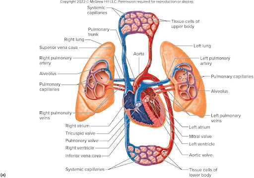
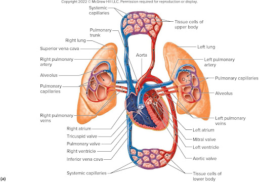
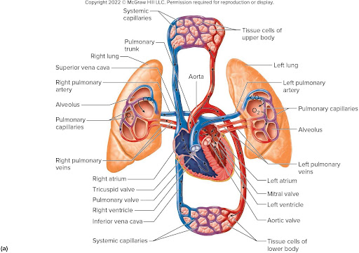
What is the pulmonary circuit?
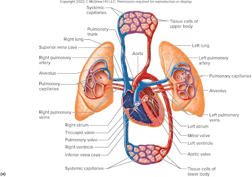
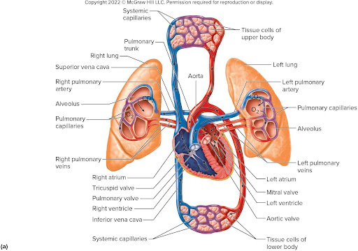
the pulmonary circuit sends oxygen poor blood to the lungs to unload carbon dioxide and pick up oxygen before returning to the heart.
what is the systemic circuit?
the systemic circuit sends oxygen rich blood and nutrients to all body cells and removes wastes and returns oxygen poor blood back to the heart.
which side of the heart pumps blood to the lungs?
right side
which side of the heart pumps blood to the body?
left side
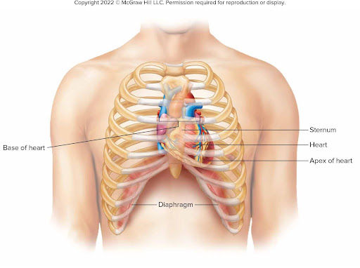
Describe the location of the heart.
located in the mediastinum of the thoracic cavity just superior to the diaphragm, the heart is bordered laterally by the lungs, posteriorly by the vertebral column, and anteriorly by the sternum.
what is the shape of the heart?
hollow, cone-shaped muscular organ
where is the base of the heart?
beneath the 2nd rib
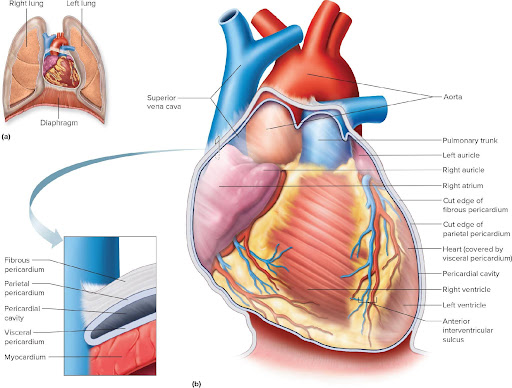
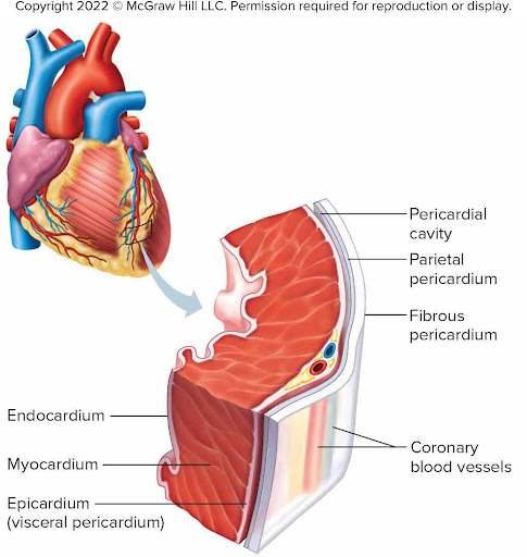
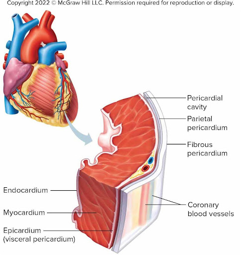
what is the pericardium?
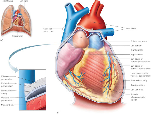
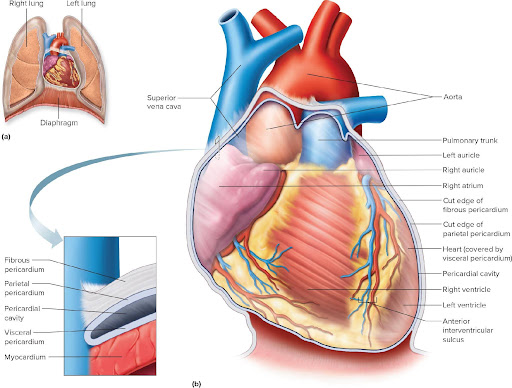
the sac surrounding the heart and the proximal ends of the large vessels to which it attaches.
what is the fibrous pericardium?
tough outer layer of the pericardium
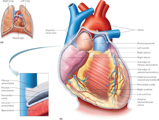
what is the parietal pericardium?
outer layer of the serous membrane lining the fibrous pericardium.
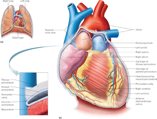
what is the visceral pericardium?
inner layer attached directly to the heart; also called the epicardium.
what is the pericardial cavity?
space between the visceral and parietal pericardium containing serous fluid (pericardial fluid) secreted by the pericardial membranes
what are the three layers of the heart wall?
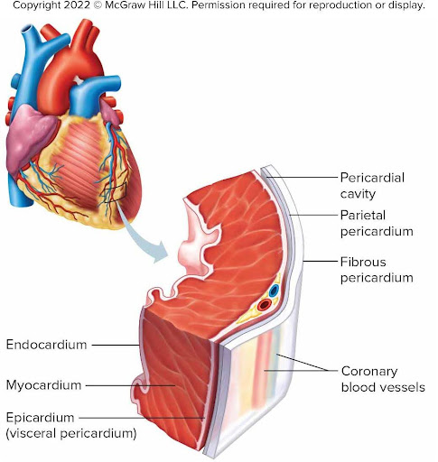
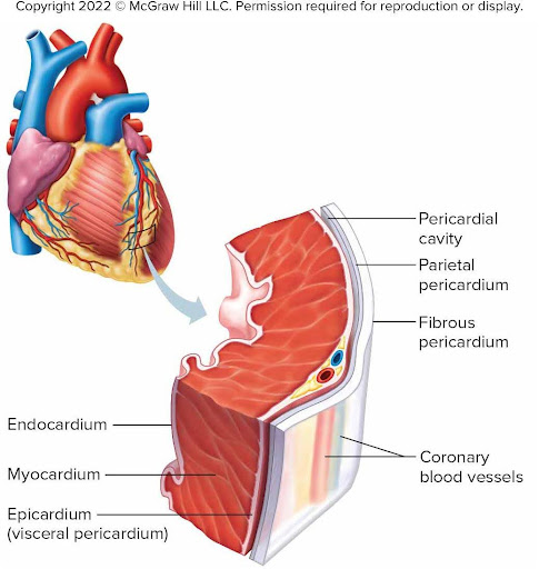
Epicardium
Myocardium
Endocardium
which heart layer is the outer layer?
epicardium
which heart layer is the thickest?
myocardium
which layer forms the inner lining of the chambers?
endocardium
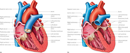
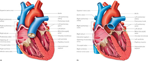
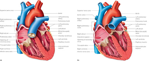
what are the atria? (singular term is atrium)
Thin-walled upper chambers that receive blood returning to the heart.
what are auricles?
Flap-like projections that allow the atria to expand.
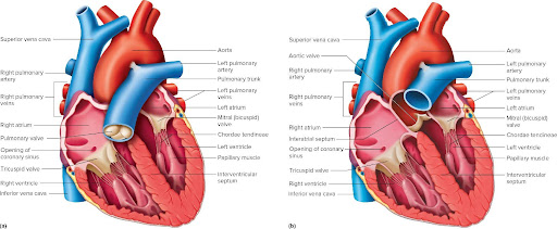
what are the ventricles?
Thick-walled lower chambers that pump blood into arteries.
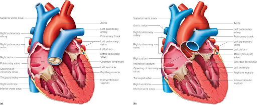
what does the right atrium recieve?
Oxygen-poor blood from:
Superior vena cava
Inferior vena cava
Coronary sinus
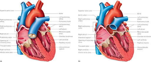
What does the right ventricle do?
Receives blood from the right atrium; pumps blood to lungs
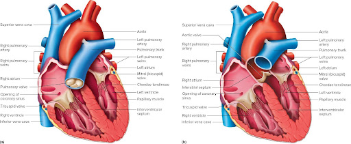
what does the left atrium receive?
Receives blood from the pulmonary veins
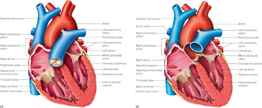
What does the left ventricle do?
Receives blood from the left atrium; pumps blood to systemic circuit
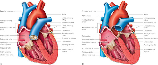
Name the four valves of the heart.
Tricuspid valve
Pulmonary semilunar valve
Mitral (bicuspid, left AV) valve
Aortic semilunar valve
Which valve is also called the right AV valve?
Tricuspid valve
Which valve prevents blood from flowing back into the right atrium?
Mitral valve
Which valve prevents blood from flowing back into the right ventricle?
Pulmonary semilunar valve
Which valve prevents blood from flowing back into the left ventricle?
Aortic semilunar valve
What separates the right and left atria?
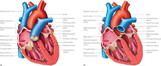
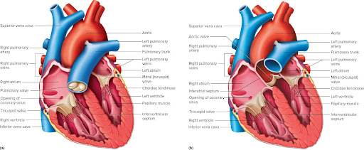
Interatrial septum
what separates the right and left ventricles?
Interventricular septum
What are chordae tendineae?
Fibrous cords that connect the AV valves to the papillary muscles.
What do papillary muscles do?
Prevent the AV valves from prolapsing during ventricular contraction.
Which arteries supply blood to the heart muscle?
Coronary arteries.
Trace blood flow through the heart.
Superior/Inferior Vena Cava
↓
Right Atrium
↓
Tricuspid Valve
↓
Right Ventricle
↓
Pulmonary Semilunar Valve
↓
Pulmonary Trunk
↓
Pulmonary Arteries
↓
Lungs
↓
Pulmonary Veins
↓
Left Atrium
↓
Mitral (Bicuspid) Valve
↓
Left Ventricle
↓
Aortic Semilunar Valve
↓
Aorta
↓
Body
what is systole?
the phase of the cardiac cycle when the heart muscle contracts and pumps blood.
what is distole?
The phase of the cardiac cycle when the heart muscle relaxes and the chambers fill with blood.
What happens during atrial systole?
The atria contract, pushing blood into the ventricles.
What happens during ventricular systole?
The ventricles contract, forcing blood into the pulmonary trunk and aorta
What happens during cardiac diastole?
Both the atria and ventricles relax and fill with blood.
what is a functional syncytium?
Cardiac muscle cells contract together as one coordinated unit because they are electrically connected.
what are intercalated discs?
Specialized junctions that connect cardiac muscle cells and allow electrical signals to pass rapidly from cell to cell.
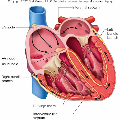
What is the correct pathway of the cardiac conduction system?
SA node → AV node → AV bundle (Bundle of His) → Right & Left Bundle Branches → Purkinje fibers
Which structure is the pacemaker of the heart?
Sinoatrial (SA) node
location: In the wall of the right atrium near the opening of the superior vena cava.
What is the function of the AV node?
Delays the electrical impulse so the atria can finish contracting before the ventricles contract.
What is the function of the Bundle of His?
Carries the electrical impulse from the AV node into the interventricular septum.
What do the bundle branches do?
Carry the electrical impulse toward the apex of the heart.
What do Purkinje fibers do?
Distribute the electrical impulse throughout the ventricular myocardium, causing ventricular contraction.
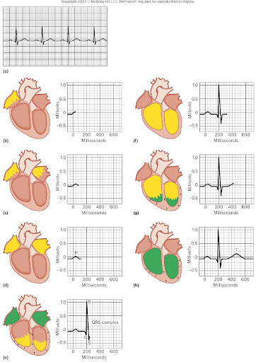
What does ECG/EKG stand for?
Electrocardiogram: A recording of electrical changes that occur in the myocardium during the cardiac cycle
Used to assess ability of heart to conduct impulses
The deflections in the normal ECG, or waves, include:
P wave: Atrial depolarization; occurs just prior to atrial contraction
QRS complex (3 waves): Ventricular depolarization; occurs just prior to ventricular contraction
Record of atrial repolarization is “hidden” in the large QRS complex, since ventricular depolarization is a much larger event
T wave: Ventricular repolarization; occurs just prior to ventricular relaxation
What causes the "lubb" (S1) heart sound?
Closure of the tricuspid and mitral (AV) valves.
What causes the "dupp" (S2) heart sound?
Closure of the aortic and pulmonary semilunar valves.
What is a heart murmur?
An abnormal heart sound caused by turbulent blood flow, often due to defective valves.
Where is the cardiac center that regulates the cardiac cycle?
In the medulla oblongata of the brainstem.
List the five types of blood vessels.
Arteries
Arterioles
Capillaries
Venules
Veins
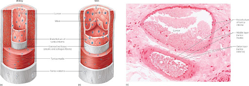
What are arteries?
thick-walled blood vessels that carry blood away from the heart under high pressure.
What are arterioles?
Small branches of arteries that regulate blood flow into capillaries.
What are capillaries?
Microscopic vessels where exchange occurs between blood and tissues.
What are venules?
Small vessels that collect blood from capillaries and carry it to veins.
What are veins?
Blood vessels that return blood to the heart under low pressure.
Name the three layers of an artery.
Tunica interna (intima)
Tunica media: thickest layer
Tunica externa (adventitia)
How do capillaries accomplish exchange?
Through diffusion, filtration, and osmosis.
What is blood pressure?
The force exerted by blood against the walls of arteries. Normal adult blood pressure is approximately 120/80 mmHg
What does the top number of a blood pressure reading represent?
Systolic pressure (pressure during ventricular contraction).
What does the bottom number represent?
Diastolic pressure (pressure during ventricular relaxation).
What is the official name of a blood pressure cuff?
Sphygmomanometer
where is radial and brachial pulse taken?
radial: at the wrist on the thumb size
brachial: inside the elbow (antecubital region)
name other common pulse sites.
Carotid
Femoral
Popliteal
Posterior tibial
Dorsalis pedis
Temporal
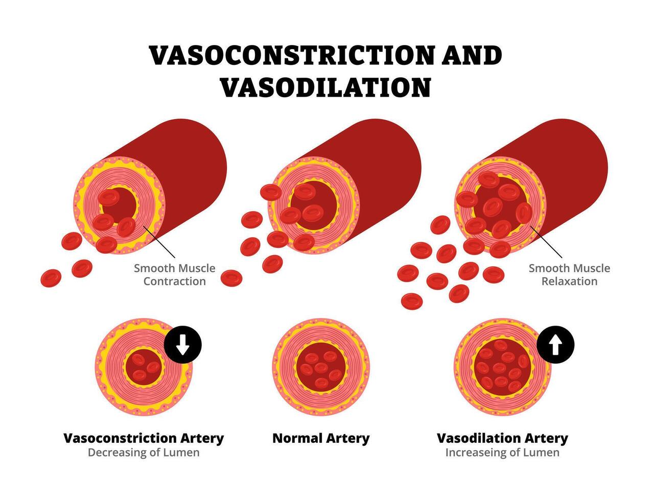
what is vasoconstriction?
Narrowing of blood vessels, increasing blood pressure.
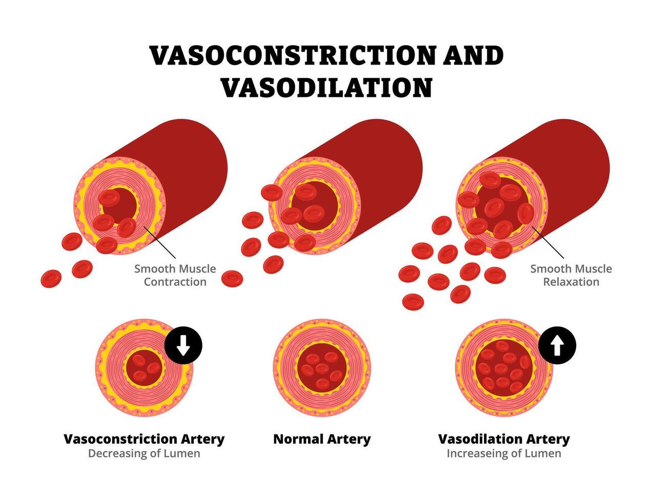
What is vasodilation?
Widening of blood vessels, decreasing blood pressure.
What is peripheral resistance?
The resistance blood encounters as it flows through blood vessels.
What is hypertension? Common causes of hypertension?
Persistently high blood pressure. common causes:
High-sodium diet
obesity
Stress
Smoking
Lack of exercise
Kidney disease
Genetics
Aging
What factors help return venous blood to the heart?
Skeletal muscle pump
Respiratory pump (breathing)
One-way valves in veins
Smooth muscle contraction in vein walls
What are the three formed elements of blood (common and scientific names)?
Red blood cells (Erythrocytes) – Transport oxygen and carbon dioxide
White blood cells (Leukocytes) – Defend against infection
Platelets (Thrombocytes) – Help blood clot
What is hematopoiesis?
The process of producing all blood cells in red bone marrow from hematopoietic stem cells.
Where do blood cells originate, and which stem cells produce each type?
All blood cells originate in red bone marrow from hematopoietic stem cells.
Myeloid stem cells: erythrocytes, platelets, neutrophils, eosinophils, basophils, monocytes
Lymphoid stem cells: B lymphocytes, T lymphocytes, and NK cells
What are erythrocytes and what are their characteristics?
Erythrocytes (red blood cells) are biconcave discs, lack a nucleus and most organelles, are flexible, and transport oxygen and carbon dioxide.
What is the function of hemoglobin?
Hemoglobin is the oxygen-carrying protein inside red blood cells that transports oxygen from the lungs to tissues and carries some carbon dioxide back to the lungs.
What hormone stimulates erythropoiesis(production of RBCs), and what triggers its release?
Erythropoietin (EPO) is released by the kidneys when blood oxygen levels are low (hypoxia).
What is the average lifespan of a red blood cell?
Approximately 120 days
What nutrients are required for erythropoiesis?
Iron
Vitamin B12
Folic acid
Amino acids
what is anemia?
A condition in which the blood has too few red blood cells or too little hemoglobin, reducing oxygen transport.
What are the two categories of white blood cells and their types?
Granulocytes
Neutrophils
Eosinophils
Basophils
Agranulocytes
Lymphocytes
Monocytes
Define diapedesis, chemotaxis, and phagocytosis.
Diapedesis: WBCs squeeze through blood vessel walls.
Chemotaxis: WBCs move toward chemicals released by damaged tissue.
Phagocytosis: Cells engulf and digest pathogens or debris.
How can you identify each type of white blood cell, and what is its function?
Neutrophils: Multilobed nucleus; most numerous; phagocytize bacteria.
Eosinophils: Bilobed nucleus; red/orange granules; fight parasites and reduce allergic reactions.
Basophils: Dark blue/purple granules; release histamine and heparin.
Lymphocytes: Large round nucleus; provide adaptive immunity (B cells, T cells, NK cells).
Monocytes: Largest WBC; kidney-shaped nucleus; become macrophages for phagocytosis.
Which white blood cells are most common and least common?
Most common → Neutrophils
Least common → Basophils
Remember: Never Let Monkeys Eat Bananas
Neutrophils
Lymphocytes
Monocytes
Eosinophils
Basophils
What are the two main types of lymphocytes?
B lymphocytes (B cells)
T lymphocytes (T cells)
(Natural Killer cells are also lymphocytes.)
What are leukocytosis and leukopenia?
Leukocytosis: Increased white blood cell count
Leukopenia: Decreased white blood cell count
What is a differential white blood cell count?
A test that measures the percentage of each type of white blood cell in the blood.
What is leukemia?
A cancer of blood-forming tissues that causes abnormal production of white blood cells.
platelets originate from what type of cell, what do they look like, and what is their function?
Platelets are fragments of megakaryocytes, appear as small cell fragments without nuclei, and function in blood clotting.
What are thrombocytosis and thrombocytopenia?
Thrombocytosis: Increased platelet count
Thrombocytopenia: Decreased platelet count
What are the components and functions of plasma?
Plasma is mostly water (about 90%) and contains:
Plasma proteins (albumin, globulins, fibrinogen)
Electrolytes
Nutrients
Hormones
Gases
Wastes
Functions:
Transports substances
Maintains blood volume and pH
Helps regulate temperature
Carries clotting proteins
What is an electrolyte, and which electrolytes are found in plasma?
Electrolytes are ions that conduct electricity in body fluids.
Examples:
Sodium (Na⁺)
Potassium (K⁺)
Calcium (Ca²⁺)
Magnesium (Mg²⁺)
Chloride (Cl⁻)
Bicarbonate (HCO₃⁻)
What is hemostasis?
The process that stops bleeding after a blood vessel is damaged.
What roles do platelets and platelet plugs play in hemostasis?
Platelets stick to damaged vessels, become activated, and form a temporary platelet plug to reduce blood loss.
What is the overall function of the clotting cascade?
It activates clotting factors that convert fibrinogen into fibrin, forming a stable blood clot.
What are clotting factors, and why is vitamin K important?
Clotting factors are proteins needed for blood clotting. Vitamin K is required by the liver to produce several clotting factors.
What is the major event in blood clotting?
The conversion of fibrinogen into fibrin, which forms the mesh that stabilizes a blood clot.