Gas exchange and animal transport, etc
1/83
There's no tags or description
Looks like no tags are added yet.
Name | Mastery | Learn | Test | Matching | Spaced | Call with Kai |
|---|
No analytics yet
Send a link to your students to track their progress
84 Terms
Why do larger organisms need circulatory systems?
They can gain reactants via specialised exchange surfaces, but as they are made up of many layers of cells, the time for substances to diffuse to every cell would be too great.
What are the types of circulatory systems?
Open, closed, single and double.
What constitutes an open circulatory system?
Where the blood does not move around the body in blood vessels, but it bathes the tissues directly while held in a cavity called the haemocoel.
What is a closed circulatory system?
Where the blood moves in blood vessels. Blood pumped by muscular heart at high pressure. Tissues are not in direct contact with blood, but bathe in tissue fluid which can exit out of walls of capillaries. Blood contains haemoglobin.
What is an insect's circulatory system?
Open, they have a long, dorsal tube-shaped heart, which runs the length of the body. It pumps blood out at low pressure into the haemocoel, where mats are exchanged, before blood goes back to the heart.
What are some advantages of closed circulatory systems?
Blood flow more rapid and efficient
Can be directed to where it is needed
More efficient delivery of oxygen and glucose.
What is a single circulation system?
Where the blood moves through the heart once.
What is the circulatory system in fish?
The ventricle pumps deoxygenated blood to the gills, where the capillaries reduce its pressure. Oxygenated blood pumped to tissues, and from there the deoxygnated blood returns to the atrium.
What is the circulatory system in an earthworm?
A single circulatory system. Blood moves forward in the dorsal vessel, and back in the ventral vessel. Five pairs of 'pseudohearts', pump the blood from the dorsal to the ventral vessel.
What is a double circulatory system?
The blood flows through the heart twice. Mammals have these. Blood is pumped by a heart, giving a rapid flow rate.
Blood pressure is reduced in capillaries of lungs for gas exchange, so pressure would be too low to reach rest of body. So, blood goes back to heart, which raises the pressure and allows it to be pumped to rest of body.
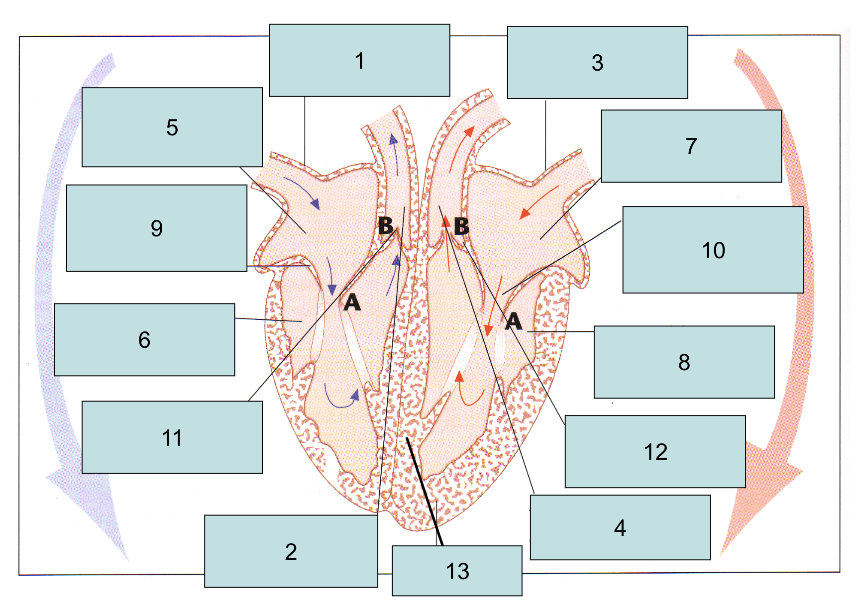
Label this image:
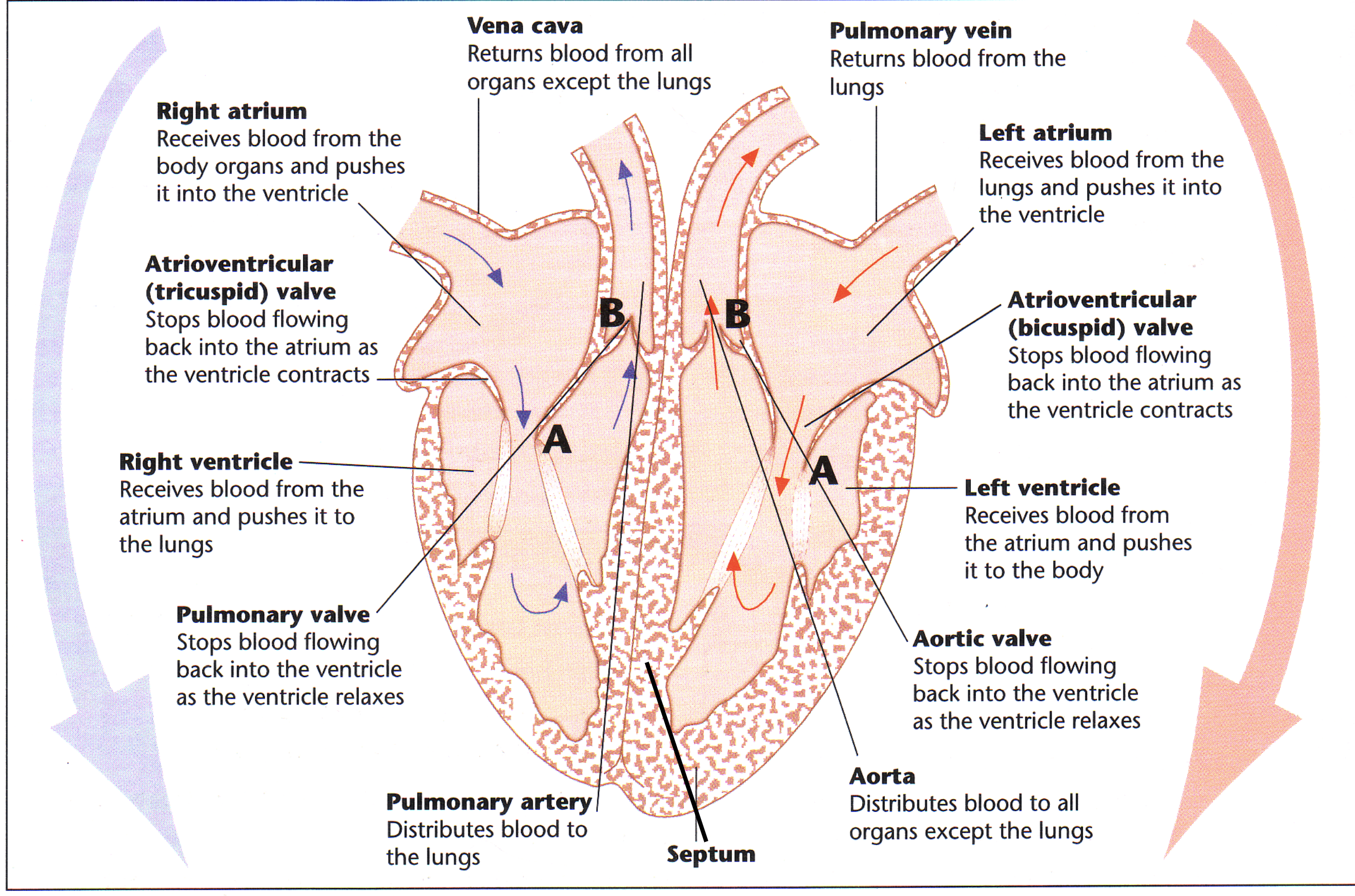
What is the function of the vena cava?
Returns deoxygenated blood from all organs except the lungs.
What is the function of the pulmonary vein?
Returns blood from the lungs.
What is the function of the left atrium?
Receives blood from the lungs and pushes it into the ventricle left ventricle.
What is the function of the Bicuspid valve?
Stops blood flowing back into the atrium as the ventricle contracts.
What is the function of the left ventricle?
Receives blood from the atrium and pushes it to the body.
What is the function of the aortic valve?
Stops blood flowing back into the ventricle as the ventricle relaxes.
What is the function of the aorta?
Distributes blood to all organs except the lungs.
What is the function of the pulmonary artery?
Distributes blood to the lungs.
What is the function of the pulmonary valve?
Stops blood flowing back into the ventricle as the ventricle relaxes.
What is the function of the right ventricle?
Receives blood from the atrium and pushes it to the lungs.
What is the function of the tricuspid valve?
Stops blood flowing back into the atrium as the ventricle contracts.
What is the function of the right atrium?
Receives blood from the body organs and pushes it into the ventricle.
How can very small organisms get nutrients from their environment?
They do not need a separate transport system, and instead they can rely on diffusion to supply the substances required.
When do organisms require a gaseous exchange system?
As soon as an organism has an anatomy more than 2 cells thick, this means that diffusion is too slow.
How does gas exchange occur in an amoeba?
It is very small therefore simple diffusion provides enough oxygen for the cell.
What is an insect's gas exchange system?
They have spiracles (openings in exoskeleton which has valves), this allows air to enter.
This then flows into tracheae, which leads to tracheoles, which then run between cells and into muscle fibres - where gas exchange occurs.
How are tracheae kept open?
There are rigid rings of chitin that keep the tracheae open.
What is the ventilation mechanism in insects?
They create a mass flow of air into the tracheal system, by closing the spiracles and using abdomen muscles to create pumping movement for ventilation.
What is the structure of gills in bony fish?
There are a series of gills on each side of the head, and each gill arch is attached to stacks of lamellae. On the surface of each filament, there are gill plates (which have a single layer of cells that cover a network of capillaries).
What do gill lamellae do?
Provide a large surface area, filled with blood for a short diffusion pathway.
What does a gill arch do?
Has a bony structure to support gill filaments and gill rakers.
What do gill rakers do?
Filter water and trap prey.
How does parallel flow function?
Water and blood flow in the same direction, deoxygenated blood meets oxygenated water until they reach equilibrium.
How does gas exchange occur in a fish (counter-current)?
The capillary system within the plates, ensured blood flow is in the opp direction to the flow of water (counter-current), which ensures the conc gradient of O2 and CO2 is maintained the whole length of the capillary (water with lowest O2 conc is found adj to most deoxy blood).
Which is more efficient, counter-current or parallel flow?
Counter-current is more efficient than parallel flow. Blood flowing through gill is always in contact with water which has a higher saturation of o2. Diffusion takes place the whole length of gill plate. Equilibrium is never reached.
How does inspiration occur in fish?
The fish opens its mouth, which lowers the floor of the buccal cavity, which increases the volume, and decreases the pressure. The pressure is higher outside the fish, so water flows into the buccal cavity.
How does expiration occur in fish?
The floor of the buccal cavity raises, which increases pressure. Water flows into gill cavity, over gills. THen the pressure in opercular cavity becomes greater, so operculum forced open and water exits.
What are the 5 common features of exchange surfaces?
Large surface area: vol (more area for diffusion)
Permeable (easy diffusion of gases)
Thin (decreased diffusion distance)
Moist (O2 and CO2 diffuse in solution)
Mechanism to maintain diff gradient.
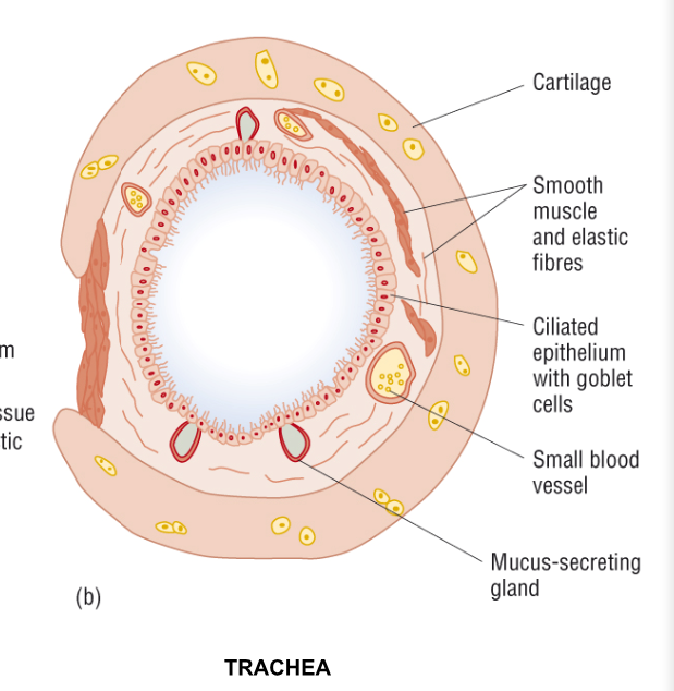
What is the structure of the trachea?
It has C-shaped rings of cartilage (c-shaped to allow for expansion of oesophagus) to ensure the channel remains open. Lined with ciliated epithelium, which sweep mucus, dust, bacteria to the mouth to stomach. Inside covered in mucus (produced by goblet cells and mucus glands). Wall contains smooth muscle and elastic fibres.
What is the function of the smooth muscle in the trachea?
It adjusts the size of the airways, can constrict to reduce lumen in trachea etc, and restrict air flow.
What is the function of the elastic fibres in the trachea?
They stretch when inhaling, and alveoli expand, increasing surface area. Fibres recoil when breathing air, forces out more air.
Helps prevent alveoli from bursting.
Describe the journey of air into the lungs.
Nose/mouth, epiglottis, trachea, bronchus, bronchioles, alveoli.
What covers the alveoli?
Surfactant - this prevents them from collapsing when exhaling by reducing surface tension.
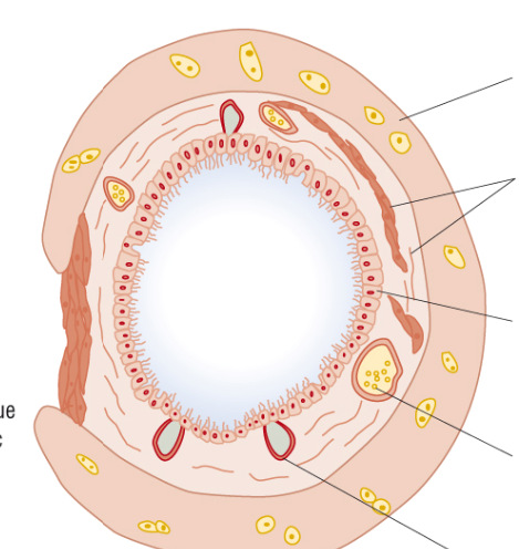
Label the diagram of the trachea.
Describe the process of inhalation (human).
External intercostal muscles contract, pulling ribcage upwards and outwards.
Pulls out outer pleural membrane, reduces pressure in pleural cavity and inner pleural membrane moves outward/ Pulls on surface of lungs and causes alveoli to expand.
Diaphragm contracts, to a flattened shape.
Thorax and lungs volume increase, pressure reduced, air enters.
Describe the process of exhalation (human).
External intercostal muscles relax and ribcage falls under its own weight.
Diaphragm relaxes and gut pressure pushes it back into dome shape.
Vol of thorax and lungs decreases, pressure increased, air forced out.
What are the 2 circulation systems in mammals?
Pulmonary - Serves lungs (pumps from right side to lungs and carries oxygenated blood back)
Systemic - Serves body tissues (left side of heart pumps oxygenated blood to tissues)
What are the 3 types of blood vessel?
Arteries, veins and capillaries.
What is the inner layer of veins & arteries and what does this consist of?
Tunica intima, single layer of endothelium (smooth lining to reduce friction).
What is the middle layer of veins & arteries and what does this consist of?
Tunica media, contains elastic fibres and smooth muscle. Contraction of smooth muscle regulates blood flow and maintains blood pressure.
What is the outer layer of veins & arteries and what does this consist of?
Tunica externa, contains collagen fibres, which resist overstretching.
How does the tunica media differ in veins & arteries?
In arteries, the elastic fibres allow stretching to accomodate changes in blood flow and pressure as blood is pumped from the heart. At a certain point, stretched elastic fibres recoil, pushing blood on through the artery. This is felt as the pulse.
What is the functions of arteries?
They carry blood away from the heart. Their thick, muscular walls withstand the blood's high pressure. They branch into smaller vessels called arterioles, which then divide into capillaries.
What is the function of capillaries?
They form a vast network that penetrates all tissues and organs of the body. Blood from capillaries collects into venules, which take blood into venules for transport back to heart.
What are the properties and function of veins?
Larger diameter lumen and thinner walls with less muscle than arteries. Blood pressure and flow rate are lower. Veins have semi-lunar valves along their length, preventing back flow.
How long does the cardiac cycle last?
0.8 seconds on average.
What are the 3 stages of the cardiac cycle?
Atrial systole, ventricular systole and diastole.
What happens in atrial systole?
The atrium walls contract, and the blood pressure in the atria increases. This pushes the blood through the tricuspid and bicuspid valves down into the ventricles, which are relaxed.
What happens during ventricular systole?
Ventricle walls contract, and increase blood pressure in ventricles. This forces blood thru SL valves, out of heart and into p artery and aorta. The blood cannot flow back from the ventricles as the AV valves close due to the rise in V pressure.
What happens during diastole?
The ventricles relax. Increases volume and decreases pressure. SL valves close due to tendency of backflow from aorta and pulmonary artery. Atria also relax, so blood from vena cava and Pulmonary vein enter the atria again.
What is the sinoatrial node?
Pacemaker. A specialised tissue that generates electrical excitations.
What happens when a signal is sent? (Pulse)
An electrical impulse travels from SAN to the walls of atria, making them contract
The impulse reaches the AVN which delayed it by 0.1s (allows ventricles to fill with blood)
Bundle branches carry signals from the AVN to the heart apex
The signal spreads through the ventricle walls, causing them to contract.
What happens when oxygen attaches to haemoglobin?
The first attachment of o2 is difficult and slow.
Hb changes shape —> next attachment of 3o2s is a lot easier.
What conditions favour the attachment of o2 to Hb? (LUNGS)
high pp of o2
low pp of co2
lower temperature
What conditions favour the disassociation of o2 to Hb? (CELLS/TISSUES)
low pp of o2
high pp of co2
higher temperature
What is the Bohr shift/effect and what happens?
It’s when more co2 is added that causes the sigmoid arc to shift to the right.
The pp of co2 increases meaning Hb has less affinity for o2 —> favours conditions in tissues.
What are the components of blood plasma?
red blood cells
white blood cells
water
plasma protein
ions
glucose
antibodies (immunoglobulin)
lipids
What are the components of tissue fluid?
white blood cells
water
ions
glucose
antibodies (immunoglobulin)
lipids
What are the components of lymph?
white blood cells
water
ions (less than tf)
glucose (less than tf)
antibodies (immunoglobulin) (more than tf)
lipids (more than tf)
Why are no red blood cells found in tissue fluid?
Because red blood cells are too large to pass through capillary walls, meaning that they can’t leak into the tissue fluid, whereas wbc are flexible.
Why is the cartilage of the trachea not in a full ring?
The oesophagus is directly beside the trachea so the muscles need to be able to stretch and expand to push food down. Needs flexibility.
What is the cartilage present in and what is the function?
trachea (ring)
bronchi (plate)
provides strength and holds open the airways for less resistance.
What is the ciliated epithelium present in and what is the function?
trachea
bronchi
bronchial
on outer membrane of cells, moves mucus up to mouth - for germs to be swallowed + killed by stomach acid
What are the goblet cells present in and what is their function?
trachea
bronchi
bronchial
(shaped like goblet)
secretes mucus (glycoprotein) and traps particles of dust, bacteria and spores.
What is the smooth muscle present in and what is the function?
trachea
bronchi
bronchial
contracts to narrow the airways which reduces water and heat loss.
What are the elastic fibres present in and what is their function?
trachea
bronchi
bronchial
alveoli
stretch when breathing in, RECOIL when breathing out to help force air out of lungs.
What happens at the P wave?
depolarisation of the atria
contraction of the atria (systole)
What happens at the QRS complex?
depolarisation of the ventricles
contraction of the ventricles (systole)
What happens at the T wave?
repolarisation of the ventricles
relaxation of the ventricles (diastole)
Name the structures which ensure that AV valves only open in one direction:
Tendinous chords.
How does increasing co2 in the plasma cause haemoglobin to release oxygen?
co2 diffuses into cytoplasm of rbc
carbonic anhydrase catalyses the conversion of co2 and h2o into carbonic acid
carbonic acid dissociates into H+ ions and hydrogen carbonate ions
hydrogen carbonate ions diffuse into plasma - chloride ions diffuse into rbc to achieve ELECTRICAL NEUTRALITY (CHLORIDE SHIFT)
H+ ions attach to Hb to form haemoglobinic acid and displaces o2
o2 diffuses out of rbc to plasma & tissues
REVERSE IN LUNGS
Why does altitude have an impact on red blood cells?
Because there’s a low partial pressure of oxygen, so more rbc need to be created in order to retain the maximum amount of o2 and respire.
Equation for oxygen dissociation?
carbon dioxide + water ———→ carbonic acid —> hydrogen ion + hydrogen carbonate ions
carbonic anhydrase