TOP 20 CAT DISEASES
 Call Kai
 Learn
Learn Practice Test
 Spaced Repetition
 Match
Match Flashcards
 Knowt Play
1/202

There's no tags or description
Looks like no tags are added yet.
Name | Mastery | Learn | Test | Matching | Spaced | Call with Kai | Chat |
|---|
No analytics yet
Send a link to your students to track their progress
203 Terms
Feline asthma classic presentation
Young adult cat with a history of coughing, wheezing, tachypnea, and +/- dyspnea or respiratory distress ("status asthmaticus").
Status asthmaticus
Dyspnea or respiratory distress in feline asthma.
Feline asthma risk factors and predispositions
Urban cats, Siamese, and Himalayans are at an increased risk.
Feline asthma thoracic radiograph findings
Bronchiolar or bronchointerstitial pattern, pulmonary inflammation, and pulmonary hyperinflation.
Feline asthma CBC findings
May show +/- eosinophilia.
Bronchoalveolar lavage with cytology +/- culture
Diagnostic test used to rule out differential diagnoses such as Mycoplasma in feline asthma workup.
Fecal Baermann
Diagnostic test used to rule out respiratory parasites in cats.
Status asthmaticus acute treatment
Oxygen and minimizing handling, bronchodilators by nebulization (albuterol) or parenteral (terbutaline), and parenteral glucocorticoids.
Chronic feline asthma therapy
Bronchodilators (theophylline, aminophylline, terbutaline), glucocorticoids (prednisolone), oral Doxycycline suspension for concurrent airway infection, and cyclosporine if there is no response to glucocorticoids.
Environmental irritants in feline asthma
Aerosols, dusty cat box filler, incense, and smoke, which should be reduced to manage feline asthma.
Feline coughing clinical pearl
Cats cough with asthma, NOT cardiac disease.
Bronchodilator monotherapy rule
Bronchodilators should NOT be used as monotherapy for feline asthma.
Beta-blockers in asthmatic cats
Medications that can worsen bronchoconstriction and should not be given to asthmatic cats.
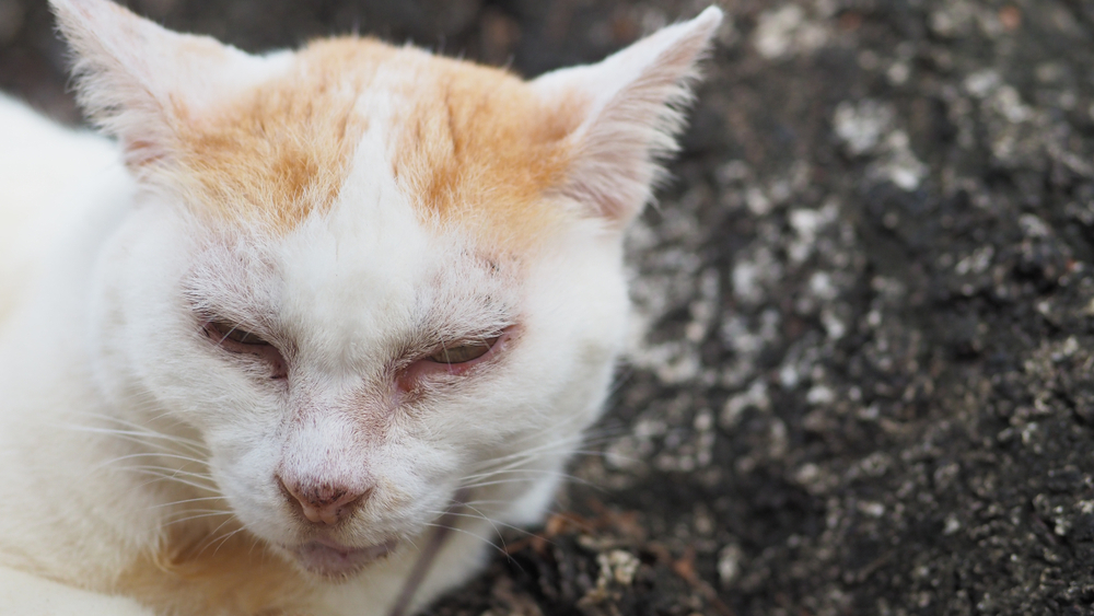
Chlamydiosis
An ophthalmic disease in young cats/kittens presenting with
unilateral then bilateral conjunctivitis (ocular discharge, redness)
with or without concurrent upper respiratory tract infection (nasal discharge, sneezing).
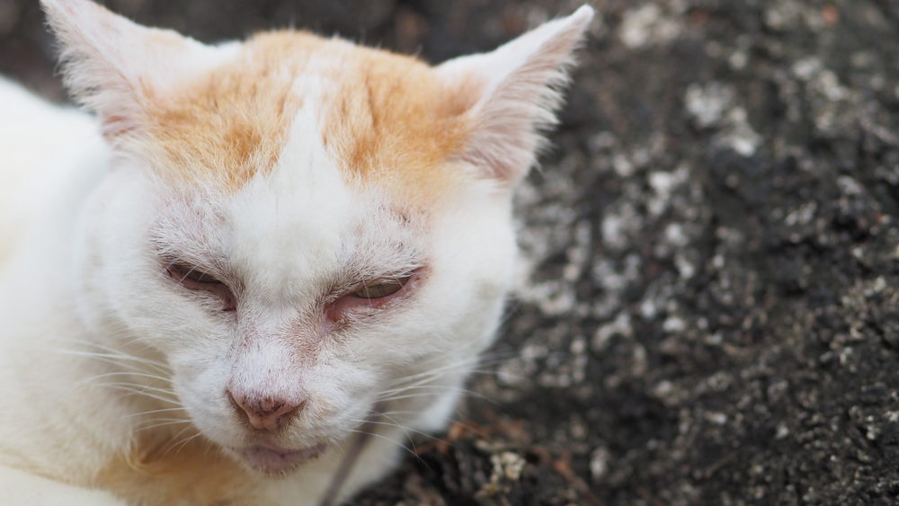
Chlamydophila felis
The etiologic infectious agent responsible for feline chlamydiosis.

FIP Uveitis
An ophthalmic condition in young, purebred cats
characterised by
systemic signs (anorexia, lethargy)
iris color change
keratitic precipitate
aqueous flare, carrying a poor prognosis.
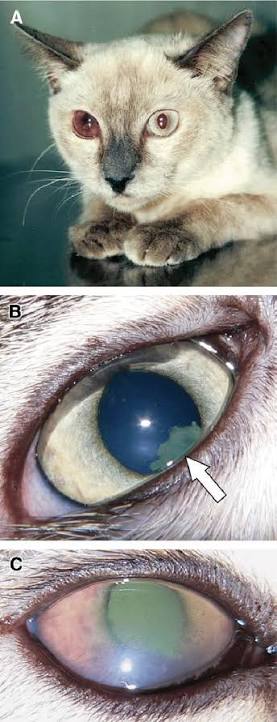
FIP Uveitis Diagnostic Findings
Ophthalmic exam showing aqueous flare and hyphema
tonometry showing typically decreased intraocular pressure
difficult to confirm antemortem due to tests indicating exposure to either benign feline coronavirus (FCoV) or mutated FIP-causing coronavirus.

Fluorescein Stain
A diagnostic stain that reveals a bright green delineation on corneal ulcers, used to confirm corneal scratches.

Descemetoceles
Deep corneal scratches that reach Descemet's membrane and characteristically test fluorescein stain negative.
Chlamydiosis Treatment
Systemic doxycycline (using liquid oral suspension to avoid esophagitis and stricture formation) and topical doxycycline, treating all household cats, and cleaning the environment.
FIP Uveitis Treatment
Topical glucocorticoids with or without topical mydriatics (1c/o atropine sulfate, specified as 1\text{\textpercent} or 1\text{\textperthousand} atropine sulfate).
Corneal Scratches Clinical Signs
Condition presenting with blepharospasm, epiphora, conjunctival hyperemia, and a corneal ulcer stained with fluorescein.
Corneal Scratches Treatment
Topical broad-spectrum antibiotic ointment (avoiding gentamicin as first choice), systemic analgesics (buprenorphine), topical mydriatics, Elizabethan collar, optional autologous serum, and keratectomy/conjunctival graft referral if deep or ruptured.
Autologous Serum
A therapeutic agent used in corneal scratch management specifically for its anticollagenase activity.
Topical Corticosteroids Contraindication
A category of medication that is strictly contraindicated in the presence of corneal scratches.
Jugular Pressure Precaution
An action to avoid during corneal scratch management because it may increase intraocular pressure.

Dermatophytosis
Also known as ringworm
a fungal skin disease common in juvenile or geriatric cats (especially Persians and Himalayans)
characterised by non-pruritic lesions, scaling, crusting, ulceration, and alopecia.
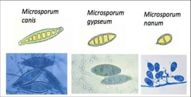
Microsporum canis
The fungal etiology responsible for 98% of ringworm cases in cats.
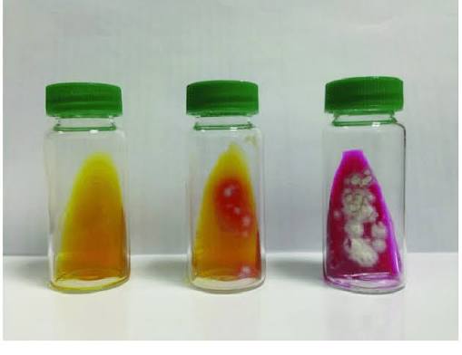
Dermatophyte Testing Media (DTM)
The best culture medium for diagnosing ringworm, collected by rubbing a new toothbrush on lesions.

Wood's lamp exam
A diagnostic screening tool for ringworm that can yield false positives and false negatives.
Ringworm Topical Treatment
Topical therapy consisting of lime sulfur or enilconazole.
Ringworm Systemic Treatment
Systemic medication consisting of intraconazole (or griseofulvin).
Environmental Ringworm Control
Decontamination process requiring hair removal and cleaning with a 1:32 bleach/water solution.
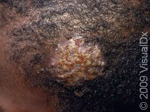
Kerion
An atypical nodular mass lesion associated with ringworm.
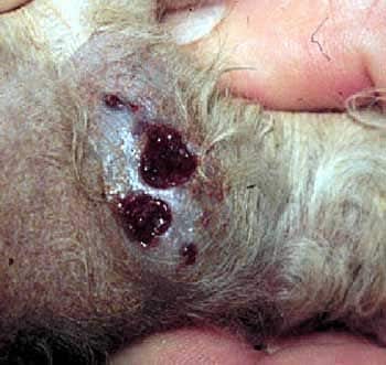
Mycetoma
An atypical pyogranulomatous nodule lesion caused by ringworm.

Ringworm Zoonotic Risk
High zoonotic potential transmitted via fomites, where around 50% of humans living with infected cats will develop lesions.
Ringworm Risk Factors
Predisposing factors including FeLV, malnutrition, stress, immunosuppression, crowding, and poor sanitation.

Otodectes cynotis
The etiology (causative agent) of ear mite infections, commonly affecting kittens and treatable with otic and systemic miticidals.
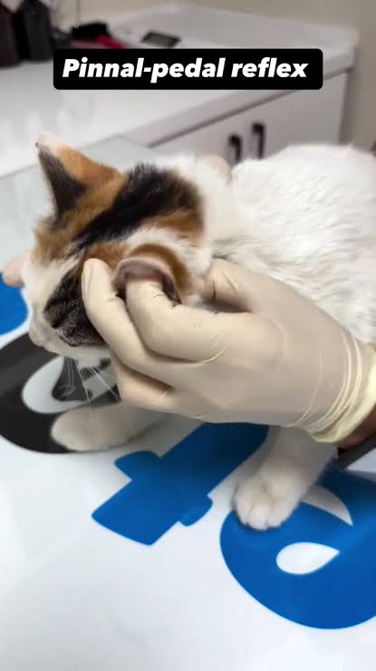
Ear-pedal reflex
A reflex present in ear mite cases where the patient scratches with their pelvic limb when the ear is rubbed.
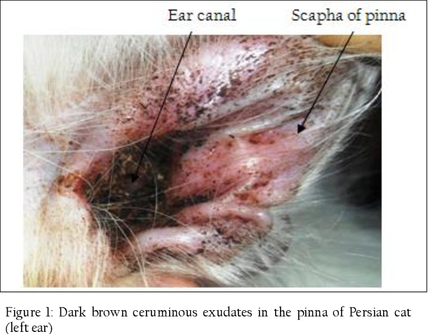
Ear mite clinical signs
Classic presentation including
head shaking
pruritus
erythematous ears
dark brown debris in ear canals
papules on the rump, tail, and neck.

Cytology of ear swab in mineral oil
A diagnostic test used to visualize Otodectes cynotis mites, eggs, and +/- secondary bacteria under a microscope.
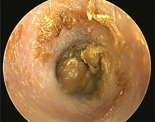
Otoscopy (Ear Mites)
A diagnostic method used to directly visualize live mites and inflammation within the ear canal.
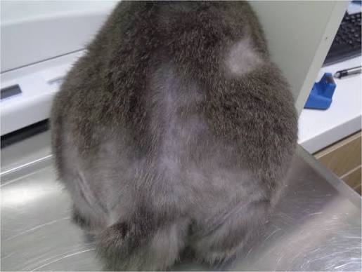
Ectopic mites
Mites located on areas outside the ear canal, such as the rump, tail, and neck, requiring systemic miticidal therapy.
Miticidal otic preparation
A topical ear treatment (such as milbemycin or ivermectin) applied after removing ear debris to eliminate ear mites in the canal.
Systemic miticidal
A systemic treatment (such as selamectin or ivermectin) used to eliminate ectopic ear mites.

Otodectes cynotis magnification
The magnification level (100x) used to identify Otodectes cynotis.
Ear mite prognosis
The overall prognosis is good, although the condition may recur or become chronic.
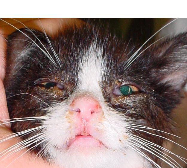
Classic Presentation of Feline URIs
Fever, lethargy, and ocular or nasal discharge seen in kittens from shelters or rescues.
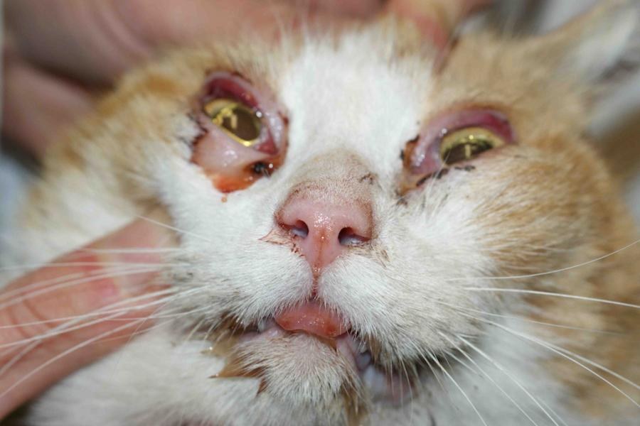
Rhinotracheitis
A respiratory disease caused by feline herpesvirus 1 (FHV-1), characterized by rhinitis, clear nasal discharge, and conjunctivitis.
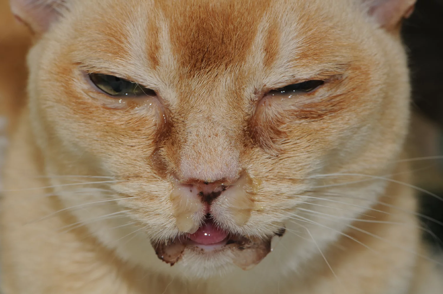
Feline Herpesvirus 1 (FHV-1)
The causative agent of rhinotracheitis which, along with calicivirus, accounts for 90% of upper respiratory infections (URIs) in cats.
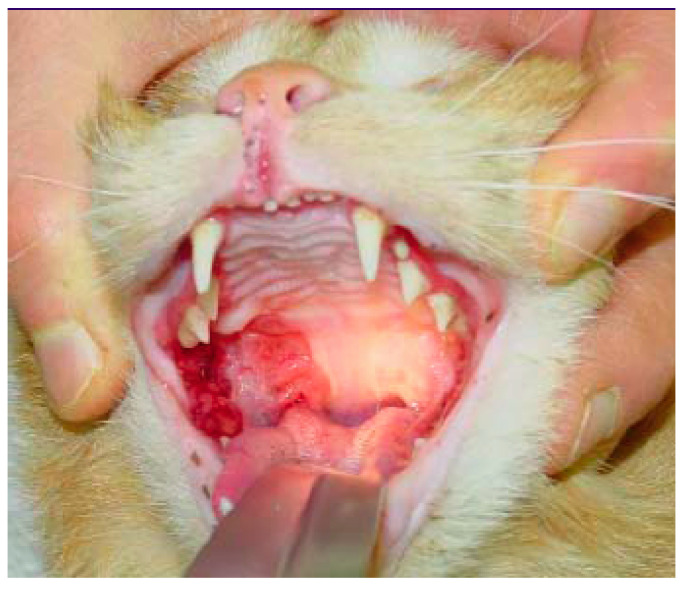
Calicivirus
A feline upper respiratory virus characterized by tongue ulceration, gingivitis, and ptyalism.

Chlamydiosis
Primarily an ocular disease in cats characterized by conjunctivitis that is first unilateral and then bilateral.
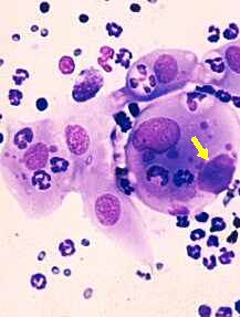
Chlamydiosis Diagnosis
Diagnosed primarily by identifying the presence of inclusion bodies in conjunctival scrapings.
FHV-1 and Calicivirus Treatment
Symptomatic and supportive care, with early oral use of antivirals (triuoridine, idoxuridine, cidofovir, and famciclovir) being safe and effective.
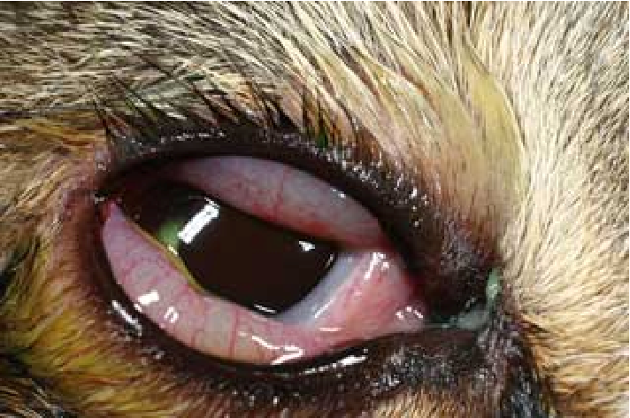
Chlamydiosis Treatment
Treatment with doxycycline.
Incubation Period
The 2 to 10 day period required for feline respiratory viruses to develop.
Viral Swabs (PCR / Culture)
Mouth or eye swabs required to determine which specific virus is affecting an individual cat.
Carrier State
The condition of most cats after recovery, wherein they remain carriers and may shed the virus intermittently.
Disinfection Solution
A 1:32 bleach solution to which feline upper respiratory viruses are susceptible.
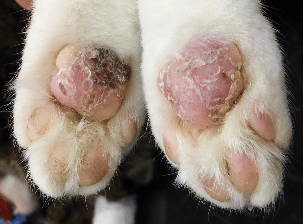
Acetaminophen Toxicosis
A toxicity caused by ingestion of at least
10mg/kg of Tylenol® (regular strength = 325mg, extra strength = 500mg)
presenting with lethargy, inappetence, weakness, depression, dyspnea, vomiting, and facial and paw edema.
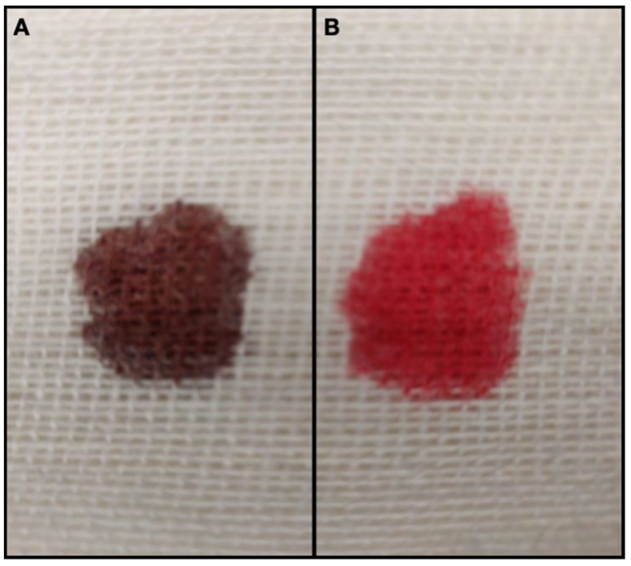
Methemoglobin
Elevated parameter in EDTA blood used as the primary diagnostic finding for acetaminophen toxicosis.

Acetaminophen Decontamination & Medical Therapy
Treatment protocol consisting of aggressive decontamination
xylazine or dexmedetomidine to induce vomiting
N-acetylcysteine (NAC, MucomystⓇ) IV or PO
S-adenosylmethionine (SAMe) PO
ascorbic acid PO or injectable
+/- oxygen support.
Ethylene Glycol (EG)
Antifreeze toxicant whose ingestion causes clinical signs of ataxia, stupor, depression, and PU/PD.

Ethylene Glycol (EG) Test Kit
Diagnostic kit for ethylene glycol toxicosis that must be used before giving activated charcoal.
Ethylene Glycol Treatment Protocol
Therapy requiring prompt action:
induce vomiting and give activated charcoal if within 30-60min
IV fluids with bicarbonate
IV fomepizole (4-MP) if within 3hrs; or ethanol in IV fluids if > 3hrs.
Fomepizole (4-MP)
An IV treatment for ethylene glycol toxicosis administered if within 3hrs of ingestion, given at a much higher dose than for dogs.
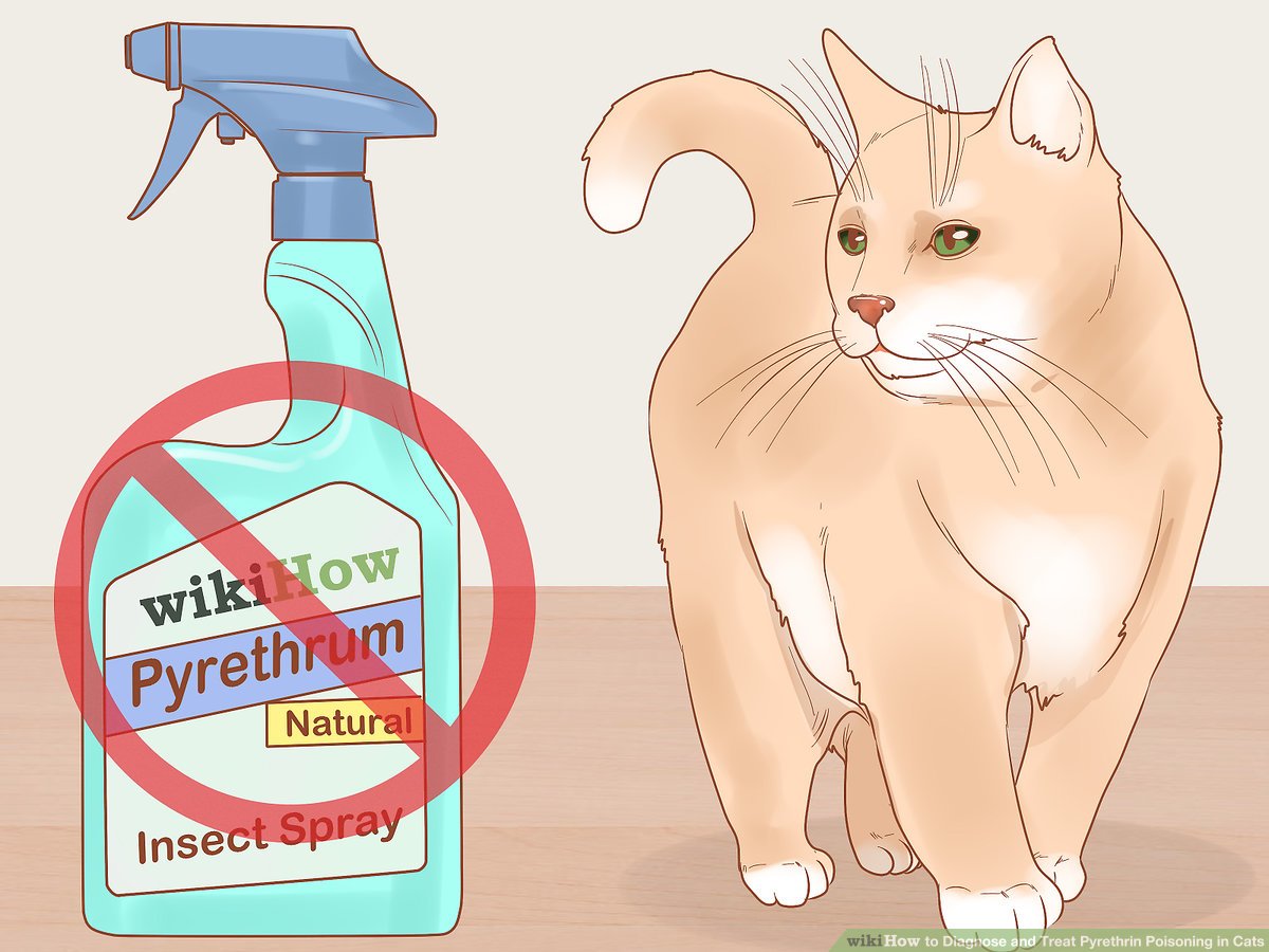
Pyrethrin / Pyrethroid Toxicosis
Toxicosis caused by over-the-counter spot-on flea control, usually with a history of a recently treated family dog or use of a dog product on a cat, presenting clinically with seizures.
Pyrethrin / Pyrethroid Diagnosis
Diagnosed based on patient history or by observing a greasy spot on the fur.

Pyrethrin / Pyrethroid Treatment
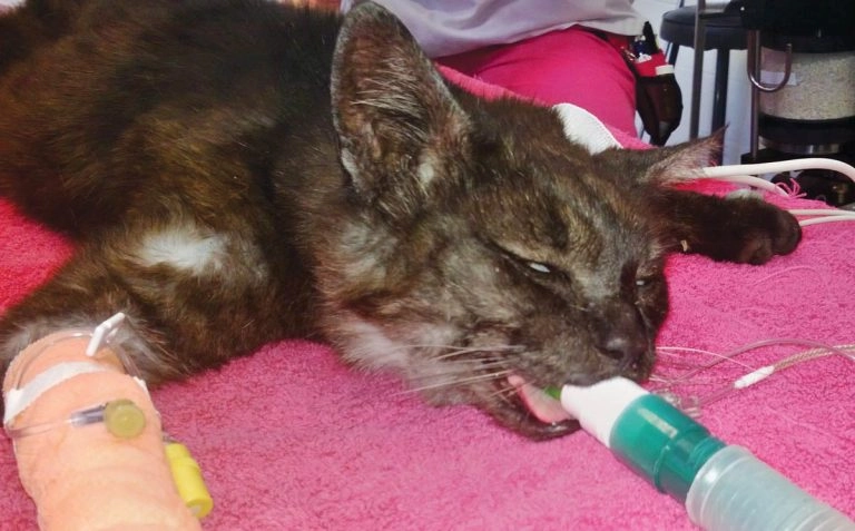
Management consisting of methocarbamol IV
+/- midazolam or propofol IV
bathing with warm water and mild dishwashing detergent if few or no signs of toxicosis.
Refractory Seizures
Severe toxicosis-induced seizures that may require gas anesthesia to control.

Eosinophilic skin disease
A condition in mature cats classically characterized by
raised, reddened, or yellow linear
Or circular plaques on the lips, footpads, ventral abdomen, and thighs.

Eosinophilic ulcer
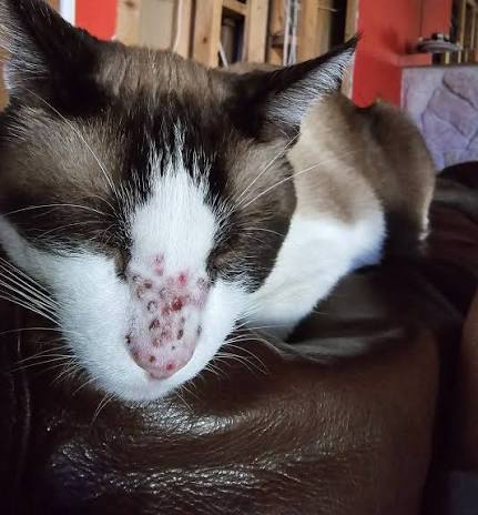
Also known as a rodent ulcer, an erythematous, nonpainful, and nonpruritic ulcer usually located on the upper lip.
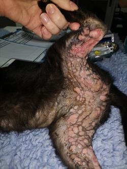
Eosinophilic plaque
Raised erythematous lesions typically found on the medial thigh and abdomen
accompanied by peripheral eosinophilia and pruritus.
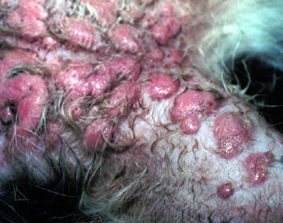
Eosinophilic granuloma
Yellow, orange, or pink lesions found anywhere but mostly on the caudal thighs (linear) and in the oral cavity; occurs more frequently in females and displays peripheral eosinophilia when oral cavity lesions are present.
Eosinophilic skin disease diagnosis
Performed using incisional or excisional biopsy.
Eosinophilic skin disease treatment
Includes anti-inflammatory therapy with prednisolone or cyclosporine, radiotherapy, laser therapy, surgical excision, and treating underlying causes like atopy or allergies.
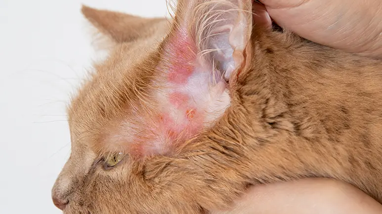
Eosinophilic ulcers and plaques associations
Conditions commonly associated with flea or food allergies and atopy.
Eosinophilic granulomas association
Condition commonly associated with mosquito bites.
Hypertrophic cardiomyopathy
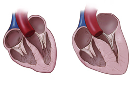
A common feline heart disease, often asymptomatic initially, characterized by
left ventricular wall thickening (6mm or greater)
atrial dilation
potential progression to congestive heart failure or aortic thromboembolism.
Maine coon cats and ragdolls
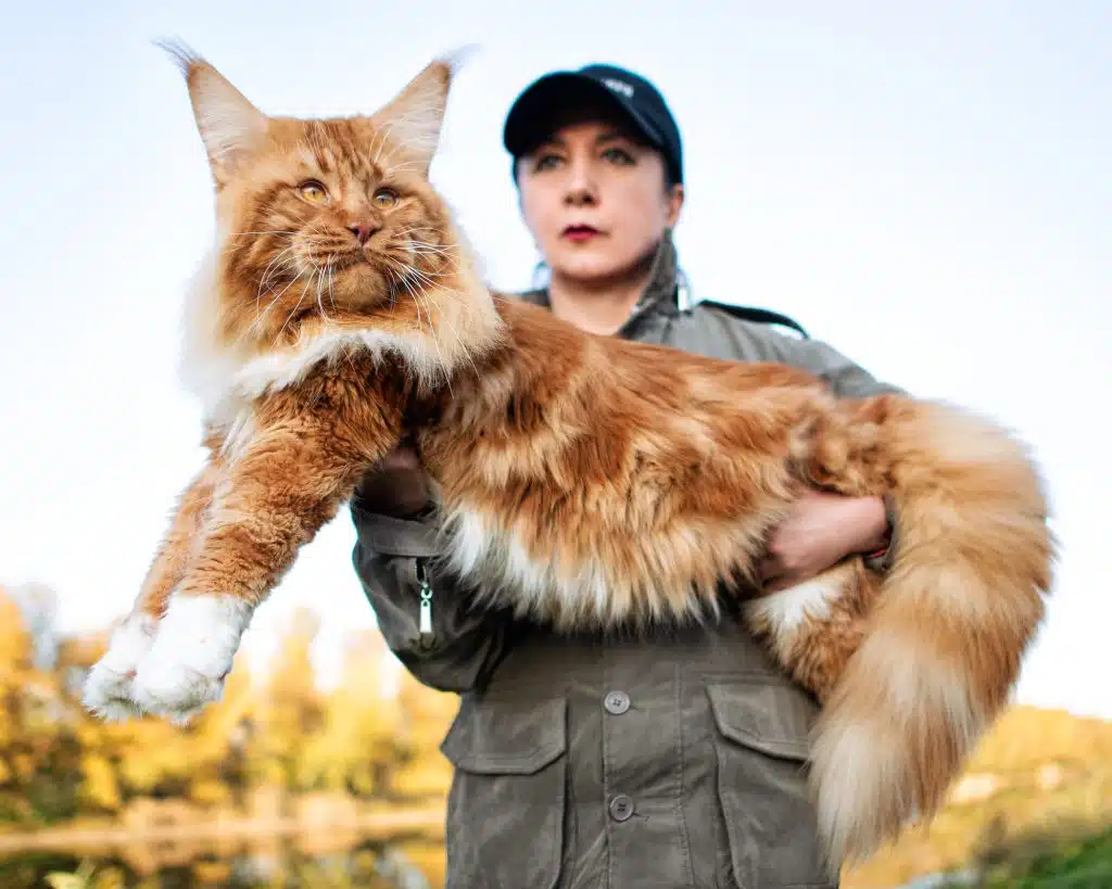
Cat breeds with a hereditary basis for hypertrophic cardiomyopathy (presents as early as 4 months)
linked to autosomal dominant mutations in the cardiac myosin-binding protein C gene.
Signs of congestive heart failure (CHF)
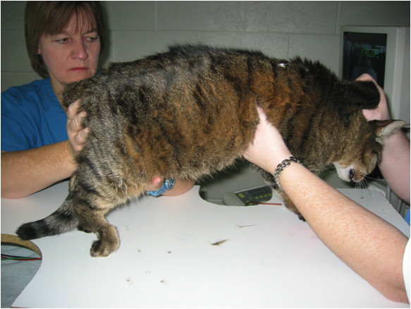
Clinical indications in cats with HCM including tachypnea, dyspnea, and open-mouth breathing.
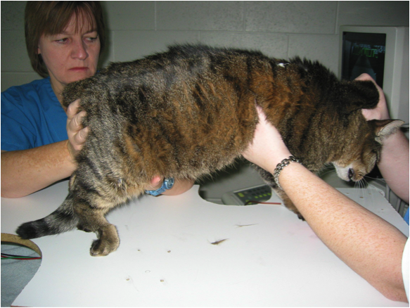
Signs of aortic thromboembolism
Clinical findings resulting from blood clot occlusion, including cyanotic nail beds, loss of pulses, and paraplegia.
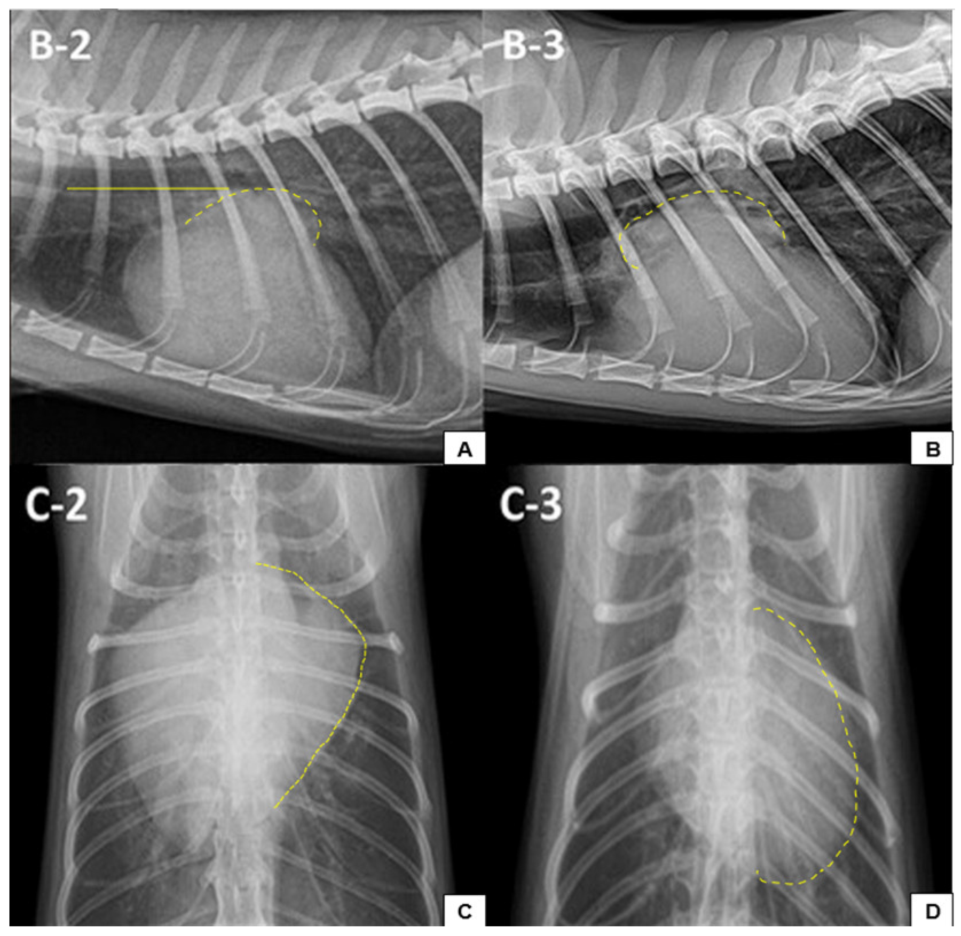
Lateral thoracic radiograph
A radiographic view demonstrating increased sternal contact of the heart as seen with hypertrophic cardiomyopathy.

Dorsoventral (DV) thoracic radiograph
The most important and better-tolerated radiographic view for feline HCM diagnosis, which may show cardiomegaly, pulmonary edema, pleural effusion, or a classic valentine shape.
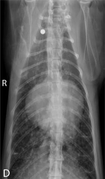
Valentine shape
The classic cardiac silhouette seen on a DV thoracic radiograph resulting from a dilated left atrium in cats with hypertrophic cardiomyopathy.
Echocardiography (M-mode) findings in HCM
Diagnostic ultrasound findings including
atrial dilation
left ventricular wall thickening (6mm or greater)
papillary muscle hypertrophy
systolic anterior motion of the mitral valve in obstructive cardiomyopathy.
Plasma brain natriuretic peptide (BNP)
A cardiac biomarker elevated in severe cases of hypertrophic cardiomyopathy that may also be high in compensated cats.
Acute CHF treatment protocol
Emergency management consisting of
supplemental oxygen
thoracocentesis if necessary
furosemide
nitroglycerin
avoiding fluid therapy and beta-blocker therapy (unless tachyarrhythmia is present).
Chronic HCM treatment protocol
Long-term medical therapy including a
beta-blocker starting at a low dose (e.g., Atenolol)
furosemide
clopidogrel (PlavixⓇ) or baby aspirin to prevent thromboembolism
potassium supplementation if hypokalemia is present.
Cardiac myosin-binding protein C gene
The gene in Maine coons and ragdolls associated with mutations causing hypertrophic cardiomyopathy through autosomal dominant inheritance with incomplete penetrance.
Triggers for CHF in compensated HCM
Interventions or events that commonly trigger congestive heart failure in previously compensated cats, specifically IV fluids, corticosteroids, or recent anesthesia.
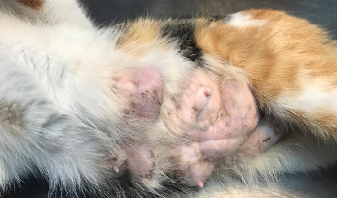
Feline Mammary Tumors
Common feline cancer (85-90% malignant) presenting in unspayed females or cats spayed after several litters as an enlarged mammary gland or rm nodule, most commonly involving anterior glands.
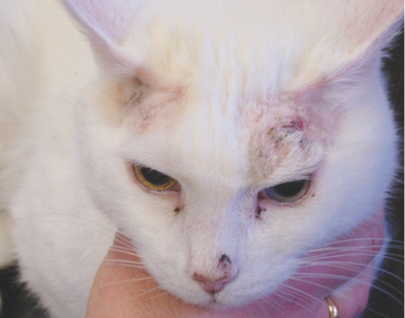
Cutaneous Squamous Cell Carcinoma (SCC)
Form of SCC presenting as small, crusty or ulcerated sores on the head (pinnae, nasal planum, eyelids) in white-haired cats.

Oral Squamous Cell Carcinoma (SCC)
Form of SCC in senior/geriatric cats affecting the tongue, gingiva, and sublingual areas; carries a very poor prognosis with survival times ranging from 3-4 months.
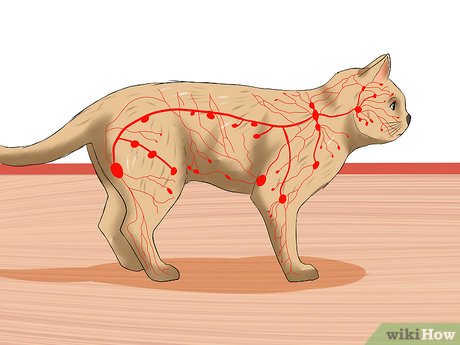
Lymphosarcoma (LSA)
Cancer accounting for 30% of all feline tumors, characterized by lymphadenopathy and thickened bowl loops.

FeLV Link to LSA
Viral infection linked with most feline lymphosarcoma cases except for the GI form.
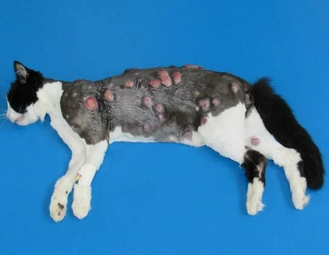
FIV Risk in LSA
Feline immunodeficiency virus infection that makes cats 5X more likely to develop LSA.
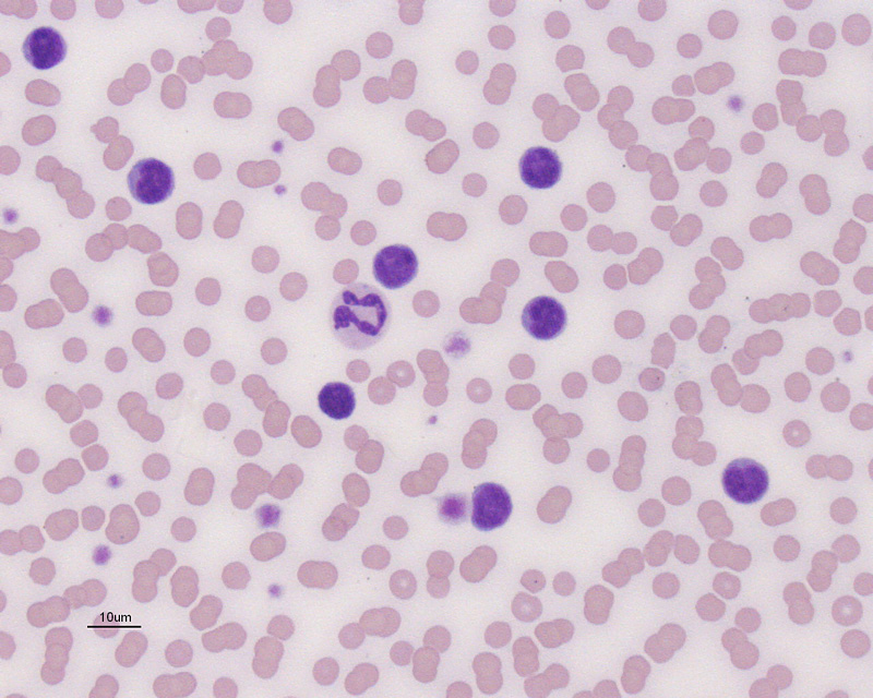
Small Cell GI LSA Treatment
Specific medical treatment for small cell gastrointestinal LSA consisting of prednisolone and chlorambucil.
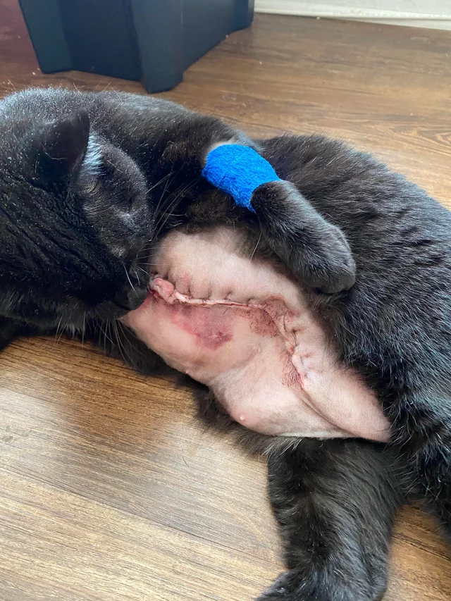
Feline Mammary Tumor Surgical Treatment
Surgical procedure consisting of unilateral mastectomy and regional lymph node removal (axillary and inguinal).
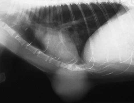
Thoracic Radiographs in Mammary Tumors
Diagnostic imaging performed to evaluate common metastases to lungs and lymph nodes.