Perio II final from study guide with explanation
 Call Kai
 Learn
Learn Practice Test
 Spaced Repetition
 Match
Match Flashcards
 Knowt Play
1/159

There's no tags or description
Looks like no tags are added yet.
Name | Mastery | Learn | Test | Matching | Spaced | Call with Kai | Chat |
|---|
No analytics yet
Send a link to your students to track their progress
160 Terms
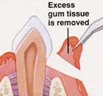
What is a gingivectomy?
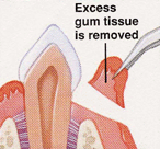
Excision of the soft tissue wall of a pathologically deepened gingival crevice or periodontal pocket.
(Explanation: A gingivectomy removes excess gingival tissue to eliminate periodontal pockets or improve aesthetics. It's typically performed when pockets are suprabony and don't involve bone loss, focusing on soft tissue reshaping. This procedure is less invasive than flap surgeries but sacrifices keratinized tissue, which is critical for gingival health.)
What are the disadvantages of a gingivectomy?
Increased post-op discomfort/bleeding, heals by secondary intention (no primary closure), no osseous recontouring (no bone access), sacrifices keratinized tissue.
(Explanation: Healing by secondary intention means the wound is left open to granulate, leading to more discomfort and slower recovery compared to primary closure. Lack of bone access limits its use in cases requiring osseous surgery, and removing keratinized tissue can compromise gingival stability, especially in patients with thin biotypes.)
What are the indications for a gingivectomy?
Suprabony pockets with firm fibrous walls/pseudo-pockets, gingival enlargement (plaque control/esthetics), patients on cyclosporine with no bone loss, patients on amlodipine (gingival hyperplasia), gingival craters, altered passive eruption, lack of bilateral symmetry.
(Explanation: Suprabony pockets are above the bone level, making gingivectomy suitable as it doesn't require bone manipulation. Gingival enlargement, often drug-induced (e.g., cyclosporine, amlodipine), or esthetic issues like uneven gingiva or altered passive eruption (where teeth appear short due to excess gingiva) are addressed to improve function and appearance.)
Why might amlodipine cause gingival hyperplasia, and what can be done?
Amlodipine (a calcium channel blocker) causes gingival hyperplasia; the MD could prescribe a different medication, unlike cyclosporine where this is not an option.
(Explanation: Amlodipine, used for hypertension, can cause gingival overgrowth due to its effect on fibroblast proliferation. Switching to another antihypertensive is feasible, but cyclosporine, an immunosuppressant for transplant patients, is often irreplaceable, making gingivectomy a primary solution for hyperplasia in these cases.)
What are the requirements for performing a gingivectomy?
Wide zone of keratinized tissue, no vertical osseous defect, no need for osseous access.
(Explanation: A wide zone of keratinized tissue ensures enough healthy gingiva remains post-surgery to maintain periodontal health. Vertical osseous defects require bone recontouring, which gingivectomy cannot address, as it only involves soft tissue excision without flap elevation to access bone.)
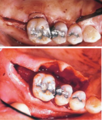
What is the purpose of a soft tissue wedge procedure?
To reduce periodontal pockets in retromolar areas and edentulous areas adjacent to teeth, used for crown lengthening, and as extensions of palatal/buccal flap incisions.
(Explanation: The distal wedge targets deep pockets in areas like the retromolar pad or edentulous ridges, where access is challenging. It's also used in crown lengthening to expose more tooth structure for restorative purposes, often integrated with flap surgeries to enhance access and outcomes.)
What are the contraindications for a soft tissue wedge procedure?
Insufficient keratinized tissue, osseous defects, flat palate, heavy external oblique ridge, proximity to the ascending ramus.
(Explanation: Insufficient keratinized tissue risks poor healing and gingival recession. Osseous defects require bone management, not just soft tissue removal. Anatomical constraints like a flat palate or prominent external oblique ridge (in the mandible) limit surgical access, and proximity to the ascending ramus risks nerve damage.)
How is the buccolingual width determined in a soft tissue wedge procedure?
Depends on probing depth and amount of keratinized tissue; should not exceed the distance between buccal and lingual line angles.
(Explanation: The width is tailored to the pocket depth and available keratinized tissue to ensure adequate tissue remains for healing. Limiting incisions to the buccal-lingual line angles prevents overextension, which could compromise blood supply or flap stability.)
How deep should the incision be in a soft tissue wedge procedure?
1/2 to 2/3 of the distal probing depth.
(Explanation: This depth ensures sufficient pocket reduction while preserving enough tissue for healing. It's calculated based on the probing depth to remove diseased tissue without overly thinning the gingiva, which could lead to recession or poor wound closure.)
What are the steps of a soft tissue wedge procedure?
1. Parallel distal internal bevel incisions (train tracks). 2. Distal transverse incision. 3. Intrasulcular incision. 4. Remove palatal collar (if pocket present). 5. Grab wedge with curved hemostat. 6. Incise root of tooth with Orban knife. 7. Remove wedge with large curette. 8. SRP proximal tooth surface. 9. Osseous recontouring/resection if needed.
(Explanation: The "train tracks" incisions create a wedge-shaped excision for precise tissue removal. The transverse and intrasulcular incisions facilitate access, while the palatal collar is removed only if a pocket exists to avoid unnecessary tissue loss. Instruments like the Orban knife and curette ensure clean removal, followed by scaling and root planing (SRP) to smooth the tooth surface. Osseous recontouring addresses any minor bone irregularities.)
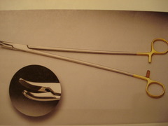
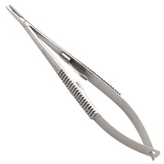
What are the differences between Crile Wood and Castroviejo needle holders?
Crile Wood is hard to open with one hand; Castroviejo is easy to operate with one hand and preferred by Gellin.
(Explanation: Castroviejo needle holders are designed for precision in periodontal surgery, allowing one-handed operation, which is critical for delicate suturing tasks. Crile Wood holders, while effective, are less ergonomic, requiring two hands to open, which can slow down procedures.)
What type of needle holder should be used for suture needles?
Needle holder (straight head), not a hemostat.
(Explanation: Needle holders are specifically designed to grasp and manipulate suture needles securely without damaging them. Hemostats, meant for clamping tissue or vessels, can crush or bend needles, compromising suturing precision.)
What are the characteristics of suture needles used in periodontics?
Stainless steel, 1/2 or 3/8 circle radius, fused to suture material, reverse cutting (cuts on outer curvature, passes easily without tearing tissue).
(Explanation: Stainless steel ensures durability and biocompatibility. The 1/2 or 3/8 circle radius allows for precise maneuvering in tight oral spaces. Reverse cutting needles have a cutting edge on the outer curve, reducing tissue trauma compared to cutting needles, which cut on the inner curve.)
Which suture needles are commonly used for soft tissue grafts?
PS-2, PS-3, C-3.
(Explanation: These needles are smaller and designed for delicate soft tissue graft procedures, such as connective tissue grafts, where precision is critical to avoid damaging thin tissues or grafts.)
Which suture needles are commonly used for flap surgery?
FS-2, X-1.
(Explanation: FS-2 and X-1 needles are slightly larger and suited for flap surgeries, where thicker tissues or larger flaps require more robust needles to penetrate and secure tissue effectively.)
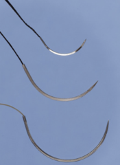
What are the ideal properties of sutures?
Ideal tensile strength (won't break), easy to handle (not too stiff/flimsy), minimal tissue reaction (heals as expected), secure knot (won't untie prematurely).
(Explanation: These properties ensure sutures can hold tissues together under tension, are easy to manipulate during surgery, cause minimal inflammation, and maintain knot integrity during healing to prevent wound dehiscence.)
How are sutures classified by duration?
Absorbable/resorbable (lasts ≤ 60 days, e.g., plain gut, chromic gut, PGA) vs. nonabsorbable (lasts > 60 days, e.g., silk, polypropylene, nylon, PTFE).
(Explanation: Absorbable sutures break down naturally, ideal for internal tissues that heal quickly. Nonabsorbable sutures are used when prolonged support is needed or for external closure, requiring removal post-healing.)
What is the difference between braided and monofilament sutures?
Braided (multifilament) is stronger but harbors debris/bacteria (wicking effect); monofilament has less friction, resistance, and bacteria/debris accumulation.
(Explanation: Braided sutures, like silk, are stronger but their multi-strand structure traps bacteria, increasing infection risk via wicking. Monofilament sutures, like PTFE, glide through tissue smoothly, reducing trauma and bacterial adherence, but may be less strong.)
Give examples of braided sutures.
Silk (natural, nonabsorbable), PGA (synthetic, absorbable).
(Explanation: Silk is used for its strength but is prone to bacterial wicking, making it less ideal in periodontics. PGA (Vicryl) is absorbable and used for internal suturing, balancing strength and biodegradability.)
Give examples of monofilament sutures.
Polypropylene, nylon, PTFE (synthetic, nonabsorbable).
(Explanation: These synthetic monofilaments are smooth, reducing tissue drag and bacterial adherence. PTFE, in particular, is favored for its biocompatibility and ability to stretch with edema, minimizing tissue trauma.)
How are synthetic and natural sutures different?
Synthetic (e.g., PTFE, nylon, PGA) cause less immune response; natural (e.g., silk, gut) cause greater immune response.
(Explanation: Synthetic sutures are engineered for consistency and minimal tissue reaction, improving healing outcomes. Natural sutures, derived from animal or plant sources, can trigger stronger immune responses, potentially leading to inflammation or delayed healing.)
How does suture size relate to the numbering system?
Higher number = thinner suture; common oral cavity sizes are 3-0 (larger), 4-0, 5-0 (smallest).
(Explanation: The numbering system (e.g., 3-0, 4-0) indicates suture diameter, with higher numbers denoting thinner sutures. Thinner sutures (e.g., 5-0) are used for delicate periodontal tissues to minimize trauma, while thicker ones (e.g., 3-0) are for stronger closures.)
What is the typical length of sutures used in periodontics?
18-27 inches.
(Explanation: This length provides enough material for multiple sutures or complex techniques in the oral cavity, allowing flexibility for knot tying and flap manipulation without needing frequent rethreading.)
Compare plain gut and chromic gut sutures.
Both are resorbable, natural, monofilament; plain gut retains strength for 3-5 days, chromic gut for 5-7 days (resistant to enzymatic resorption).
(Explanation: Plain gut, derived from sheep or cow intestines, degrades quickly via enzymatic breakdown, suitable for short-term closure. Chromic gut is treated with chromium salts to slow resorption, extending its strength for slightly longer healing periods.)
What are the characteristics of PGA 910 (Vicryl) sutures?
Synthetic, braided, absorbable; tensile strength for 10-14 days, resorbs in ~60 days.
(Explanation: PGA (polyglycolic acid) is widely used in periodontics for its predictable resorption and adequate strength during early healing. Its braided nature enhances strength but may increase bacterial adherence compared to monofilaments.)
Why is PTFE preferred by Gellin?
Synthetic, monofilament, nonresorbable, stretches with edema.
(Explanation: PTFE's smooth, nonabsorbable nature minimizes tissue trauma and bacterial adhesion. Its elasticity accommodates post-operative swelling (edema), reducing tension on the wound and improving patient comfort.)
Why is black silk not used in periodontics?
Major plaque trap, loosely braided, wicks bacteria.
(Explanation: Black silk's loose braiding traps plaque and bacteria, promoting infection via wicking (bacteria traveling along the suture into deeper tissues). This makes it unsuitable for the oral cavity, where hygiene is critical.)
What are the principles of suturing in periodontics?
Hold needle at center at 90° angle, start/tie on facial (not lingual), place 3 mm from flap edge, tie firmly without blanching, use 2-1-2 modified surgeon's knot.
(Explanation: Holding the needle at 90° ensures control and prevents breakage. Facial tying avoids lingual discomfort for patients. The 3 mm margin prevents flap tearing, and avoiding blanching (tissue whitening) prevents necrosis. The 2-1-2 knot balances security and tissue health.)
Describe the 2-1-2 modified surgeon's knot.
First throw: away, double loop; second throw: toward, single loop; third throw: away, double loop; grab tail (1.5 in) with needle holder, tie firmly without blanching.
(Explanation: This knot ensures secure closure with minimal tissue trauma. The double loops in the first and third throws add strength, while the single loop in the second alternates direction for stability. The 1.5-inch tail allows easy manipulation without excess suture.)
What is the alternative knot some surgeons use?
2-1-1 knot.
(Explanation: The 2-1-1 knot is a simpler variation, with a single loop in the third throw, used by some surgeons for faster tying or in less tension-heavy closures, though it may be less secure than the 2-1-2 knot.)
When is a loop interrupted suture used?
For primary closure of buccal and lingual flaps; holds flaps more coronally than apically.
(Explanation: This suture is ideal for aligning flaps tightly at the coronal level, ensuring close adaptation for healing, especially in flap surgeries where primary closure (edge-to-edge contact) is achievable.)
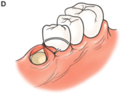
Describe the loop interrupted suture technique.
Pierce facial keratinized tissue → toward lingual under contact → pierce lingual from underside → back toward facial under contact → tie on facial.
(Explanation: This technique loops the suture under the contact point to secure both flaps, pulling them coronally for tight closure. Tying on the facial side improves patient comfort and accessibility for suture removal.)
What suture materials are used for a distal wedge?
Chromic gut or Vicryl; no suture placed between buccal and lingual flaps.
(Explanation: Chromic gut and Vicryl are absorbable, suitable for distal wedge areas where sutures don't need to hold flaps together tightly, as the procedure often leaves an open wound that heals by secondary intention.)
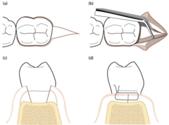
When is an interrupted figure eight suture used?
When there is no primary closure of flaps; holds flap more apically than coronally.
(Explanation: The figure eight suture is used when flaps cannot be fully approximated, such as in apically positioned flaps, securing them lower on the tooth to maintain position during healing.)
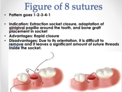
Describe the interrupted figure eight suture technique.
Pierce facial keratinized tissue → toward lingual under contact → pierce lingual from keratinized side → toward facial under contact → tie on facial.
(Explanation: This technique crosses the suture in a figure-eight pattern, providing apical tension to stabilize flaps without forcing primary closure, ideal for procedures like crown lengthening.)
When is a single tooth sling suture used?
When flap is elevated on only one side of the tooth or when facial/lingual flaps are positioned at different levels.
(Explanation: This suture stabilizes a single flap or accommodates uneven flap positioning, often used in localized procedures where only one side of the tooth requires surgical intervention.)
Describe the single tooth sling suture technique.
Pierce facial keratinized tissue → sling around lingual → toward facial under contact → pierce facial keratinized tissue → sling around lingual → tie on facial.
(Explanation: The sling technique wraps the suture around the tooth, securing the facial flap without involving the lingual side, useful for single-sided flaps or when maintaining different flap heights.)
How are mattress sutures classified?
External or internal; horizontal or vertical.
(Explanation: Mattress sutures distribute tension evenly across larger areas. External mattress sutures pull tissue toward bone, while internal ones evert tissue edges. Horizontal and vertical refer to the direction of tissue piercing, affecting tension distribution.)
What is the purpose of external mattress sutures?
Provide tension toward the crest of bone; used for crown lengthening and apically positioned flaps.
(Explanation: These sutures anchor flaps apically to expose more tooth structure (e.g., for restorations) or position gingiva lower on the bone, ensuring stable healing in the desired position.)
What is the purpose of internal mattress sutures?
Evert flaps with connective tissue surfaces touching; used to preserve papilla height.
(Explanation: Internal mattress sutures roll flap edges inward, promoting connective tissue contact and minimizing papilla recession, critical for esthetic outcomes in anterior regions.)
When is a horizontal mattress suture used?
For large alveolar ridge grafts; piercing is positioned mesiodistally ~3 mm apart.
(Explanation: Horizontal mattress sutures distribute tension across wider areas, ideal for stabilizing large grafts on the alveolar ridge, ensuring even pressure and graft immobility.)
When is a vertical mattress suture used?
To preserve papilla height; piercing is positioned apicocoronally ~3 mm apart.
(Explanation: Vertical mattress sutures align tissue vertically to maintain papilla height, critical for esthetics in areas where interdental papilla loss would be noticeable.)
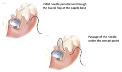
Describe the internal vertical mattress suture technique for narrow papilla.
Start facial 6 mm apical to coronal flap → pierce keratinized tissue → pierce lingual underside 6 mm from coronal → pierce lingual keratinized side 3 mm from coronal → pierce facial underside 3 mm from coronal → tie.
(Explanation: This technique everts narrow papillae, maximizing height preservation by suturing deeper (6 mm) and closer (3 mm) to the coronal edge, ensuring connective tissue contact without exposing the suture coronally.)
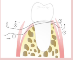
When is an internal horizontal mattress suture used?
For wide papilla.
(Explanation: Wide papillae require broader tension distribution, which horizontal mattress sutures provide by piercing tissue mesiodistally, stabilizing larger interdental areas.)
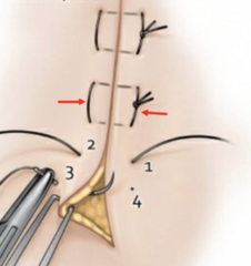
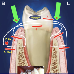
Describe the external vertical mattress suture technique.
Start facial 8 mm apical to papilla → pierce keratinized tissue → pierce facial underside 3 mm from coronal → go under contact to lingual → pierce lingual keratinized side 8 mm from coronal → pierce lingual underside 3 mm from coronal → back to facial under contact → tie.
(Explanation: This technique pulls flaps apically, anchoring them to bone for procedures like crown lengthening, with deeper (8 mm) and coronal (3 mm) piercings to secure tissue under tension.)
What is the post-op schedule after periodontal surgery?
First visit: 1-2 weeks for post-op; second visit: 2-3 months for second post-op; additional visits as needed.
(Explanation: The 1-2 week visit assesses initial healing and suture removal, while the 2-3 month visit evaluates tissue maturation and bone remodeling. Follow-ups ensure long-term stability and address complications.)
What post-op care is provided after periodontal surgery?
Remove dressing and sutures, deplaque with curettes/proxabrush, reinforce oral hygiene (toothbrush, floss, proxabrush), use chlorhexidine (reduce inflammation) and Stella Life (kill bacteria).
(Explanation: Dressings protect wounds, and suture removal prevents irritation. Deplaquing ensures a clean healing environment. Chlorhexidine reduces bacterial load and inflammation, while Stella Life, a natural antimicrobial, supports infection control.)
What is the primary mechanism of wound healing?
Restoration of tissue integrity upon injury.
(Explanation: Wound healing aims to repair or regenerate damaged tissues to restore function and structure, involving complex cellular processes like inflammation, proliferation, and remodeling.)
What forms immediately after periodontal surgery?
A blood clot.
(Explanation: The blood clot acts as a scaffold for healing, stabilizing the wound and providing a matrix for cell migration. A thinner clot is preferred as it's replaced faster by granulation tissue.)
What are the two overlapping phases of wound healing?
Soft tissue healing and osseous healing.
(Explanation: Soft tissue healing (gingiva, connective tissue) and osseous healing (bone remodeling) occur simultaneously but at different rates, with soft tissue healing faster than bone regeneration.)
What happens during the inflammatory phase of soft tissue healing?
Occurs 0-3 days; involves neutrophils, monocytes, and blood clot formation (thinner clot is better for faster replacement).
(Explanation: Neutrophils clear debris and bacteria, while monocytes differentiate into macrophages to orchestrate healing. A thin clot minimizes the barrier to new tissue formation, speeding up subsequent phases.)
What happens during the granulation phase of soft tissue healing?
Occurs 12 hours-10 days; involves macrophages, fibroblasts, endothelial cells, and smooth muscle cells.
(Explanation: Macrophages remove debris, fibroblasts produce collagen, endothelial cells form new blood vessels, and smooth muscle cells support vascular structure, creating granulation tissue as a foundation for repair.)
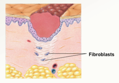
When does epithelialization begin, and at what rate do epithelial cells migrate?
Begins ~24 hours post-wounding; epithelial cells migrate at 0.5 mm/day.
(Explanation: Epithelial cells migrate from wound edges to cover the surface, restoring the barrier function. The 0.5 mm/day rate is consistent across oral tissues, critical for calculating healing timelines.)
What is contact inhibition in epithelialization?
Epithelial cells stop migrating when surrounded by like cells.
(Explanation: Contact inhibition prevents over-migration, ensuring epithelial cells form a single layer, halting once they meet adjacent cells to create a uniform surface.)
How long does it take for epithelialization of a 2x2 mm wound?
5 days (Day 1: 0 mm, Day 2: 0.5 mm, Day 3: 1 mm, Day 4: 1.5 mm, Day 5: 2 mm).
(Explanation: This calculation assumes bilateral migration at 0.5 mm/day per side, covering 2 mm in 5 days. It's a critical clinical benchmark for assessing healing progress in small wounds.)
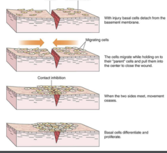
How does connective tissue healing lag behind epithelialization?
Connective tissue (fibroblasts, endothelial cells) starts healing at ~5 days.
(Explanation: Connective tissue healing involves collagen deposition and angiogenesis, which are slower processes than epithelial migration, requiring a stable epithelial cover first.)
How long does it take for a gingivectomy to heal?
3-5 weeks.
(Explanation: Gingivectomy healing is slower due to secondary intention (open wound), requiring granulation tissue formation and epithelialization over a larger area compared to flap surgeries.)
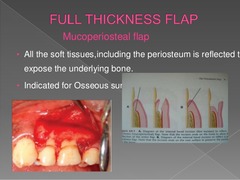
How long does it take for a full-thickness flap surgery to be fully epithelialized?
1 month.
(Explanation: Full-thickness flaps, involving primary closure, heal faster than gingivectomies due to direct flap apposition, with epithelialization completing in about 4 weeks.)
Why is probing delayed for 6 weeks post-periodontal treatment?
To allow full epithelialization (takes ~1 month).
(Explanation: Probing too early can disrupt fragile epithelial and connective tissue healing, potentially causing bleeding or detachment of the new junctional epithelium.)
What happens during the differentiation/maturation phase of soft tissue healing?
Occurs 2.5 weeks to 4-6 months; involves fibroblasts (ECM production) and endothelial cells (angiogenesis).
(Explanation: Fibroblasts produce extracellular matrix (collagen, elastin) for tissue strength, while endothelial cells form new capillaries (angiogenesis), maturing the tissue to restore function and resilience.)
What are the phases of osseous healing?
Osteoclastic phase (2-10 days) and osteoblastic phase (10-28 days).
(Explanation: Osteoclasts resorb damaged bone, clearing the site, while osteoblasts deposit new bone matrix, which mineralizes over weeks to restore bone structure.)
What is the average healing time for bone?
3 months, but remodeling may continue for 18 months.
(Explanation: Bone healing involves initial matrix deposition (3 months), but remodeling, where bone adapts to functional stresses, can extend to 18 months, especially in complex cases.)
How does thick bone heal compared to thin bone?
Thick bone heals without deformity due to abundant marrow cells and vasculature.
(Explanation: Thick bone, with more marrow and blood supply, supports robust osteoblast activity, preventing deformities. Thin bone, with less vascularity, is more prone to resorption or irregular healing.)
What is the net bone loss after a full-thickness flap alone?
0.6 mm.
(Explanation: Elevating a full-thickness flap exposes bone, leading to minor resorption (0.6 mm) due to osteoclastic activity triggered by surgical trauma and exposure.)
How does a partial-thickness flap differ from a full-thickness flap in terms of bone loss?
Partial-thickness flap avoids bone exposure, preventing 0.6 mm bone loss.
(Explanation: Partial-thickness flaps leave the periosteum intact, protecting the bone from exposure and resorption, making them preferable when bone preservation is critical.)
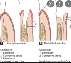
What factors influence the final shape of bone after surgery?
Type of bone and amount of trauma, more than the amount of bone reshaping.
(Explanation: Bone type (thick vs. thin) and surgical trauma (e.g., excessive manipulation) significantly affect healing outcomes, often overshadowing deliberate reshaping efforts.)
What factors negatively affect healing?
Bone dehydration (long surgery), poor flap adaptation (thick clot), bone necrosis (motor use without constant irrigation).
(Explanation: Dehydration from prolonged exposure, thick clots from poor flap fit, and necrosis from heat (e.g., from drills without cooling) impair cellular activity and healing, leading to bone loss or delayed recovery.)
What is repair in the context of periodontal healing?
Healing that does not fully restore architecture/function; only epithelial cells repopulate, forming a long junctional epithelium.
(Explanation: Repair results in a functional but suboptimal outcome, with a long junctional epithelium forming a weak attachment that doesn't restore the original periodontal ligament or bone.)
What is the most common healing outcome after periodontal procedures?
Repair with a long junctional epithelium.
(Explanation: Most periodontal treatments (e.g., SRP) result in repair rather than regeneration due to the faster migration of epithelial cells, which outpace periodontal ligament or bone cells.)
Which procedures commonly result in repair?
Scaling and root planing (SRP), osseous surgery.
(Explanation: SRP and osseous surgery primarily clean and reshape tissues, promoting epithelial healing but rarely achieving full regeneration without additional regenerative techniques.)
What is regeneration in periodontal healing?
Restoration of all architecture and function; epithelial, connective tissue, PDL, and alveolar cells repopulate.
(Explanation: Regeneration recreates the original periodontal structures (gingiva, PDL, cementum, bone), restoring full function, unlike repair, which only partially restores tissue.)
How can regeneration be confirmed?
Requires histological analysis to identify cell types (not possible clinically).
(Explanation: Only microscopy can confirm the presence of new cementum, PDL, and bone. Clinical assessments (e.g., probing) can suggest regeneration but aren't definitive.)
What is equivalent to new attachment in regeneration?
Newly regenerated fibers embedded in new cementum on a root surface previously affected by disease.
(Explanation: New attachment signifies successful regeneration, with new periodontal ligament fibers anchored in newly formed cementum, restoring the tooth's support system.)
Which cells repopulate first in periodontal regeneration?
Periodontal ligament (PDL) cells.
(Explanation: PDL cells have progenitor potential, rapidly migrating to the root surface to initiate regeneration, outpacing epithelial cells if guided tissue regeneration is used.)
What evidence supports PDL's role in regeneration?
Strong evidence that PDL contains progenitor cells for periodontal regeneration.
(Explanation: Studies show PDL cells can differentiate into cementoblasts, osteoblasts, and fibroblasts, critical for regenerating cementum, bone, and ligament, respectively.)
What is reattachment in periodontal healing?
Reattachment of gingiva to areas mechanically removed.
(Explanation: Reattachment occurs when healthy gingiva re-adheres to a cleaned or surgically altered tooth surface, often seen in flap surgeries where tissue is repositioned.)
What is resorption in periodontal healing?
Loss/blunting of the root due to various factors; connective tissue repopulates first.
(Explanation: Root resorption can result from trauma, inflammation, or surgical complications, with connective tissue forming a fibrous attachment instead of cementum, weakening the tooth.)
What is ankylosis in periodontal healing?
Fusion of tooth to bone; alveolar bone repopulates first, may lead to root resorption.
(Explanation: Ankylosis occurs when bone directly contacts the root without PDL, leading to rigid fusion. This can cause root resorption over time, compromising tooth viability.)
What type of bone forms post-extraction?
Provisional matrix and woven bone, with some lamellar bone.
(Explanation: Post-extraction, a provisional matrix forms first, followed by woven bone (immature, disorganized), which partially matures into stronger lamellar bone during socket healing.)
How does healing occur after scaling and root planing (SRP)?
Through long junctional epithelium and recession.
(Explanation: SRP removes plaque and calculus, allowing epithelial cells to form a long junctional epithelium, which seals the pocket but doesn't regenerate lost bone or PDL, often leading to gingival recession.)
How does the cervical line and interdental bone shape differ in anterior teeth?
Cervical line is more concave; interdental bone is very convex.
(Explanation: The concave cervical line and convex interdental bone in anteriors reflect their scalloped gingival contour, affecting surgical planning to preserve esthetics and bone support.)
How does the cervical line and interdental bone shape differ in premolars?
Cervical line is slightly concave; interdental bone is slightly convex.
(Explanation: Premolars have a less pronounced scallop than anteriors, with flatter bone contours, influencing flap design and defect management strategies.)
How does the cervical line and interdental bone shape differ in molars?
Cervical line is nearly flat; interdental bone is nearly flat.
(Explanation: Molars' flat contours simplify osseous surgery but increase the risk of furcation involvement, requiring careful defect assessment.)
Why should prominent teeth with thin alveolar plates not be root planed?
To avoid further bone loss due to thin alveolar plates.
(Explanation: Thin alveolar plates, common in prominent teeth, are prone to resorption if root planed aggressively, as the bone lacks sufficient thickness to withstand additional stress.)
Does the shape of gingiva indicate underlying osseous contour?
No, gingival shape gives little hint of underlying osseous contour.
(Explanation: Gingival contours are influenced by soft tissue thickness and inflammation, not bone shape, necessitating direct visualization or imaging for accurate osseous assessment.)
What is the difference between horizontal and vertical bone loss?
Horizontal: uniform bone loss; Vertical: irregular, can be treated with bone grafts.
(Explanation: Horizontal bone loss affects all teeth evenly, often managed with scaling. Vertical bone loss forms angular defects, amenable to regenerative techniques like grafting.)
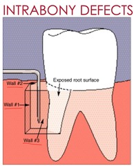
What are intrabony osseous defects?
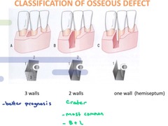
Affects at least one tooth surface within alveolar bone, surrounded by 1, 2, or 3 bony walls or a combination.
(Explanation: Intrabony defects are localized, with varying bony walls determining the potential for regeneration (more walls = better regenerative potential).)
What is a hemiseptum defect?
Intrabony defect with 1 bony wall.
(Explanation: Hemiseptum defects, with only one supporting wall, have limited regenerative potential due to minimal bone support, often requiring grafts or membranes.)
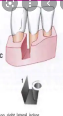
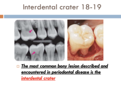
What is a crater defect?
Intrabony defect with 2 bony walls.
(Explanation: Crater defects, with two walls, offer moderate regenerative potential, commonly treated with guided tissue regeneration to support bone fill.)
What is a circumferential defect (moat)?
Intrabony defect with 3 bony walls.
(Explanation: Circumferential defects, surrounded by three walls, have the highest regenerative potential due to greater bone support, ideal for grafting and GTR.)
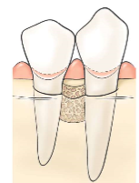
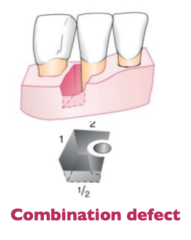
What is a combination defect?
Intrabony defect with varying walls (e.g., 2 walls coronally, 3 walls apically).
(Explanation: Combination defects have complex anatomy, requiring tailored regenerative approaches based on the number of walls at different levels of the defect.)
What are other types of osseous defects?
Reverse architecture, osseous ledge, furcations (I, II, III), dehiscence, fenestration.
(Explanation: Reverse architecture (bone higher interdentally than marginally), ledges (irregular bone protrusions), furcations (bone loss in multi-rooted teeth), dehiscence (bone loss exposing root), and fenestration (window-like bone loss) complicate surgical management.)
What is the only reliable method to determine the extent of osseous defects?
Direct visualization.
(Explanation: Probing and radiographs can suggest defects, but only surgical exposure allows precise assessment of defect shape, size, and wall number, guiding treatment planning.)
What are the etiologies of osseous defects?
Faulty dentistry (over-contoured restorations, open margins, insufficient embrasure space, overhangs); plaque within 2 mm of bone causing resorption.
(Explanation: Poor restorations trap plaque, while plaque within 2 mm of bone triggers osteoclastic resorption, leading to bone loss and defect formation.)
What is the purpose of osseous surgery?
To provide a sound, solid base for gingiva without excessive sulcular depth, recontouring bone to normal topography, minimizing post-op sulcus depth.
(Explanation: Osseous surgery reshapes bone to mimic healthy anatomy, reducing pocket depths and creating a stable gingival foundation, often for restorative or periodontal health.)
When might osseous surgery be necessary?
For crown lengthening.
(Explanation: Osseous surgery exposes more tooth structure for restorations by reshaping bone, ensuring adequate crown height and biologic width.)
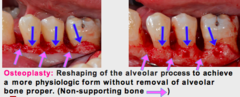
What is osteoplasty?
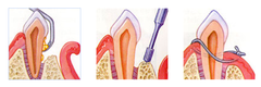
Removal of non-supporting bone.
(Explanation: Osteoplasty removes bone not critical to tooth support (e.g., ledges) to create a smooth contour, improving gingival adaptation and reducing pocket depths.)
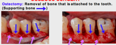
What is ostectomy?
Removal of supporting bone.
(Explanation: Ostectomy removes bone that supports the tooth, used sparingly to correct defects or achieve desired bone contours, but it risks compromising tooth stability.)
What treatments are used for severely decayed teeth or short anatomical crowns?
Resection or resection with orthodontic extrusion.
(Explanation: Resection removes compromised tooth structure, while orthodontic extrusion moves the tooth coronally to expose healthy structure, both aiding restorative access.)
What is used to repair osseous defects with bone fill?
Bone or Alloplast grafts.
(Explanation: Bone grafts (autogenous, allogenic) provide a scaffold and cells for bone regeneration, while Alloplasts (synthetic) act as fillers, supporting bone fill.)