Chapter 19, Lesson 1: Overview of the Cardiovascular System
1/20
Earn XP
Description and Tags
Flashcards from Chapter 19, Lesson 1 of McGraw Hill Anatomy and Physiology, Tenth Edition, by Kenneth S. Saladin.
Name | Mastery | Learn | Test | Matching | Spaced | Call with Kai |
|---|
No analytics yet
Send a link to your students to track their progress
21 Terms
Cardiology
The study of the heart and its disorders
Cardiovascular system
Consists of the heart and blood vessels, where the heart keeps pumping blood
Arteries
Vessels that carry blood away from the heart
Veins
Vessels that carry blood toward the heart
Capillaries
Microscopic vessels connecting small arteries and veins
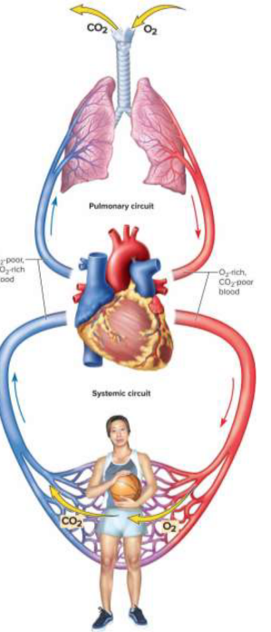
Cardiovascular circuits
Divided into the pulmonary (lung-bound) and systemic (body-bound) circuits
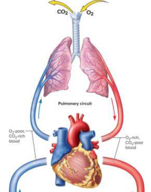
Pulmonary circuit
Carries blood to lungs for gas exchange and back to the heart; present on the anatomical right side
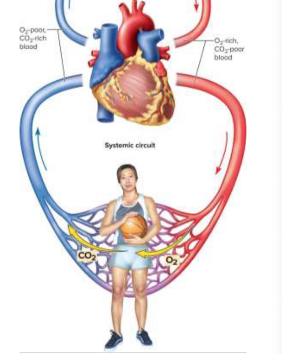
Systemic circuit
Supplies oxygenated blood to bodily tissues for cycle return; present on the anatomical left side
Pulmonary circuit process
Deoxygenated blood arrives at circuit
Blood is sent to lung alveoli by trunk and arteries
Blood is returned
Systemic circuit process
Oxygenated blood is sent via aorta, branching off
Blood releases oxygen at tissues
Deoxygenated blood returns via superior and inferior vena cava

Great vessels
The major arteries and veins entering and leaving the heart
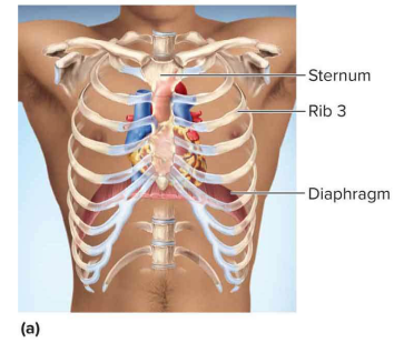
Mediastinum
The space between the lungs where the heart is located
Base
The wide, superior portion of the heart with great vessels
Apex
The tapered inferior end of the heart that tilts to the left
Heart size
Adults have a weight of ~10 ounces; heart size is proportional to body (size of fist)
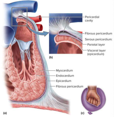
Pericardium
A double-walled sac that allows the heart to beat and expand without friction
Fibrous pericardium
The outermost layer of the pericardium, visible
Parietal pericardium
Below the fibrous pericardium as a liner
Epicardium
Adheres to heart surface and outermost heart layer
Pericardial cavity
The space between the visceral and parietal layers of the serous pericardium, filled with 5 to 30 mL of pericardial fluid
Pericarditis
The inflammation of the pericardium which may result in friction rub