Nucleic Acid Part 3
 Call Kai
 Learn
Learn Practice Test
 Spaced Repetition
 Match
Match Flashcards
 Knowt Play
1/19

There's no tags or description
Looks like no tags are added yet.
Name | Mastery | Learn | Test | Matching | Spaced | Call with Kai | Chat |
|---|
No analytics yet
Send a link to your students to track their progress
20 Terms
Infor card: what this lecture is about
In this lecture, we explore the higher-order organization, chemical stability, and experimental manipulation of nucleic acids, with a focus on both their biological roles and practical applications in molecular biology.
DNA Supercoiling and Topoisomerases
Packaging of large DNA molecules inside cells requires supercoiling.
Linking number (L), twist (T), and writhe (W) describe DNA topology.
Topoisomerases are key enzymes that regulate DNA supercoiling by introducing reversible breaks.
Chromatin and Nucleosomes
In eukaryotes, DNA is packaged into chromatin, with nucleosomes as the fundamental unit.
Histones (H2A, H2B, H3, H4) form a histone core, while H1 clamps the DNA to stabilize the nucleosome.
Higher-order chromatin structure involves coiling into fibers and attachment to nuclear scaffolds.
DNA Sequencing – Sanger Method
The chain termination method (dideoxy method) uses DNA polymerase, primers, and labeled ddNTPs to generate DNA fragments of varying lengths.
Fragments are separated by gel electrophoresis to read DNA sequences.
DNA Duplex Stability
DNA can denature under heat or chemical conditions and renature upon cooling.
Melting temperature (Tm) depends on GC content, with higher GC regions being more stable.
Hybridization is a key technique for measuring sequence similarity between DNA molecules.
RNA and DNA Hydrolysis
RNA is more susceptible to hydrolysis due to its 2′-OH group.
DNA, lacking this group, is significantly more stable.
Restriction Enzymes
Type II restriction enzymes cut DNA at specific palindromic recognition sequences.
Examples include EcoRI, BamHI, HpaI, and TaqI.
They generate either sticky ends or blunt ends, enabling precise DNA manipulation in recombinant DNA technology.
Key Takeaway:
This lecture highlights how nucleic acids are packaged, stabilized, sequenced, and enzymatically manipulated—knowledge that underpins modern genetics, genomics, and biotechnology.
Info card: what is covered in this lecture
This slide outlines the key topics to be discussed in the third lecture of the nucleic acids series, focusing on advanced DNA and RNA concepts and experimental methods. The main areas include:
DNA Supercoiling and Topoisomerases
Introduction to how DNA is compacted through supercoiling.
Role of topoisomerases in relieving torsional stress and enabling replication and transcription.
Chromatin and Nucleosomes
Structure and organization of eukaryotic DNA into chromatin.
Nucleosome composition and its role in DNA packaging and regulation.
Sanger Sequencing Method
Overview of the classic DNA sequencing technique.
Principles of chain termination and reading sequence data.
Thermostability of the DNA Duplex
Factors influencing DNA melting temperature (GC content, length, ionic conditions).
Importance in PCR and hybridization experiments.
Hydrolysis of RNA and DNA
Chemical stability differences between RNA and DNA.
Mechanisms of hydrolysis and their significance in research and diagnostics.
Restriction Digestion of DNA
Introduction to restriction endonucleases and their recognition sites.
Applications in cloning and molecular biology workflows.
Key message:
This lecture expands from fundamental nucleic acid structures to how they are manipulated and studied in the lab, connecting biochemical properties to molecular biology tools.

What is DNA supercoiling?
This slide introduces the concept of DNA supercoiling, a critical process for DNA packaging and function inside cells.
What is supercoiling?
Supercoiling refers to the coiling of the DNA double helix upon itself.
It is necessary to compact large DNA molecules so they fit within cells and to facilitate processes like replication and transcription.
Key points:
Outside the cell:
Only circular DNA (e.g., bacterial plasmids) can exist in a supercoiled state.
Inside the cell:
Both linear and circular DNA can adopt a supercoiled shape due to interactions with DNA-binding proteins (e.g., histones in eukaryotes).
Supercoiling regulates accessibility of DNA and influences gene expression.
Diagram explanation:
Relaxed linear DNA: A straight double helix with no supercoiling.
Relaxed circular DNA: A closed loop with no twists beyond the double helix.
Supercoiled circular DNA: The DNA helix coils upon itself, forming a more compact structure.
Key message:
Supercoiling is an essential biological strategy for organizing DNA and controlling its use in replication, transcription, and recombination.

What is L= T+ W?
This slide explains the mathematical relationship used to describe DNA supercoiling and introduces key parameters: linking number (L), twist (T), and writhe (W).
Key terms and concepts:
Linking number (L):
Represents the total number of times one strand of DNA winds around the other.
Can only be changed by breaking one or both DNA strands (e.g., by topoisomerases).
Twist (T):
Number of helical turns in the DNA molecule.
For B-form DNA, 10 base pairs per turn are assumed in this course, so T = (number of base pairs) / 10.
Always considered positive here.
Writhe (W):
Measures the degree of supercoiling, or how the axis of the DNA helix coils in space.
Left-handed (positive W): overwound.
Right-handed (negative W): underwound.
Relationship:
L = T + W
This equation describes the sum of twist and writhe that gives the total linking number.
Diagram explanation:
Middle: relaxed circular DNA with T = 20, W = 0, so L = 20.
Top: overwound DNA with W = +2, so L = 22.
Bottom: underwound DNA with W = -2, so L = 18.
Other points:
Circular duplex DNA will supercoil if it has more or fewer than 10 bp per turn.
Key message:
The linking number is constant unless DNA is cut, and supercoiling is reflected in changes to writhe while twist remains proportional to base pairs. Understanding this relationship is critical for studying DNA topology.

What does Topoisomerase?
This slide explains the role of topoisomerases, enzymes that control DNA topology by modifying the degree of supercoiling.
What are topoisomerases?
A class of enzymes that alter the supercoiling state of DNA.
They work by making transient cuts in one or both DNA strands, allowing the DNA to unwind or overwind before resealing the breaks.
This process is reversible and essential for replication, transcription, and recombination.
DNA gyrase:
A specialized type of topoisomerase (found in bacteria) that introduces negative supercoils (right-handed).
Uses ATP to add twists to DNA, reducing strain.
Diagram explanation:
Left (a): Relaxed circular DNA (T = 20, W = 0, L = 20).
Middle (b): Action of gyrase adds negative supercoils, resulting in underwound DNA (T = 20, W = -4, L = 16).
Right (c): Shows the effect of unwinding 4 helical turns—a strained state with T = 16, W = 0, L = 16. The molecule will supercoil to reach the middle configuration.
Key points:
Topoisomerases are essential for DNA metabolism and chromosome structure.
They relieve torsional stress and maintain proper DNA superhelical density.
Key message:
Without topoisomerases, DNA processes would stall due to excessive twisting or tangling. These enzymes act as nature’s tools for maintaining DNA integrity and accessibility.

What are chromatin and nucleosomes?
This slide introduces chromatin structure and nucleosomes, essential elements for packaging and organizing eukaryotic DNA.
Chromatin:
Found in eukaryotic chromosomes, which are large and linear.
Chromatin refers to the complex of DNA and associated proteins, mainly histones, that allow DNA to be compacted yet accessible for transcription, replication, and repair.
Nucleosomes:
The fundamental structural unit of chromatin.
Consist of DNA wrapped around a histone core in a left-handed supercoil.
The histone core is made of two copies each of four histone proteins: H2A, H2B, H3, and H4.
The DNA-histone interaction helps neutralize negative DNA charges and stabilize higher-order folding.
Histone structure:
Each histone contains a conserved histone fold domain, which forms the central core.
The histone fold consists of three α-helices separated by two unstructured elements, allowing tight packing and dimerization.
Histones also have N-terminal tails, which are sites for chemical modifications (e.g., acetylation, methylation) that regulate gene expression.
Visual elements:
Electron micrograph: Shows chromatin as beads-on-a-string, with nucleosomes as “beads.”
Diagram below: Shows the organization of the four core histones, their histone folds, and N-terminal tails.
Key message:
Nucleosomes are the building blocks of eukaryotic chromosomes, balancing DNA compaction with accessibility. Histone structure and modifications play crucial roles in regulating chromatin dynamics.
Chromatin is DNA packaged with proteins called histones, and its basic building block is the nucleosome, where DNA wraps around 8 histones (2 each of H2A, H2B, H3, and H4) like thread around a spool. Each histone has a histone fold, which helps the histones fit together to form the core, and an N-terminal tail that sticks out and can be chemically modified to help regulate how tightly DNA is packaged and how accessible it is for transcription, replication, and repair.

Describe the composition and organization of the histone core
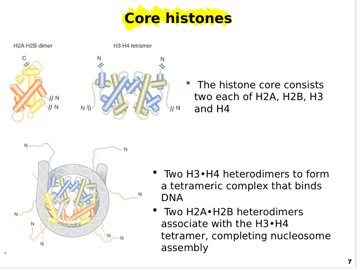
This slide details the composition and organization of the histone core, which is central to nucleosome structure.
Histone core composition:
The histone octamer contains eight histone proteins:
Two copies each of H2A, H2B, H3, and H4.
These histones are highly conserved and rich in positively charged residues, allowing strong interactions with the negatively charged DNA backbone.
Subunit organization:
Two H3–H4 heterodimers pair to form a stable H3–H4 tetramer.
Two H2A–H2B heterodimers then associate with the tetramer to complete the octamer structure.
This arrangement creates a core particle around which ~147 bp of DNA are wrapped.
Diagram explanation:
Top: Separate views of the H2A–H2B dimer and H3–H4 tetramer, highlighting their α-helical folds.
Bottom: The assembled histone core, showing the arrangement of dimers and tetramers and the protruding N-terminal tails.
Functional note:
The histone core provides a scaffold for DNA compaction while maintaining accessibility for gene regulation and chromatin remodeling.
Key message:
The precise assembly of histone
What is the composition and organization of the histone core?
This slide details the composition and organization of the histone core, which is central to nucleosome structure.
Histone core composition:
The histone octamer contains eight histone proteins:
Two copies each of H2A, H2B, H3, and H4.
These histones are highly conserved and rich in positively charged residues, allowing strong interactions with the negatively charged DNA backbone.
Subunit organization:
Two H3–H4 heterodimers pair to form a stable H3–H4 tetramer.
Two H2A–H2B heterodimers then associate with the tetramer to complete the octamer structure.
This arrangement creates a core particle around which ~147 bp of DNA are wrapped.
Diagram explanation:
Top: Separate views of the H2A–H2B dimer and H3–H4 tetramer, highlighting their α-helical folds.
Bottom: The assembled histone core, showing the arrangement of dimers and tetramers and the protruding N-terminal tails.
Functional note:
The histone core provides a scaffold for DNA compaction while maintaining accessibility for gene regulation and chromatin remodeling.
Key message:
The precise assembly of histone dimers and tetramers is critical for nucleosome formation and overall chromatin structure.
Scaffold basically means a supporting structure or framework.
In this context, the histone core acts like a scaffold for DNA because it provides a structure that the DNA can wrap around and be organized on.
Think of it like a spool:
Histone core = spool/scaffold
DNA = thread
DNA wraps around the histone core → making it compact and organized
So when the slide says:
“The histone core provides a scaffold for DNA compaction”
it means:
The histone core provides a stable structure that DNA wraps around, allowing the long DNA molecule to be packed into a smaller, organized space.
What does histone H1 do?
This slide explains the role of Histone H1, also called the linker histone, in chromatin structure and stability.
Role of Histone H1:
H1 binds to the outside of the nucleosome, specifically at the site where DNA enters and exits the histone octamer.
It acts as a clamp, securing the DNA wrap and preventing premature unwrapping.
H1 helps establish the higher-order structure of chromatin by stabilizing nucleosome interactions.
DNA contact points:
Each nucleosome core particle has approximately 145 base pairs of DNA wrapped around the histone octamer, corresponding to 1.8 turns of the DNA helix.
Linker DNA, about 60 base pairs, connects one nucleosome to the next. This linker region is where H1 primarily binds, facilitating chromatin compaction.
Diagram explanation:
Top: Open nucleosome without H1, showing exposed DNA ends.
Middle: H1 protein approaching the nucleosome.
Bottom: H1 bound, clamping the DNA securely onto the histone core.
Key message:
While H2A, H2B, H3, and H4 form the nucleosome core, H1 is essential for locking the DNA in place and promoting chromatin fiber formation.

How is DNA packaged and organized?
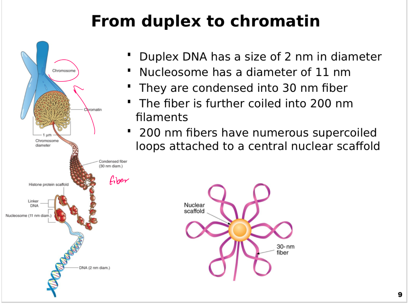
This slide illustrates how DNA is packaged and organized into progressively higher-order structures to fit inside a eukaryotic nucleus.
Levels of compaction:
DNA duplex: The simplest form, a double helix with a diameter of 2 nm.
Nucleosome: The first level of packaging, DNA wrapped around a histone core, forming particles with a diameter of 11 nm.
30 nm fiber: Nucleosomes are coiled and stacked into a solenoid or zig-zag arrangement, forming a thicker fiber.
200 nm filaments: The 30 nm fiber is further folded and coiled into larger chromatin fibers, about 200 nm in diameter.
Nuclear scaffold: Chromatin fibers form supercoiled loops attached to a central protein scaffold, providing structural support and further compaction.
Key visual elements:
Left panel: Shows the progression from naked DNA to nucleosomes to 30 nm fiber to chromatin filaments.
Right panel: Highlights the nuclear scaffold with chromatin loops attached, creating a radial structure.
Key points:
Each level of folding reduces DNA length and increases density.
The nuclear scaffold plays a critical role in organizing chromatin into chromosomes during cell division.
Why it matters:
Understanding chromatin structure explains how long DNA molecules fit into microscopic nuclei while remaining accessible for replication and transcription.
What are the importantly components of the Sanger Sequencing method?
This slide introduces the Sanger sequencing method, also known as the dideoxy chain termination method, and highlights the role of DNA polymerase in sequencing.
Sanger method overview:
Developed by Frederick Sanger, it is one of the most widely used DNA sequencing techniques.
Relies on chain-terminating nucleotides to generate fragments that can be read to determine the sequence.
Key components:
DNA polymerase (DNAP): Catalyzes the synthesis of a new DNA strand complementary to a template.
Template DNA: The strand to be sequenced.
Primer: A short oligonucleotide that anneals to the template and provides a starting point for DNAP.
dNTPs (deoxynucleotide triphosphates): The standard nucleotides (A, T, G, C) incorporated into the growing strand.
Mechanism:
DNAP extends the primer in the 5’ to 3’ direction, adding nucleotides complementary to the template.
The incorporation of dideoxynucleotides (ddNTPs), which lack a 3’-OH group, causes chain termination, producing fragments of varying lengths.
By separating these fragments and detecting the terminal base, the DNA sequence can be determined.
Diagram explanation:
Shows a DNA template, primer, and DNAP adding nucleotides.
Highlights the chemical structure of a nucleotide and its 3’-OH group necessary for chain elongation.
Key message:
Sanger sequencing was a groundbreaking method for decoding DNA and remains foundational for modern sequencing technologies.

How does Sanger sequencing method works
This slide explains how the Sanger sequencing reaction generates DNA fragments of varying lengths by terminating DNA synthesis at specific bases.
Key concept:
During DNA synthesis, occasional incorporation of a dideoxynucleotide (ddNTP) halts strand elongation because ddNTPs lack the 3’-OH group needed to form a phosphodiester bond.
This produces a collection of DNA fragments, each ending at a different nucleotide position.
Reaction components:
DNA polymerase, template, primer, and normal dNTPs (dATP, dGTP, dCTP, dTTP).
A small proportion of each ddNTP (ddATP, ddGTP, ddCTP, ddTTP), each labeled with a distinct fluorescent dye.
Process outcome:
DNA polymerase synthesizes new strands from the primer.
Whenever a ddNTP is incorporated, the chain stops, creating fragments of different lengths.
The mixture of labeled DNA molecules reflects the entire sequence, with each fragment’s terminal base identified by its fluorescent tag.
Diagram explanation:
Top: Shows primer annealed to a template.
Right: Displays a series of DNA fragments ending with different ddNTPs, color-coded for each base (A, T, G, C).
Bottom left: Chemical structure of a dideoxynucleotide emphasizes the missing 3’-OH group.
Key message:
Sanger sequencing works by controlled chain termination, allowing researchers to read the DNA sequence base by base when fragments are separated and analyzed.

How is DNA fragments generated during Sanger sequencing?
This slide explains how DNA fragments generated during Sanger sequencing are separated and read to determine the DNA sequence.
Separation principle:
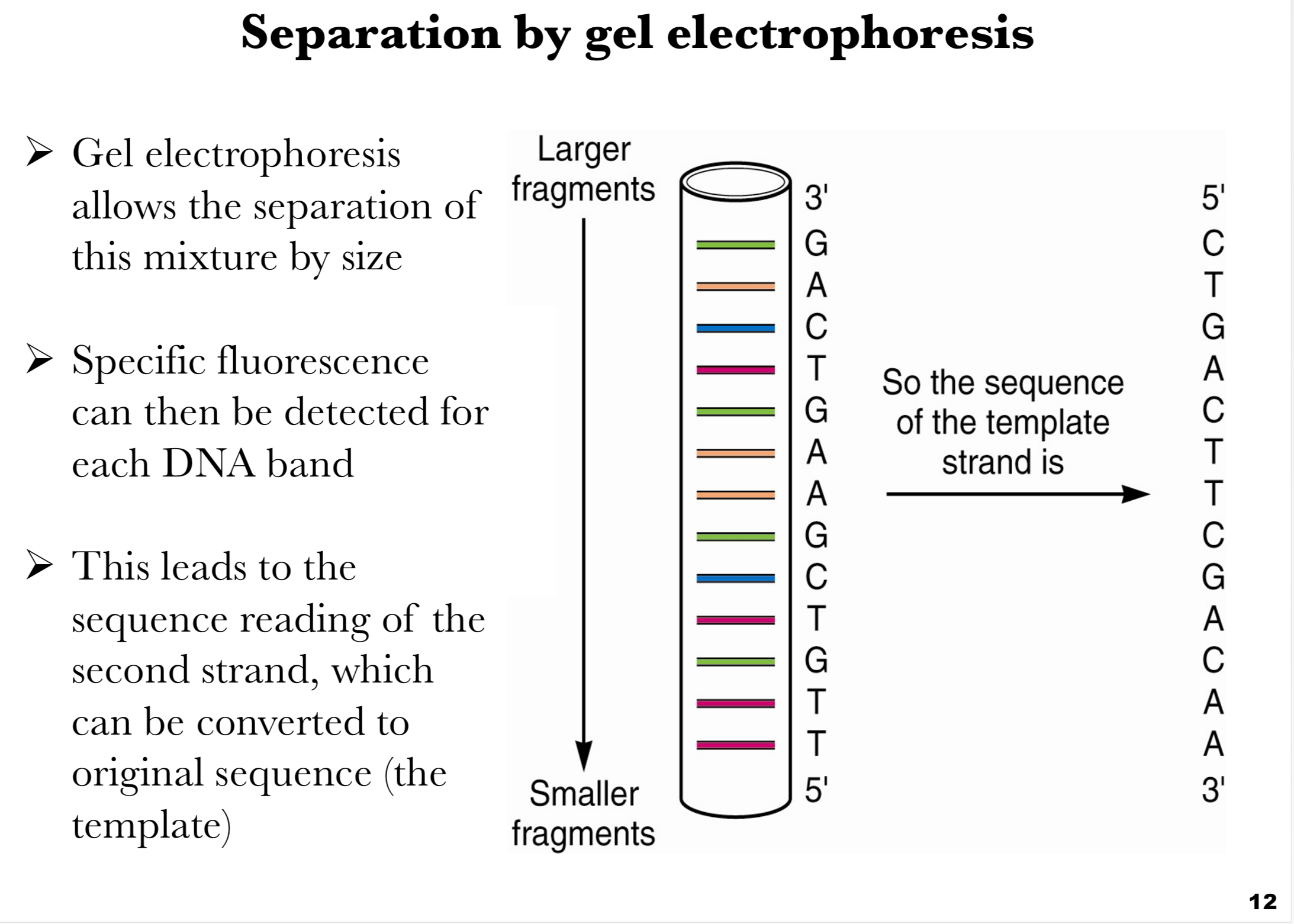
Gel electrophoresis separates DNA fragments based on size.
Fragments move through a gel matrix when an electric current is applied; smaller fragments travel farther and faster, while larger fragments remain closer to the starting point.
Detection:
Each fragment ends with a fluorescently labeled ddNTP, allowing visualization of each terminated fragment.
Specialized detectors read the fluorescence, identifying the terminal nucleotide at each length.
Sequence reading:
The gel produces a ladder of bands, each representing a fragment ending at a particular base.
By reading from smallest to largest fragments, the sequence of the synthesized strand is revealed.
This sequence can be converted to the original template strand by applying base-pairing rules.
Diagram explanation:
Left: Gel electrophoresis column with fragments sorted by size.
Right: Sequence of the template strand derived from the gel pattern (5’ to 3’).
Key takeaway:
Gel electrophoresis transforms a mixture of DNA fragments into a readable sequence, forming the core of the Sanger sequencing technique.
How can DNA be Denatured and Reannealed?
This slide explains how DNA strands can be separated and re-joined under different conditions.
Denaturation:
DNA can be “denatured,” meaning the two strands of the double helix separate.
This occurs when hydrogen bonds between complementary bases are broken.
Common methods: heat or chemicals like urea.
Denatured DNA is shown as single strands no longer paired.
Reannealing (Renaturation):
When the disruptive conditions are removed (e.g., cooling), the strands can realign and re-form the original duplex.
This process is called renaturation or reannealing.
Complements must be present and conditions must be right for hydrogen bonds to reform.
Why it matters:
This principle underlies many molecular biology techniques, including PCR, hybridization assays, and DNA melting analysis.
Diagram explanation:
Left: Native DNA helix.
Middle: Heat applied, strands separate.
Right: Cooling allows complementary strands to re-pair.
Key takeaway:
Denaturation and reannealing are reversible physical changes essential for DNA manipulation in the lab.

How can how DNA duplex stability changes with temperature and how it can be quantified?
This slide explains how DNA duplex stability changes with temperature and how it can be quantified.
UV absorbance at 260 nm:
Nitrogenous bases absorb ultraviolet light strongly at 260 nm.
Single-stranded DNA absorbs more strongly at this wavelength than double-stranded DNA due to unstacked bases.
Effect of temperature:
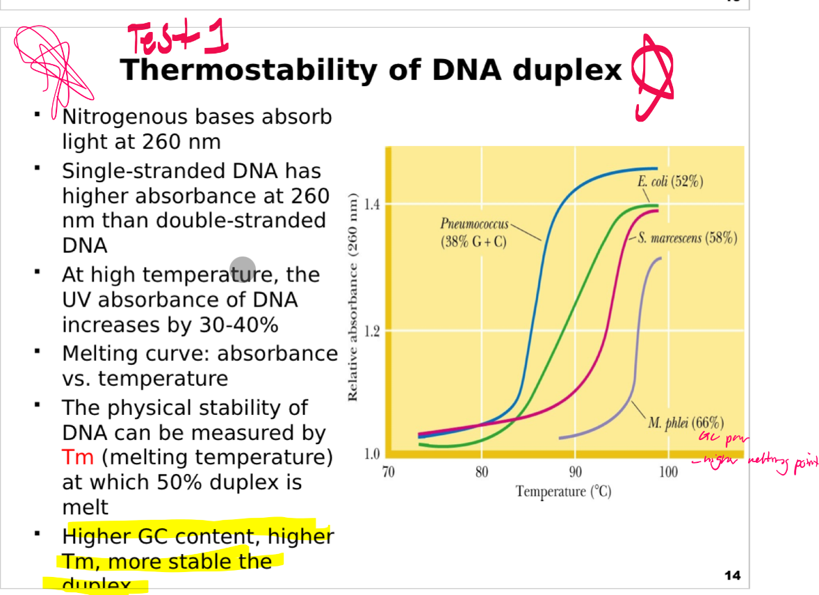
As temperature increases, DNA strands denature (melt), causing a 30–40% increase in absorbance (hyperchromic effect).
A melting curve plots absorbance versus temperature to monitor this process.
Melting temperature (Tm):
Defined as the temperature at which 50% of the DNA duplex is denatured.
Higher Tm indicates greater thermal stability.
Tm is influenced by sequence composition; GC-rich DNA is more stable because GC pairs have three hydrogen bonds versus two in AT pairs.
Graph description:
Shows melting profiles of three organisms:
Pneumococcus (38% GC) melts at a lower temperature.
E. coli (52% GC) intermediate.
M. phlei (66% GC) highest Tm, reflecting greatest stability.
Key takeaway:
DNA stability is measurable and sequence-dependent; higher GC content leads to higher Tm and more stable duplexes.
What is Nucleic acid hybridization?
This slide explains the concept and applications of nucleic acid hybridization in molecular biology.
Principle:
When two different DNA samples are mixed, denatured to single strands, and then reannealed, hybrid DNA duplexes may form if there is sequence similarity.
Complementary strands from different sources can pair, forming mixed double helices.
Key points:
The degree of hybridization (extent of duplex formation) reflects sequence similarity between the two DNA sources.
Higher hybridization indicates greater genetic relatedness.
Applications:
Commonly used to study evolutionary relationships, gene identification, and comparative genomics.
Fundamental to techniques like Southern blotting, microarrays, and FISH (fluorescence in situ hybridization).
Illustration:
Shows two DNA solutions (red and blue strands) mixed, heated to denature, and cooled to allow reannealing.
Hybrid duplexes (red/blue pairs) form where sequences are complementary.
Key takeaway:
Hybridization is a powerful tool for detecting sequence similarity and evolutionary links.

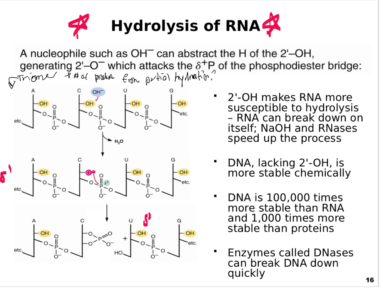
Why is RNA chemically less stable than DNA and how does it go under hydrolysis?
This slide explains why RNA is chemically less stable than DNA and how it undergoes hydrolysis.
Key Concepts
Chemical Susceptibility:
RNA contains a 2’-OH group on the ribose sugar, absent in DNA.
Under alkaline conditions (basic) or in the presence of RNases, the 2’-OH can be deprotonated, forming a reactive nucleophile.
Mechanism of Hydrolysis:
Base attack: OH⁻ abstracts the hydrogen from the 2’-OH.
The resulting 2’-O⁻ attacks the phosphate backbone, breaking the phosphodiester bond.
This results in cleavage of the RNA strand into fragments.
DNA Stability:
DNA lacks the 2’-OH group, making it much more chemically stable.
DNA is estimated to be 100,000 times more stable than RNA and about 1,000 times more stable than proteins.
Enzymatic Degradation:
RNA can self-cleave or be degraded by RNases.
DNA is resistant to self-cleavage but can be degraded rapidly by enzymes called DNases.
Visuals
Shows stepwise mechanism: deprotonation of 2’-OH, nucleophilic attack on the phosphate, and cleavage producing fragmented RNA.
Key Takeaway:
RNA’s chemical lability is due to its 2’-OH group, making it less suitable for long-term information storage compared to DNA.
What is restriction enzymes?
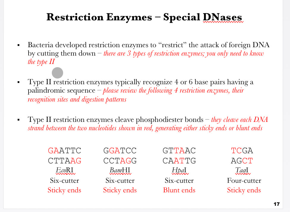
This slide introduces restriction enzymes (restriction endonucleases), which are specialized DNases that bacteria use as a defense mechanism against invading foreign DNA (e.g., bacteriophages).
Key Concepts
Function of Restriction Enzymes:
Bacteria developed these enzymes to “restrict” foreign DNA by cutting it into fragments.
Restriction enzymes recognize specific DNA sequences and cleave at or near these sites.
Types of Restriction Enzymes:
There are 3 types of restriction enzymes, but this course focuses on Type II.
Type II restriction enzymes:
Recognize specific short palindromic DNA sequences (often 4–6 base pairs).
Cut at defined positions within or near these recognition sites.
Cleavage Patterns:
Type II enzymes cut the phosphodiester bonds of DNA, producing either:
Sticky ends (overhanging single-stranded DNA ends).
Blunt ends (no overhangs, straight cuts).
Examples of Restriction Enzymes:
EcoRI
Recognition site: GAATTC
Produces sticky ends
Six-cutter enzyme
BamHI
Recognition site: GGATCC
Produces sticky ends
Six-cutter enzyme
HpaI
Recognition site: GTTAAC
Produces blunt ends
Six-cutter enzyme
TaqI
Recognition site: TCGA
Produces sticky ends
Four-cutter enzyme
Visuals
Illustrates palindromic recognition sequences for EcoRI, BamHI, HpaI, and TaqI.
Highlights cleavage positions (in red) showing how sticky or blunt ends are generated.
Key Takeaway:
Type II restriction enzymes are powerful molecular biology tools because they cut DNA at precise palindromic sequences, enabling cloning, mapping, and recombinant DNA technology.

How does EcoRi restriction enzyme recognize and cut DNA
This slide provides a detailed look at how the restriction enzyme EcoRI recognizes and cuts DNA.
Key Concepts
Recognition Sequence:
EcoRI recognizes the palindromic DNA sequence:
5′-GAATTC-3′
3′-CTTAAG-5′
Cleavage Site:
EcoRI cleaves between G and A on both DNA strands.
This produces sticky ends with a 5′ overhang.
Example cleavage:
5′-G ↓ AATTC-3′
3′-CTTAA ↑ G-5′
Resulting Ends:
After cleavage, EcoRI generates single-stranded overhangs:
5′-AATT overhang on one strand.
Complementary 5′-AATT overhang on the other.
These sticky ends are useful for DNA ligation during cloning, because complementary overhangs from different DNA molecules can anneal.
Visuals
Top panel: Shows intact recognition site with arrows marking EcoRI cut sites.
Middle panel: Demonstrates how the DNA is cleaved to produce two fragments.
Bottom panel: Highlights the palindromic recognition site again, emphasizing the cut site and sticky end formation.
Key Takeaway:
EcoRI is a Type II restriction enzyme that produces sticky ends from palindromic sequences, making it one of the most widely used tools in recombinant DNA technology.

How does the restriction enzyme EcoRI cleaves DNA at its recognition site (chemical level view)?
This slide provides a chemical-level view of how the restriction enzyme EcoRI cleaves DNA at its recognition site.
Key Concepts
Recognition Sequence:
EcoRI targets the palindromic sequence:
5′-GAATTC-3′
3′-CTTAAG-5′
Cleavage Chemistry:
EcoRI cleaves the phosphodiester bond between the G and A in the recognition site.
Specifically, the enzyme hydrolyzes the bond between the phosphate group and the sugar backbone.
Products of Cleavage:
The cleavage produces DNA fragments with:
A 5′ phosphate group (5′-P) at one end.
A 3′ hydroxyl group (3′-OH) at the other end.
This generates the characteristic sticky ends with a 5′ overhang (AATT).
Visual Explanation:
Top panel: Shows the intact phosphodiester bonds (red arrows mark EcoRI cleavage sites).
Bottom panel: Demonstrates the hydrolysis reaction, producing:
5′-phosphate termini (circled in red).
3′-hydroxyl termini (circled in red).
Biological and Experimental Importance
These sticky ends produced by EcoRI allow DNA fragments from different sources to anneal via complementary base pairing.
DNA ligase can then seal these fragments, making EcoRI one of the most powerful tools for recombinant DNA technology.
Key Takeaway:
EcoRI cleaves DNA by hydrolyzing phosphodiester bonds, generating fragments with 5′ phosphate and 3′ hydroxyl sticky ends, which are crucial for DNA manipulation in molecular cloning.
