Module 5: Cartilage and Bone
 Call Kai
 Learn
Learn Practice Test
 Spaced Repetition
 Match
Match Flashcards
 Knowt Play
1/50

There's no tags or description
Looks like no tags are added yet.
Name | Mastery | Learn | Test | Matching | Spaced | Call with Kai | Chat |
|---|
No analytics yet
Send a link to your students to track their progress
51 Terms
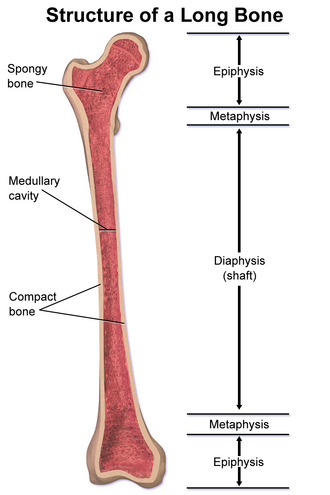
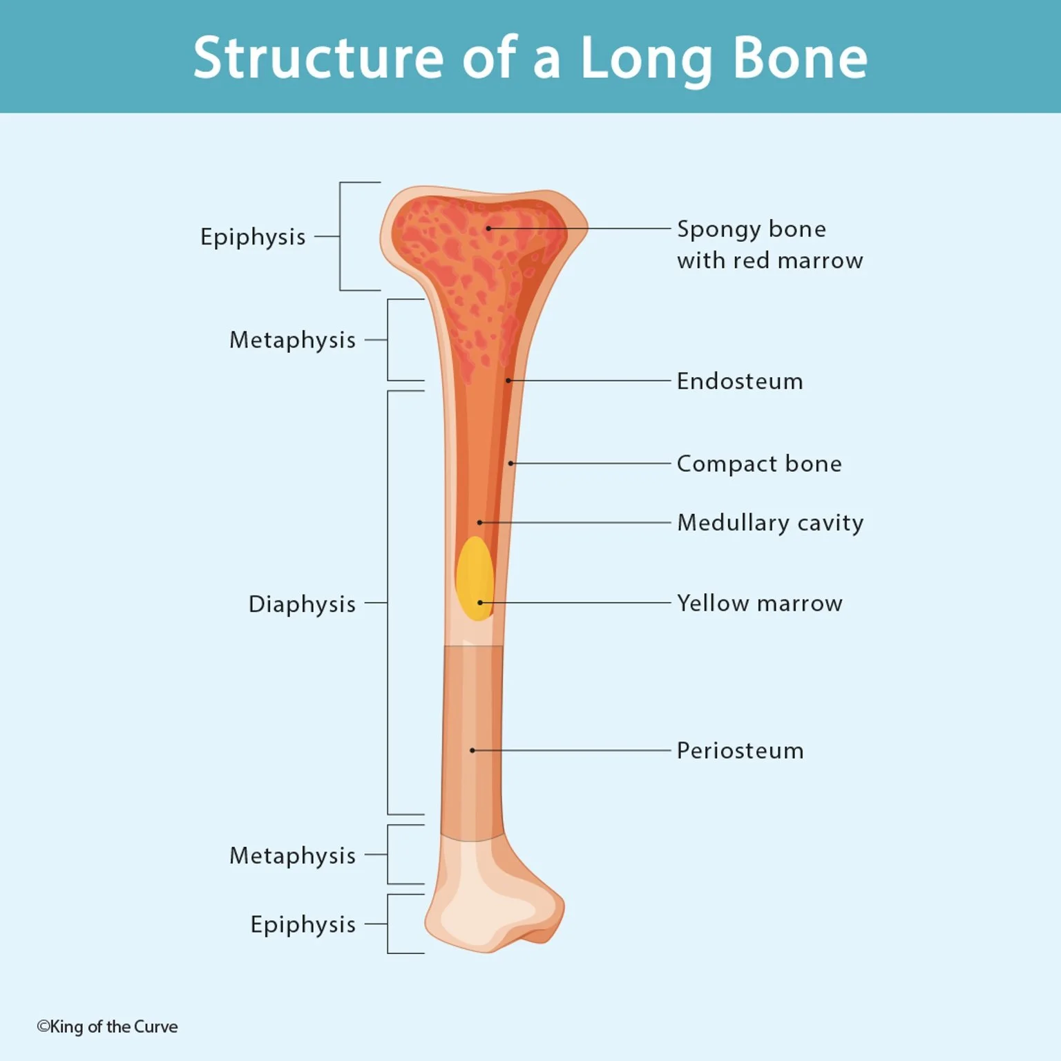
diaphysis
tubular shaft that runs between proximal and distal ends of the bone

epiphysis
layer of hyaline cartilage in growing bone
filled with spongy bone & red marrow
metaphysis
narrow area that contains epiphyseal plate
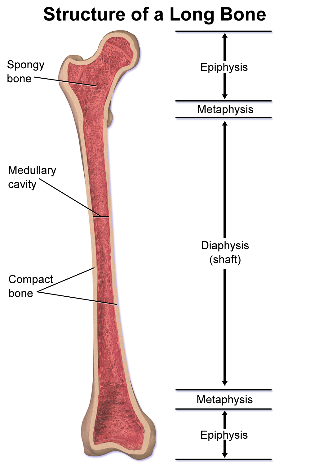
medullary cavity
hollow region in diaphysis
filled with yellow marrow

spongy bone
cancellous bone, contains osteocytes housed in lacunae
trabeculae
provide balance to compact bone to make them a little lighter and easy to move
lattice-like trix spikes
compact bone
provides support and protection
denser, stronger of the bone tissue types

nutrient artery
vein that supplies the diaphysis enters through nutrient foramen

epiphyseal arteries
supplies blood to/from the epiphysis

periosteum
fibrous membrane outer surface of bone

edosteum
membranous lining of medullary cavity where bone growth, repair, and remodeling occur

osteoclast
bone resorption
found on bone surfaces & sites of old/injured bone
osteoblast
bone formation
found in growing portions of bone (& periosteum and endosteum)

osteocyte
primary cell of mature bone and most common type of bone cell
found in lacunae/matrix

osteogenic cells
undifferentiated with high mitotic activity
only bone cells that divide to form osteoblasts
red marrow
hematopoiesis takes place (production of red blood cells)
yellow marrow
adipose tissue; source of energy

osteon
microscopic structural unit of compact bone
central canal
contains blood vessels, nerves, lymphatic vessels
runs down the center of each osteon


concentric lamellae
provide structural integrity to individual osteons to help compact bone resist twisting and compression forces along its axis
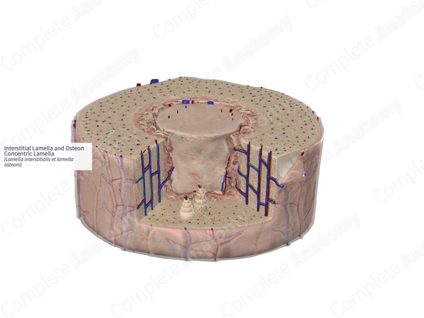

circumferential lamellae
resist overall twisting of entire bone and play a key role in appositional growth
lacunae
osteocytes located inside these spaces
found at borders of adjacent lamellae

canaliculi
allows nutrients to transport to osteocytes and wastes to be removed
connects with canaliculi of other lacunae and eventually the central canal

perforating canal
when vessels and nerves branch off to extend to periosteum and endosteum
intramembranous ossification
compact and spongy bone develop directly from sheets of mesenchymal CT

endochondral ossification
bone develops by replacing hyaline cartilage
cartilage = template
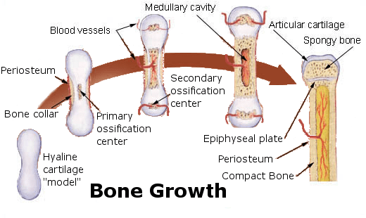
periosteal bone collar
wraps around diaphysis of developing cartilage model during fetal long bone growth
structural support, cartilage death, ossification trigger

intersitial bone growth
process by which long bones lengthen from within division, growth, and ossification of cartilage and epiphyseal plate

epiphyseal plate
area of growth in long bone
layer of hyaline cartilage where ossification occurs in immature bones

appositional bone growth
bones increase in diameter while they increase in length
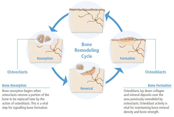
bone remodeling
continuous process where mature old bone is broken down and replaced with new bone tissue

rickets
bone disorder: bones are soft and weak and prone to deformities
osteomalacia
metabolic bone disease that causes bones to soften and weaken due to lack of proper mineralization
osteoarthritis
wear-and-tear of cartilage protecting bones breaks down
osteopenia
mineral density is deficient—warning sign for progression to osteoporosis
osteoporosis
bone density decreases making bones fragile
long bone
function as levers; they move when muscle contracts
found in arms, legs, fingers, toes


short bone
provide stability, support, limited motion
found in carpals of wrists and tarsals of ankles

flat bone
point of attachment for muscles and often protect internal organs
found in skull, scapulae

irregular bone
complex shape i.e. spinal vertebrae

sesamoid bone
protect tendons by helping overcome compressive forces & mechanical lever to allow muscle/tendon to greater torque about a joint

pneumatized bone
decrease weight of skull & for sinuses
hollow/contains numerous air pockets

chondrocytes
cartilage cells that occupy lacuna spaces

chondroblasts
young, active cells that build cartilage by secreting extracellular matrix made of collagen
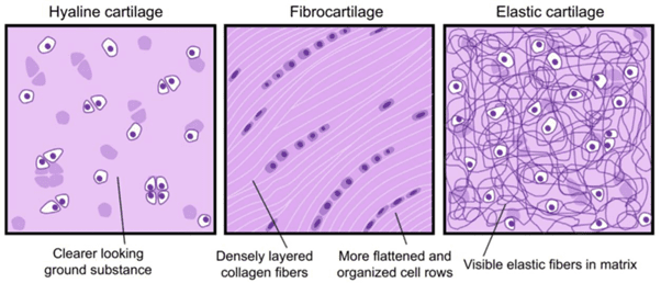
hyaline cartilage
most common type of cartilage
short & dispersed collagen fibers
strong & flexible

fibrocartilage
thick bundles of collagen fibers dispersed through matrix
found in menisci in knee joint

elastic cartilage
elastic fibers & collagen
supports and provides elasticity


periochondrium
dense irregular CT that encapsulates cartilage
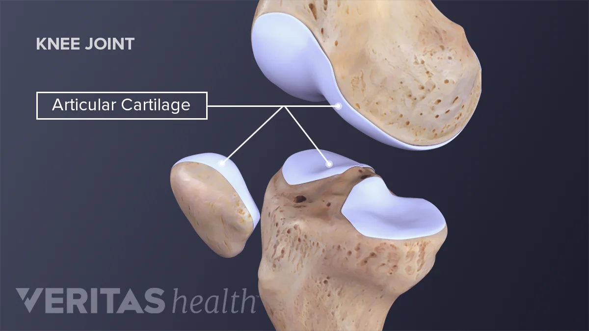
articular cartilage
thin layer of cartilage that reduces friction and acts as shock absorber

interstitial cartilage growth
expansion of cartilage from within tissue through division of existing cells and formation of new matrix

appositional cartilage growth
new cartilage is added to surface of existing tissue increasing width/thickness