Chromosomes
 Call Kai
 Learn
Learn Practice Test
 Spaced Repetition
 Match
Match Flashcards
 Knowt Play
1/32

Earn XP
Description and Tags
Chromosomes
Name | Mastery | Learn | Test | Matching | Spaced | Call with Kai | Chat |
|---|
No analytics yet
Send a link to your students to track their progress
33 Terms
Chromosome and its 3 essential parts:
Primarily consist of DNA and Protein distinguished by size and shape:
Telomeres
Origins of replication sites
Centromere
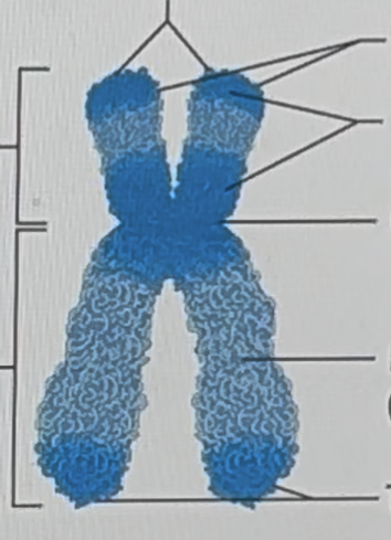
Six
P short arm
q long arm
Telomeres
Centromere
Heterochromatin (dark)
Euchromatin (light)
Centromere
Largest constriction of the chromosomes and where spindle fibers attached
Bases that form the centromere are repeats of a 171-base DNA SEQUENCE
Replicated at the end of S PHASE.
Facilitated by centromere protein A (CENP-A) is passed to next generation
Example of an EPIGENETIC CHANGE
Subtelomere
Chromosome region between the centromere and telomeres
Consist of 8000 to 300,000 bases
Near telomere the repeats are similar to the telomere sequence
Contains at least 500 protein encoding genes
About 50% are multigene families that include pseudogenes
Karyotype
A lab test where you need to get the chromosome of the cell of the patient to assess its quality.
A chromosome chart
Major clinical tool
That displays chromosomes arranged by size and structure
Humans have _____ chromosomes types
24 chromosomes
Germ cell or sex chromosomes are _______ and _______.
X and Y.
Autosomes are numbered _____ by size.
1-22
Karyotype benefits:
Confirms clinical diagnosis
Reveal effects of environmental toxins (abnormal chromosome due to triggers)
Clarify evolutionary relationships
Centromere positions:
Metacentric
Submetacentric
Acrocentric
Telocentric
In visualizing chromosomes, tissue is obtained from person: what are those:
Fetal tissue
Amniocentesis
Chorionic villi sampling
Fetal cell sorting
Chromosome microarray analysis
Adult tissue
White blood cells
Skin-like cells from cheek swab
Generally, in karyotyping, once chromosomes are extracted what is done next to visualize chromosomes?
Stained with a combination of dyes and DNA probes.
Amniocentesis
A procedure to get a sample of amniotic fluid
Detects about 1000 of the more than 5000 known chromosomal and biochemical problems
Ultrasound is used to follow needle’s movement
Chorionic Villus Sampling
10th-12th week of pregnancy (Early, 3 months of pregnancy)
Chorionic villi, found in the placental tissue. (cells of the chorion are the samples)
Provide earlier results than amniocentesis but does not detect metabolic problems
Has GREATER RISK OF SPONTANEOUS ABORTIONS
Flourescence In Situ Hybrydrization (FISH)
DNA PROBES to bind the type of chromosome, they need to paired or targeted.
DNA PROBE (reagents) labeled with flourescing dye bind complementary DNA
Flourescent dots correspond to three copies for chromosome 21
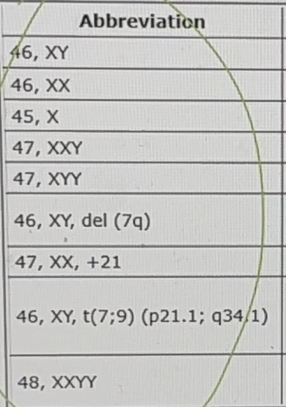
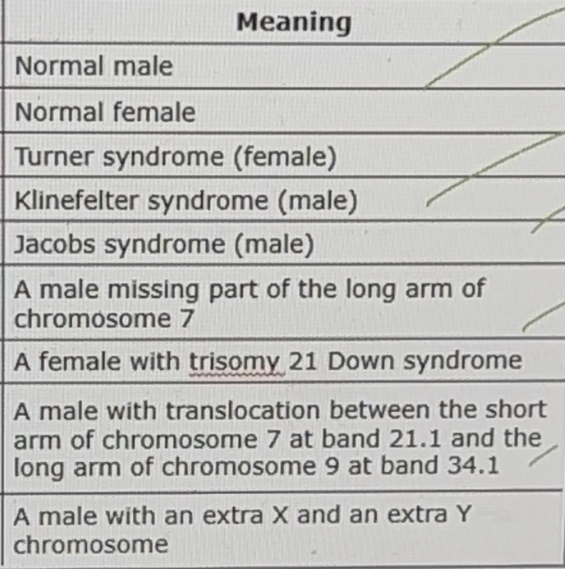
46 chromosomes: 23 pairs. _____ pair of autosome chromosome, ______ pair of sex chromosomes.
22 pairs, and 1 pair.
Ideogram
schematic chromosome map
Study the individual regions or bands located on the long arm (q) and the short arm (p)
Karyotype may be abnornal in ______ and _________?
Number
Structure
Polyploidy
Extra chromosome set
Aneuploidy
An extra or missing chromosome
Monosomy
One chromosome is absent
Trisomy
One chromosome is extra
Deletion
A part of chromosome is missing.
Duplication
Part of a chromosome is present twice.
Translocation
Two chromosomes join the long arms or exchange parts.
Inversion
A segment of chromosome is reversed.
Isochromosome
A chromosome with identical arms.
Ring chromosome
A chromosome that forms a ring due to deletion in telomeres, which causes ends to adhere.
Chromothripsis
One or more chromosome shatters.
Maternal serum markers for Trisomy 21
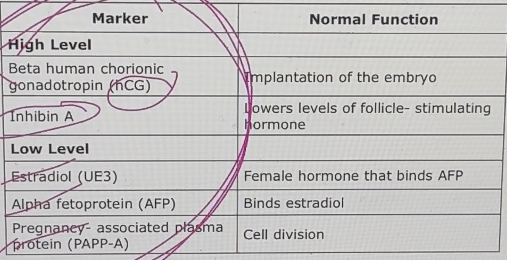
Cell - Free DNA testing
This looks at the maternal blood pieces of DNA from the fetus.
up to 20% of these pieces comes from the placenta, and thus represents the fetal genome.
Testing DNA can detect certain fetal chromosomal abnormalities like some of the trisomy conditions.
The test can be performed at 10 weeks into pregnancy.
Indirect Detection of chromosomes:
Maternal Serum Screening
Cell - Free DNA testing