2130 unit 6
1/140
There's no tags or description
Looks like no tags are added yet.
Name | Mastery | Learn | Test | Matching | Spaced | Call with Kai | Chat |
|---|
No analytics yet
Send a link to your students to track their progress
141 Terms
What are 6 basic functions of the respiratory system?
Gas exchange (simple diffusion)
O2 diffuse out → pulmonary arteries
Co2 diffuse out pulmonary capillaries → lungs
regulation blood pH
speech
host defense
trapping and dissolving small blood clots
changing chemical messenger [] s
lungs
soft, light & elastic
take in & fill air
in thoracic cavity
divided into lobes → R = 3, L = 2
How many lobes does each lung have?
R → 3 (superior, middle, inferior)
L → 2 (superior, inferior)
L smaller to accommodate heart positing
lobes get indpt supplies of air from bronchi → split into aveolis
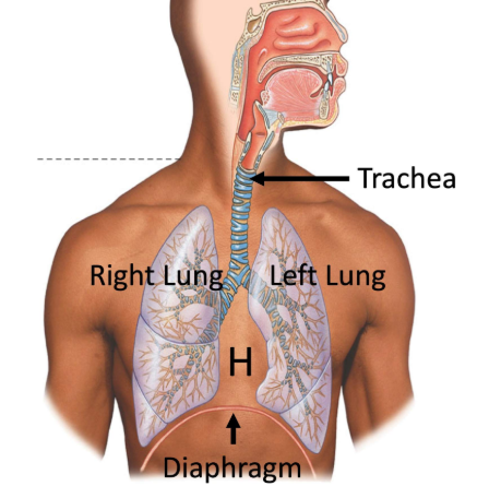
lung anatomy
in thoracic cavity → under ribs & separated by diaphragm (away from abdominal cavity)
TRACHEA
#1 airway bring air from mouth & nose
cartilage + smooth muscle
divided into bronchi
BRONCHI
large airways, subdivided into 1st, 2nd, 3rd, etc.
smooth muscle + cartilage
BRONCHOLES
X cartilage, only smooth muscle
sub divide into further terminal bronchioles
AVELOLI (gas exchange)
alveolis
LOTS of capillaries 4 gas exchange & respiration occur
INHALE → O2 pass & absorbed by blood cells in capillaries
EXHALE → CO2 waste sent in bloodstream pass thru alveolar walls
respiration
exchange of inhale & exhale w/ gas exchange
occur w/ movement of diaphragm & intercostal muscles (in btwn rib bones)
what are the different areas of the lungs?
(1) conducting zone
(2) respiratory zone
gas exchange
What is the path of the airways?
Trachea --> primary bronchi --> secondary bronchi --> tertiary bronchi --> terminal bronchioles --> respiratory bronchioles --> alveoli
What anatomical feature disappears from the conducting to respiratory zone?
Cartilage.
What feature makes terminal and respiratory bronchioles different?
Respiratory bronchioles have the odd alveolus attached to them.
What is the pathway of the pulmonary circuit?
Right ventricle --> pulmonary arteries --> pulmonary arterioles --> pulmonary capillaries (gas exchange with alveoli) --> pulmonary venules --> pulmonary veins --> left atria
What are two functions of the conducting zone?
Conducting air and preventing illness.
What is the function of the respiratory zone?
Gas exchange.
What two airway structures are found in the respiratory zone?
Respiratory bronchioles and alveoli.
What method of transport does oxygen and carbon dioxide move across the BGB?
Simple diffusion
What direction does oxygen move at the BGB?
From the alveoli to capillaries.
What direction does carbon dioxide move at the BGB?
From the capillaries to alveoli.
What is pulmonary ventilation (VE)?
The volume of air that enters the lungs.
What is tidal volume (VT)?
The volume of air that enters the lungs in a certain breath.
What is dead space ventilation (Vd)?
The volume of air remaining in the conducting zone.
How is pulmonary ventilation calculated?
VE = VT x respiratory rate
How is body weight related to dead space ventilation?
Each lb of weight is about 1 mL of dead space ventilation.
How is dead space ventilation calculated?
Vd = BW x respiratory rate
What is alveolar ventilation (VA)?
The volume of air that reaches the respiratory zone (alveoli).
How is alveolar ventilation calculated?
VA = VE - Vd = (VT x RR) - (BW x RR)
What are the two pleural membranes?
Visceral and parietal.
Where is the parietal pleura found?
Adhered to the top of the diaphragm and the inside of the rib cage.
Where is the visceral pleura found?
Adhered to the lungs and continuous with the parietal pleura.
What is the intrapleural space?
The fluid filled space in between the two pleura.
What is intrapulmonary pressure?
The pressure of air found inside the lungs.
What happens to the intrapulmonary pressure during inhalation at rest?
The intrapulmonary pressure is lower than atmospheric pressure (760 mmHg).
What happens to the intrapulmonary pressure during exhalation at rest?
The intrapulmonary pressure is higher than atmospheric pressure (760 mmHg).
Which muscles are activated during inhalation and exhalation at rest?
External intercostals and diaphragm.
What do the external intercostals do during inhalation at rest?
Contract and move the ribs up and out.
What does the diaphragm do during inhalation at rest?
Contracts and moves itself down.
What do the external intercostals do during exhalation at rest?
Relax and move the ribs down and in.
What does the diaphragm do during exhalation at rest?
Relaxes and moves itself up.
What kind of process is exhalation at rest?
A passive process.
How does inhalation during exercise differ than during rest?
There is a bigger contraction and change in diaphragm movement as well as more contractions.
What five muscles are involved in exhalation during exercise?
Diaphragm, external intercostals, internal intercostals, obliques, and rectus abdominus.
What do the internal intercostals do during exhalation during exercise?
Contract and move the ribs down and in.
What do the obliques and rectus abdominis do during exhalation during exercise?
Contract to force the diaphragm back up.
What kind of process is exhalation during exercise using the internal intercostals, obliques, and rectus abdominus?
An active process.
What is transpulmonary pressure?
The difference between pressure inside the lungs and inside the intrapleural space.
How is transpulmonary pressure calculated?
Intrapulmonary pressure - intrapleural pressure
What happens when transpulmonary pressure becomes zero?
The lungs collapse.
What is pneumothorax?
The collapse of the lungs.
What would happen if the parietal pleura was punctured?
Atmospheric air would rush into the intrapleural space, making its pressure equal to the intrapulmonary pressure and the transpulmonary pressure to be zero.
What would happen if the visceral pleura was punctured?
Air from the lungs would fill the intrapleural space, causing its pressure to be the same as the intrapulmonary pressure.
What occurs physically during a pneumothorax?
The lungs spring inward and the chest wall springs outward.
What two factors contribute to the recoil of the lungs?
Elastin (1/3) and surface tension (2/3).
What is recoil?
The lungs ability to deflate after being inflated and stretched.
What is elastin?
Elastic protein found in the walls of the alveoli.
What is the function of pulmonary surfactant?
Lies over the air-liquid interface of the alveoli and balances out attraction forces between water molecules. Decreases surface tension and prevents lungs from collapsing.
What is lung compliance?
The measure of the stretchability of the lungs, the easier a lung can stretch upon inhalation, the more compliant it is.
How is compliance calculated?
Change in volume / change in pressure
What do lungs with low compliance require upon inhalation?
A much larger change in intrapulmonary pressure.
What would happen to compliance if there was an increase in surface tension or elastin?
Too much surface tension and elastin --> too much recoil --> too much resistance to stretch --> lower lung compliance
What is neonatal respiratory distress syndrome?
Respiratory distress that occurs in premature infants due to a lack of mature surfactant system. This causes too much surface tension and alveolar collapse.
How is neonatal respiratory distress syndrome treated?
By administering surfactant.
How does a traditional spirometer work?
A subject inserts a mouthpiece that is attached to an inverted bell filled with air or oxygen that is suspended in water. As the subject breathes in and removes air, the bell sinks. As the subject breathes out and adds air, the bell floats.
What three lung volumes can a spirometer measure?
Tidal volume, inspiratory reserve volume, and expiratory reserve volume.
What lung volume can a spirometer not measure?
Residual volume.
What is the typical value for tidal volume?
500 mL.
What is inspiratory reserve volume?
How much extra air can be taken into the lungs after normal inhalation.
What is expiratory reserve volume?
How much extra air can be blown out of the lungs after normal exhalation.
How do inspiratory and expiratory reserve volume compare?
Inspiratory reserve volume is greater than expiratory.
What is residual volume?
The volume left over in the lungs that can't be exhaled.
How is total lung capacity calculated?
RV + ERV + TV + IRV
What is vital capacity?
The greatest volume of air that can be expelled from the lungs after taking the deepest possible breath.
How is vital capacity calculated?
ERV + TV + IRV
What is forced vital capacity?
The volume of air that can be forcibly exhaled after full inspiration.
How do vital capacity and forced vital capacity compare?
Both have the same volume but forced vital capacity is with a faster time frame.
How is FVC calculated from an FVC test?
The top of the IRV - the bottom of the ERV
What is FEV1?
The forced expiratory volume after 1 second of expiration.
How is FEV1 calculated from an FVC test?
Top of IRV - V at 1 second of expiration
What does the FEV1/FVC ratio show?
How much percent of volume is exhaled in 1 second, typically 80%.
How to obstructive and restrictive lung diseases differ?
Obstructive: affects exhalation
Restrictive: affects inhalation
How does the FEV1/FVC ratio for obstructive lung disease compare to healthy lungs?
Lower in obstructive lung disease due to slower and more difficult exhalation, affects FEV1.
How does the FEV1/FVC ratio for restrictive lung disease compare to healthy lungs?
Higher in restrictive lung disease due to more difficult inhalation, affects FVC.
What are two obstructive lung diseases?
Emphysema and asthma.
What causes emphysema?
Alveolar wall destruction due to the loss of elastin. As elastin breaks down it results in fewer, larger alveoli and makes it more difficult to recoil during exhalation to force air out.
How would emphysema affects someone's blood oxygen levels and lung compliance.
Lower blood oxygen levels (hypoxemia) and higher lung compliance.
What causes asthma?
Airway constriction and inflammation, causing them to become narrow due to smooth muscle contraction. Causes a difficulty in exhaling air quickly and hyperresponsiveness in airways.
What are some triggers for asthma?
Allergens, pollution, exercise, cold air, and viruses.
What kind of lung disease is pulmonary fibrosis?
Restrictive lung disease.
What are the characteristics of pulmonary fibrosis?
Scarring in the soft tissue of the alveoli, causing lower compliance as the scar tissue can't stretch as well. Causes the walls of alveoli to thicken and less gas exchange to occur.
What are the causes of pulmonary fibrosis?
Chronic inhalation of asbestos, coal dust, and pollution.
What is the PO2 of atmospheric air?
160 mmHg (21% x 760 mmHg)
What is the PCO2 of atmospheric air?
0.3 mmHg (0.04% x 760 mmHg)
Why do the alveoli have a different PO2 from the air inhaled?
The alveoli never completely empty so there is "stale air" with a lower PO2 and higher PCO2 than atmospheric air.
What are three ways that make the BGB so efficient for gas exchange?
Large concentration (pressure) gradient, lots of membrane area, and thin membrane thickness.
What are three characteristics of gas exchange?
Moves down pressure gradient, occurs at capillaries, and has a goal to reach equilibrium.
What are the different levels of PO2 in the body (from alveoli to pulmonary artery)?
Alveoli 100 mmHg --> pulmonary vein 100 mmHg --> systemic artery 100 mmHg --> rest tissue 40 mmHg --> systemic vein 40 mmHg --> pulmonary artery 40 mmHg
What would happen to the PO2 in the systemic arteries if alveolar ventilation decreased?
The PO2 in systemic arteries would be lower (less than 100 mmHg) because there is less fresh air entering the alveoli.
What would happen to the PO2 in the systemic arteries if alveolar ventilation increased?
The PO2 in systemic arteries would be higher (more than 100 mmHg) because there is more fresh air entering the alveoli.
What is hemoglobin?
A protein inside red blood cells responsible for carrying oxygen.
How much oxygen is carried by plasma?
1.5%
How much oxygen is carried by hemoglobin?
98.5%
What is the structure of hemoglobin?
Four globin side chains and four heme groups.