Thoracic Cage and Pectoral Girdle
 Call Kai
 Learn
Learn Practice Test
 Spaced Repetition
 Match
Match Flashcards
 Knowt Play
1/14

There's no tags or description
Looks like no tags are added yet.
Name | Mastery | Learn | Test | Matching | Spaced | Call with Kai | Chat |
|---|
No analytics yet
Send a link to your students to track their progress
15 Terms
Thoracic Cage
The sternum, 12 pairs of ribs, and thoracic vertebrae
Form peripheral boundary of thoracic cavity
Protect the heart, lungs, and great blood vessels

Sternum
includes 3 portions
manubrium, body, xiphoid process
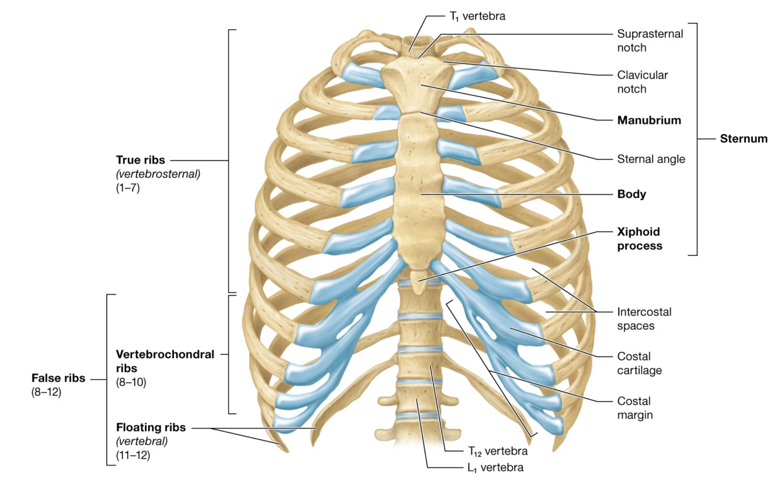
Manubrium
Superior Suprasternal Notch with lateral Clavicular Notches that articulate with clavicles
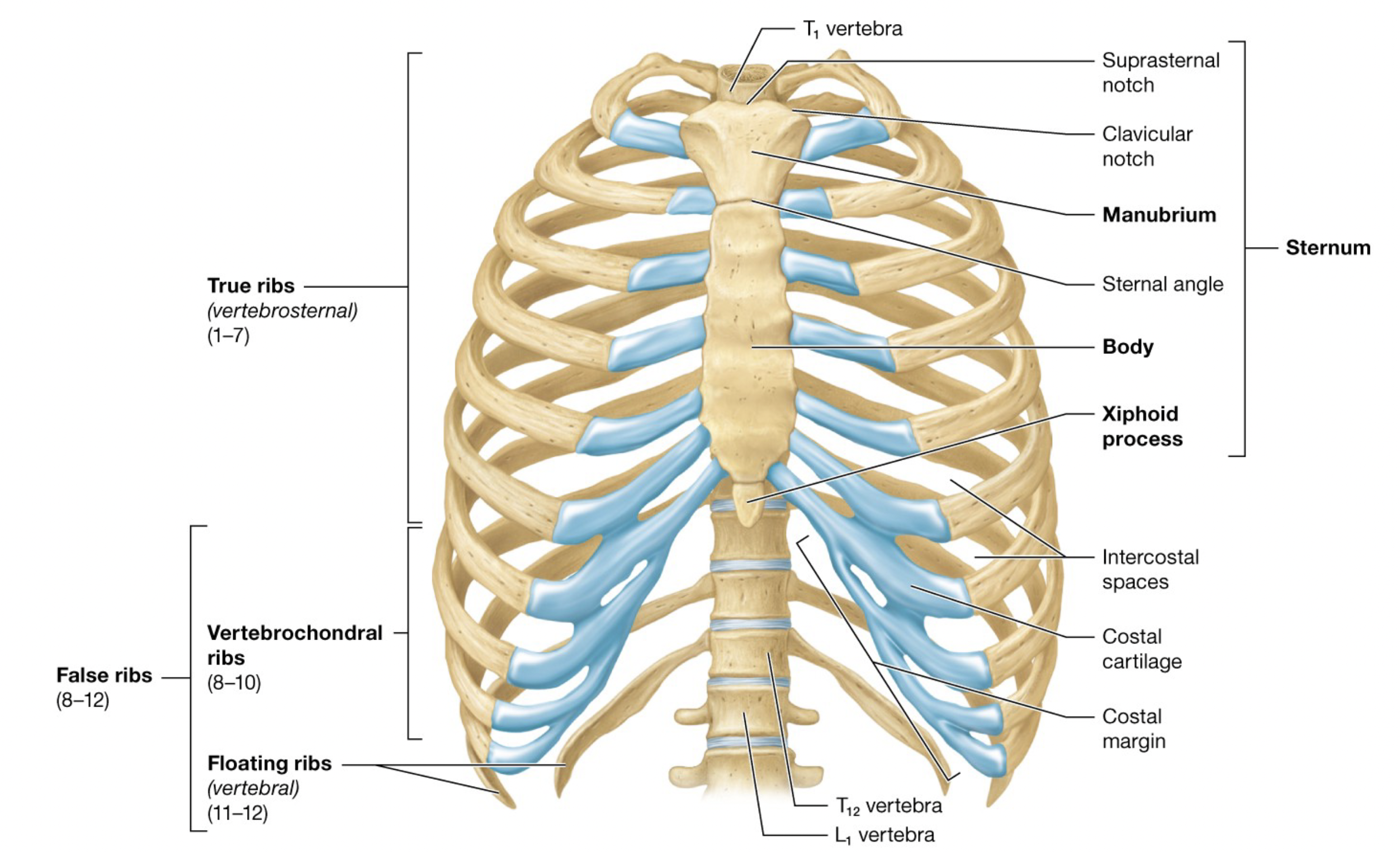
Sternum Body
Sternal Angle is the attachment site for the second set of ribs
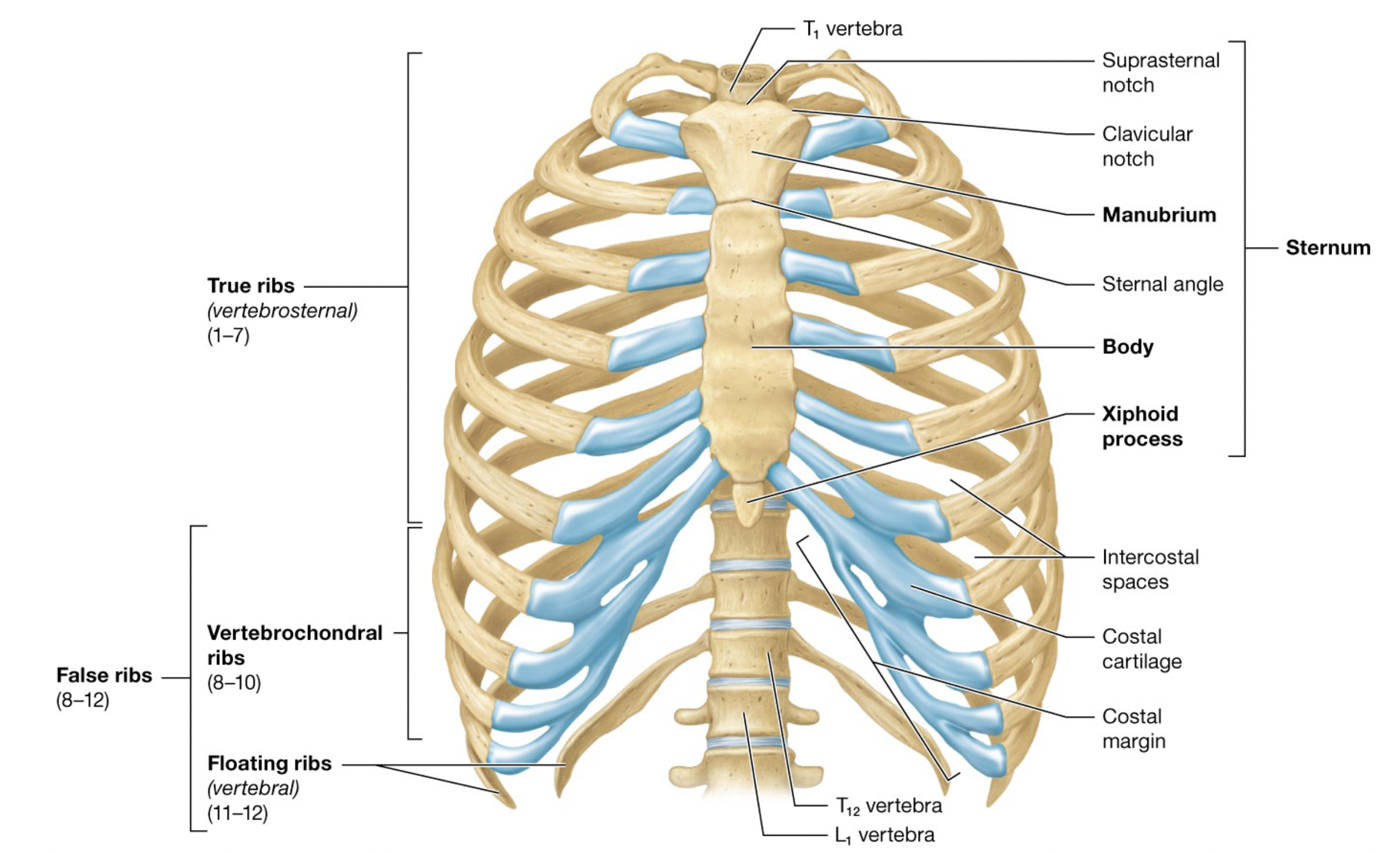
Xiphoid Process
Site of attachment for some abdominal muscles
Composed of hyaline cartilage initially, it is generally ossified by age 40

Ribs
Includes 12 pairs of ribs and Costal Cartilages, made of hyaline cartilage which provides flexibility
Spaces between ribs are the Intercostal Spaces
True ribs and false ribs

True (Vertebrosternal) Ribs
ribs 1-7
Attach to the sternum via their own costal cartilage

False Ribs
ribs 8-12
Do not attach to the sternum directly
includes vertebrochondral ribs and floating (vertebral) ribs
Vertebrochondral ribs
ribs 8-10
Costal cartilages attach to the cartilage of rib 7 as the Costal Margin
Floating (Vertebral) Ribs
ribs 11-12
do not attach to the sternum

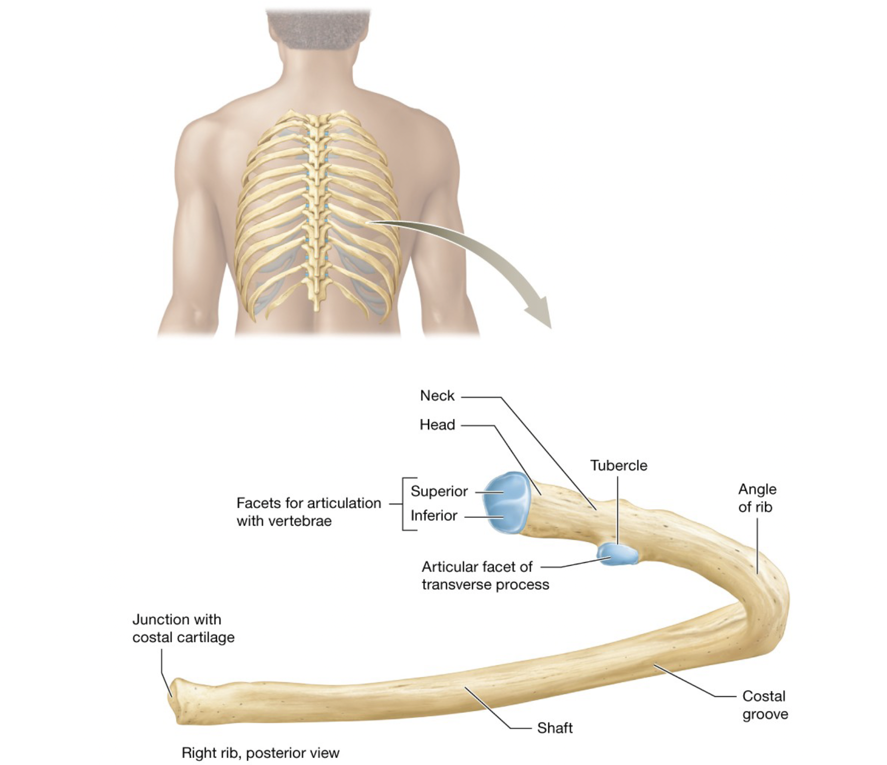
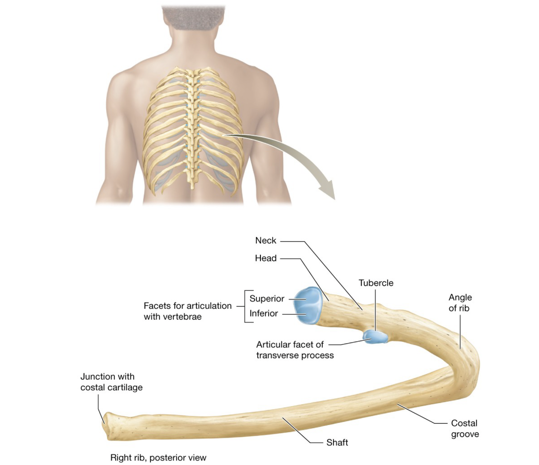
Structure of Ribs
– Rounded Head articulates with the bodies of thoracic vertebrae through Inferior and Superior Articular Facets
– The head connects laterally to a Neck, then a Tubercle, that articulates with the transverse process of the vertebra
– Rib curves at an Angle, into the Shaft (Body) with an internal costal groove for blood vessels and nerves
▪ Ribs 1–10 have square ends for costal cartilage
▪ Ribs 1 and 10–12 lack separate facets because they only attach to the body of 1 thoracic vertebra
▪ Floating ribs lack tubercles

Cardiopulmonary Resuscitation (CPR)
Life-saving technique administered when an individual’s heart has stopped functional contractions and/or breathing has stopped
CPR may restore circulation to the body by the application of repeated compressions to the chest over the sternum
Hands must be placed over the center of the sternum, and not over the xiphoid process, which may break and damage underlying organs, particularly the liver
CPR may still break the sternum and ribs, especially in elderly individuals with low bone mass
Pectoral Girdle
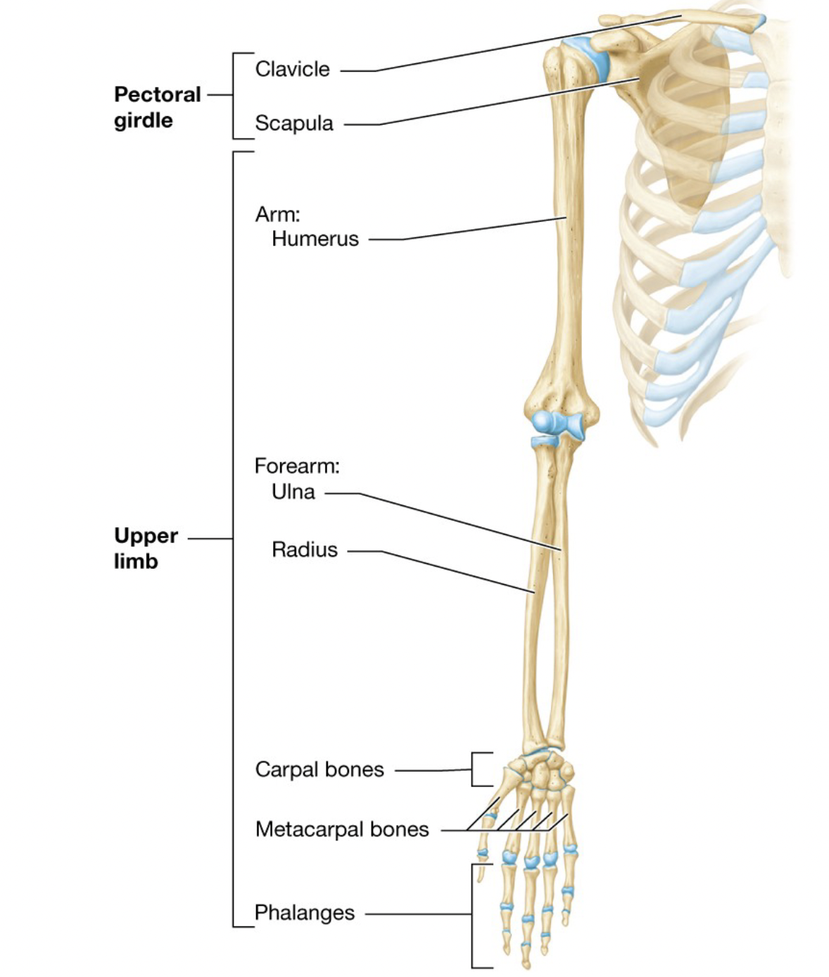
Consists of two bones, the Clavicle and Scapula, that support the upper limb
Clavicle
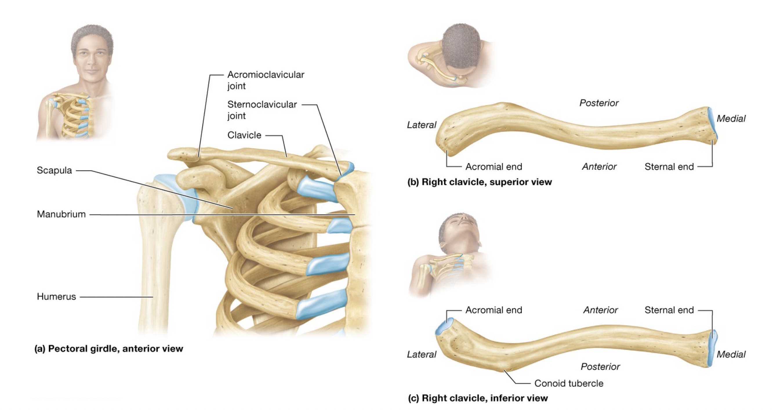
S-shaped bone
Medial Sternal End articulates with the manubrium forming the sternoclavicular joint
Lateral Acromial End articulates with the acromion process of the scapula forming the acromioclavicular joint
Conoid Tubercle is the site of ligament attachment
Functions like a brace for the upper limb so that it rests laterally to the trunk
Scapula
Sits on the posterosuperior rib cage
Body has Medial, Lateral, and Superior Borders and forms superior, inferior, and lateral angles
Coracoid Process sits over the Subscapular Fossa
Laterally, the Glenoid Cavity is visible
The posterior Spine separates the superior Supraspinous Fossa from the Infraspinous Fossa and terminates in the Acromion, which forms the acromioclavicular (AC) joint
– Injuries to the A C joint are common and are referred to as a separated shoulder
