BIOS 372 - UNIT 1
 Call Kai
 Learn
Learn Practice Test
 Spaced Repetition
 Match
Match Flashcards
 Knowt Play
1/70

There's no tags or description
Looks like no tags are added yet.
Name | Mastery | Learn | Test | Matching | Spaced | Call with Kai | Chat |
|---|
No analytics yet
Send a link to your students to track their progress
71 Terms
4 different types of pathogens
bacteria, viruses, fungi, and parasites
Why is sanitation and clean water important to prevent infection?
Dirty water can cause chronic, low grade gut inflammation and reduce the efficacy of oral vaccines
How does nutrition fuel the immune system?
Vitamin A = mucosal architect, Proteins build antibodies, Micronutrients aid in catalysis
Innate vs. Adaptive immune system
Innate - nonspecific, fast-acting
Adaptive - specific to antigen, slow-acting, retains memory of infection
Humoral immunity - B cells produce antibodies
Cell-mediated immunity - T cells kill infected cells or recruit other cells to bolster immune response
Active vs. Passive immunity
Active - individual produces their own antibodies after pathogen exposure
Passive - individual receives antibodies from another source
Types of immune dysfunction
Hypersensitivity (e.g. allergies, asthma)
Autoimmune (body attacks self)
Immunodeficiency (weakened immune system)
Primary (genetic)
Secondary (acquired)
Immune imbalance (uncontrolled inflammation)
What cell types are in the myeloid vs. lymphoid lineages?
Myeloid lineage: RBCs, platelets, monocytes/macrophages, granulocytes, dendritic cells
Lymphoid lineage: T cells, B cells, innate lymphoid cells, dendritic cells
Key transcription factors in hematopoiesis
GATA 1 - promotes erythrocyte (RBC) and megakaryocyte (platelet) development
PU.1 - initiates myeloid lineage
IKAROS - initiates lymphoid lineage
Neutrophils
Phagocytic
Release AMPs to destroy bacterial pathogens
Promote tissue remodeling after damage (collagenase)
Most numerous WBC
Eosinophils
Phagocytic
Destroy large parasites
Destroys viruses via ribonucleases
Attract other WBCs by releasing cytokines and chemokines
Basophils
Target parasites
Modulate adaptive immune response via release of cytokines
Promote vasodilation and inflammation via release of histamine
Mast cells
Target parasites
Promote vasodilation and inflammation via release of histamine
Difference between basophils and mast cells
Mast cells reside in the body’s tissues and have a longer lifespan than basophils, which circulate in the bloodstream and have a very short lifespan
Monocytes
Migrate into tissues and differentiate into a diverse array of phagocytic cells
Two categories:
Inflammatory - rapidly enter tissues to respond to infection
Patrolling - crawl along inside of blood vessels to clear cellular debris and repair tissue
Macrophages
Can form from monocytes or progenitor cells
Specialize according to the tissue they reside in
Express receptors for antibodies
High phagocytic activity
Dendritic cells
Functions are still being clarified
Can be antigen-presenting or antigen-capturing depending on location
Two types:
Conventional dendritic cells (cDCs) - present antigens to T cells to bridge the innate and adaptive immune systems
Plasmacytoid dendritic cells (pDCs) - produce large amounts of antiviral cytokines
How do we differentiate between B and T cells?
Appear identical under a microscope - must identify pattern of proteins on cell surface (CD proteins)
B cells
Mature in bone marrow
Functions: antibody production, antigen presentation, immunological memory
Types of B cells
Naive B cells, plasma cells, memory B cells
T cells
Originate in bone marrow, mature in thymus
Functions: kill infected cells, coordinate other immune cells, immunological memory
Types of T cells
Helper T cells (CD4, recognizes antigen-MHC II)
Coordinate other immune cells
4 types: Th1, Th2, Th17, Thf
Cytotoxic T cells (CD8, recognizes antigen-MHC I)
Kill infected cells
Regulatory T cells
Suppress excessive immune response
Memory T cells
Innate lymphoid cells
Lack antigen specific receptors
Three subtypes based on varying cytokine secretion:
ILC1 (includes NK cells)
Defends against intracellular pathogens, tumor surveillance
Secretes IFN-y
Similar to Th1
ILC2
Responds to parasites and allergens, repairs tissue
Secretes IL-4, IL-5, IL-13
Similar to Th2
ILC3
Responds to extracellular bacteria and fungi, maintains gut barrier integrity
Secretes IL-17, IL-22
Similar to Th17
Functions of the lymphatic system
Keeps fluid levels balanced
Transports immune cells
Monitors body for potential threats (pathogen detection occurs at lymph nodes, where immune cells reside)
How does the lymphatic system keep fluid levels balanced?
As blood moves through the circulatory system, blood capillaries leak fluid
Most interstitial fluid is pulled back into blood capillaries, remaining fluid is absorbed by lymphatic capillaries and called lymph
Lymph cannot get out once it is in (flaps of endothelial cells snap shut)
Movement of lymph
Lymphatic capillary → Vessel → Trunk → Duct → Circulatory system
Uneven drainage into right duct (right upper quadrant of body) and thoracic duct (rest of body)
Powered by:
Skeletal muscle contractions
Pressure changes from breathing
Valves prevent backflow
Primary lymphoid organs
Include bone marrow and thymus
Boot camps where immune cells develop and learn the rules (attack invaders, but don’t touch the body’s cells)
Secondary lymphoid organs
Include lymph nodes and spleen
Filter stations, where lymph and blood are exposed to immune cells that respond to potential pathogens
Components of bone marrow
Specialized cells create a niche (highly organized environment) that influences blood cell differentiation:
Perivascular cells - release cytokines, growth factors, display surface molecules that influence stem cell behavior
Osteoblasts provide a niche for developing B cells
Structure of thymus
Organized into lobes, each with specialized microenvironments that guide T cell development
Microenvironments:
Cortex: densely populated with immature T cells
Medulla: sparsely populated with mature T cells
Corticomedullary junction: entry and exit point for T cells as they move to and from the bloodstream
Main vessels that serve the lymph nodes
Afferent lymphatic vessels: entrance for lymph, antigens, some immune cells
High endothelial venules (HEV): site where naive lymphocytes circulating in the blood enter lymph node
Efferent lymphatic vessels: carry cells out of lymph nodes
Three main regions of lymph nodes
Cortex: B cells reside in follicles and germinal centers, with macrophages and dendritic cells helping trap antigens
Paracortex: rich in T cells and antigen presenting cells
Medulla: contains antibody producing plasma cells, exit point for cells via the efferent lymphatic vessel
Flow of blood through spleen
Splenic artery → Arterioles → Vascular sinusoids (where immune cells survey blood for pathogens) → Splenic vein
Structure of spleen
Red pulp: rich in RBCs, surrounds sinusoids and removes old RBCs
White pulp: forms periarteriolar lymphoid sheath (PALS) around the arterioles, populated by T cells
Contains lymphoid follicles, which are rich in B cells
Marginal zone: boundary between red and white pulp, contains specialized macrophages and B cells, traps antigens
What are MALT?
Mucosa Associated Lymphoid Tissue
Contain secondary lymphoid tissue (e.g. T cell zones and lymphoid follicles) outside the lymph nodes and spleen
Found in barrier tissues (e.g. skin, mucosal membranes of the digestive, respiratory, urogenital tracts) that represent the main entry points for pathogens
What are tertiary lymphoid organs?
Form in non-lymphoid tissues in response to chronic inflammation
Resemble secondary lymphoid organs in structure and function
Can be simple clusters or highly organized with B/T compartments, germinal centers, lymphatic vessels, and HEV
Enable localized immune responses at site of injury
Sequence of development from HSCs
Long term HSCs → Short term HSCs → Multipotent progenitors → Myeloid/Lymphoid lineages
Physical barriers of the innate immune system
Skin
Covers outside of body
Contains multiple layers of epithelial cells
Mucous membranes of the respiratory, digestive, and urogenital tracts
Single layer of epithelial cells covered with mucus
Can be enhanced by chemical additions like low pH, AMPs, mucus.
Commensal microbes functions
Use up metabolic resources/occupy binding sites to prevent other organisms from inhabiting the human body
Produce AMPs to kill invading microbes
Aid in digestion
Synthesize vitamins and neurotransmitters
Structure of the skin
Epidermis
Stratum basale (innermost layer): source of dividing keratinocytes
Stratum spinosum: cells appear spiny due to keratin deposits, houses Langerhans cells
Stratum granulosum
Stratum corneum (outermost layer): sheds layers of dead cells, keratin deposits create dry and impermeable surfaces
Dermis
Contains structural molecules like elastin and collagen
Contains macrophages and mast cells
Contains sweat glands, sebaceous glands
Chemical barriers of the skin
Sweat glands: release acidic electrolytes, flush out dirt/debris, produce dermcidin (AMP)
Sebaceous glands: release sebum, maintain acidic environment of skin, deliver AMPs
Keratinocytes and other immune cells produce defensins and cathelicidins (AMPs)
Lacrimal glands: rinse the eyes, loaded with lysozyme
Structure of mucous membranes
Can only be a single layer of epithelial cells (need to be thin in order to also handle the functions of absorption and secretion)
Coated in mucus - a sticky, protein rich layer made by goblet cells
Traps pathogens
Contains commensal microbes that produce AMPs and compete with invaders
How does the structure of mucus vary by location?
Single, thin layer in small intestine to avoid blocking absorption of nutrients
Stomach and colon have two layers:
Dense inner layer for protection against pathogen invasion
Loose outer layer that provides nutrients and attachment sites for commensal microbes
What is the complement system?
2nd line of defense in the innate immune system
Collection of proteins produced by the liver, circulate in an inactive form, unless activated by a pathogen or antigen-antibody complex
How is the complement system activated?
Signaling cascade
Signal is amplified with each step in the pathway

Outcomes of the complement system
C5b → creates membrane attack complex (MAC), which creates a pore in pathogen cell membrane, causing cellular contents to leak out
C3b → opsonizes (coats) pathogen to make phagocytosis easier
Activates itself, generating a positive feedback loop that amplifies the complement response
C3a + C5a → acts as chemoattractants that recruit neutrophils and monocytes
How does the complement system avoid destroying host tissues?
Classical pathway and lectin pathway are only initiated by binding to markers on the surface of a pathogen
C4b can bind to surface proteins or carbohydrates indiscriminately, but if it doesn’t bind to the pathogen surface, it is inactivated
What happens to complement proteins that don’t become a part of the convertase enzymes?
Bind to receptors on phagocytes to enhance phagocytosis
Bind to receptors on endothelial cells → increase production of adherins → cause immune cells to stick to blood vessels, also increase permeability of vessels to transport immune cells to infection site
Bind to receptors on mast cells to promote release of inflammatory molecules
Convergence point for complement pathways
Creation of C5 convertase, which cleaves C5 into C5a and C5b
Final steps involve C6 - C9
Ultimately results in formation of membrane attack complex

Classical Pathway

Lectin Pathway
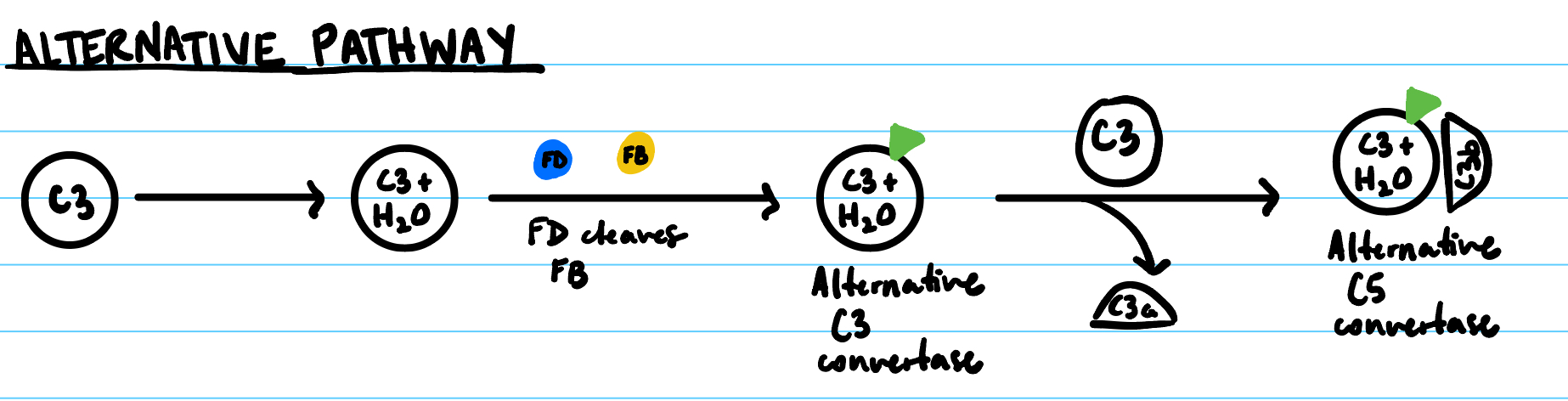
Alternative Pathway
What are cytokines?
Small proteins, released by both immune and non-immune cells
Act as signals to inform cells regarding the location, strength of the immune response
IL1 family of cytokines
Secreted by dendritic cells, monocytes, macrophages
Proinflammatory, regulate inflammation
Class 1 (hematopoietin cytokine)
Secreted by a diverse array of cells
Regulates hematopoiesis and antibody secretion
Class 2 (interferon cytokines)
Secreted by activated macrophages, DCs, activated T cells, NK cells, virally infected cells
Antiviral, immune modulation
Tumor Necrosis Factor
Secreted by activated macrophages, non-immune cells
Immune system development, effector function, homeostasis
IL17 family of cytokines
Secreted by activated T cells
Proinflammatory, promote neutrophil accumulation
Chemokines
Direct migration of other immune cells (function as chemoattractants) by acting on G protein coupled receptors
Direct other cells via concentration gradients (concentration of chemokines is highest at the site of infection)
5 properties of cytokines
Pleiotropy: single cytokine can act on multiple cell types and have different effects
Redundancy: multiple cytokines perform the same function
Ensures immune system can adapt if one pathway is disrupted
Synergy: two or more cytokines combine their actions to enhance an immune response
Antagonism: one cytokine can inhibit the effect of another
Cascade induction: one cell releases cytokines to stimulate a target cell to release additional cytokines, creates a domino effect to amplify the immune response
JAK-STAT pathway
Intracellular signaling pathway activated by cytokines
Ligand binds to cell surface receptor
JAK adds Pi to receptor
Two STAT proteins bind to the Pi on the receptor and are thus phosphorylated
STAT proteins dimerize, enter nucleus to act as transcription factors
MAP-K pathway
Intracellular signaling pathway activated by cytokines
Cytokine binds to its receptor, receptor is phosphorylated and proteins are recruited → Ras is activated → Raf (MAPKKK) is activated → MEK (MAPKK) is activated → Erk (MAPK) is activated → Erk enters nucleus to phosphorylate transcriptional regulators of cell proliferation, differentiation, and survival
NFkB signaling pathway
Intracellular signaling pathway activated by cytokines
Cytokine binds to receptor → Adaptor proteins bind to receptor to form a signaling complex → Signaling complex activates IKK complex → IKK phosphorylates IkB, marking it for degradation and releasing active NFkB → Active NFkB translocates to nucleus
GPCR pathway
Intracellular signaling pathway activated by cytokines
Ligand binds to GPCR, which undergoes a conformational change
Associated G protein exchanges GDP for GTP on alpha subunit
Alpha-GTP subunit dissociates from GBy dimer
Ga and GBy interact with downstream effectors
Ga hydrolyzes GTP to GDP, Ga reassociates with GBy to terminate the signal
What are PAMPs?
Pathogen Associated Molecular Patterns
Include molecules shared by many microbes, but not produced by host cells
Allow innate cells to quickly recognize microbes as a non-self threat
What are DAMPs?
Damage Associated Molecular Patterns
Found in the intracellular compartments of host cells and released due to damage caused by infection
What are DAMPs and PAMPs recognized by?
Pattern Recognition Receptors (PRRs) on immune cells
What are TLRs?
Toll-Like Receptors are a type of PRR that can detect many different kinds of pathogens at the cell surface or intracellularly (in endosomes/lysosomes)
Ligand binding domain contains LRRs (leucine rich repeats)
Generalized signaling pathway: TLR binds ligand → Receptor dimerization → Recruit adaptor proteins → Activation of kinase cascade → Nuclear translocation
Different types of PRRs
TLRs - recognize extracellular pathogens on cell surface/extracellular pathogens that have been endocytosed
Localized to cell surface and endosomes
CLRs - recognize only extracellular pathogens (fungal glucans)
Localized to cell surface
RLRs - recognize only intracellular pathogens (viral dsRNA)
Localized to mitochondria membrane
STING & cGAS - recognize only intracellular pathogens (DNA)
Localized to endoplasmic reticulum
ALRs - recognize only intracellular pathogens (DNA & RNA)
Localized to cytoplasm
NLRs - recognize extracellular pathogens that have been endocytosed
Localized to cytoplasm
What is extravasation?
WBCs leave blood vessels to fight infection at the site of injury
Residents DCs and macrophages release cytokines that promote vasodilation
Endothelial cells produce adherins that tether WBCs to blood vessel walls, slowing the cells to initiate rolling
WBCs squeeze through gaps between endothelial cells and leave the blood vessel in a process called diapedesis
Process of phagocytosis
PRR binds PAMP or opsonin receptors detect opsonin-coated extracellular pathogen → internalization creates phagosome → phagosome fuses with lysosome → microbe is killed → contents are released or displayed on surface
Process of autophagy
Isolation membrane forms around intracellular pathogen to form an autophagosome → fusion with lysosome → breakdown of autolysosome contents
Apoptosis vs. Necrosis
Apoptosis - noninflammatory, controlled cell death
Cell contents are neatly enclosed and disposed of through phagocytic digestion
Necrosis - inflammatory, uncontrolled cell death
Abrupt loss of membrane integrity and discharge of cell contents into the environment
What is NETosis?
A form of neutrophil cell death that also serves to trap and kill pathogens
Neutrophils are activated by the binding of cytokines/other chemical signals → NADPH oxidase produces ROS and intracellular membranes break down → Chromatin decondenses, AMPs are released from granules → Plasma membrane ruptures and releases a web of DNA and AMPs into the extracellular space